Ultrasound Elastography in the Evaluation of Venous Thrombosis - try to apply in clinical practice презентация

What is Elastography? Elastography is an imaging technique to measure the stiffness of tissues Images are acquired before and after soft compression of tissues and the deformation is evaluated

Слайд 1Ultrasound Elastography in the Evaluation of Venous Thrombosis - try to

apply in clinical practice

TADEUSZ WILKOSZ, TOMASZ DRĄŻKIEWICZ, GRZEGORZ LEWIŃSKI
JOHN PAUL II HOSPITAL, CRACOW - POLAND


VARADY’S 30TH INTERNATIONAL WORKSHOP FOR PHLEBOLOGY, LYMPHOLOGY AND ANGIOLOGY FRANKFURT 17-18.04.2015
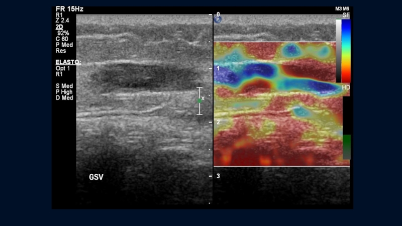
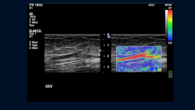
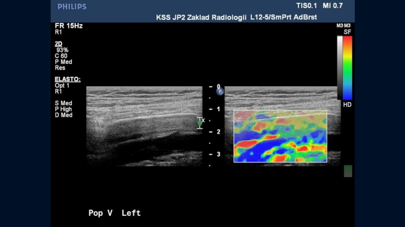
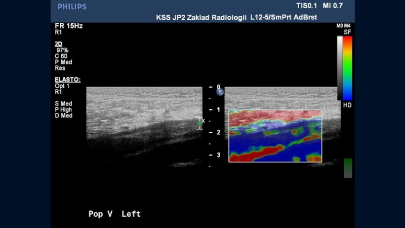
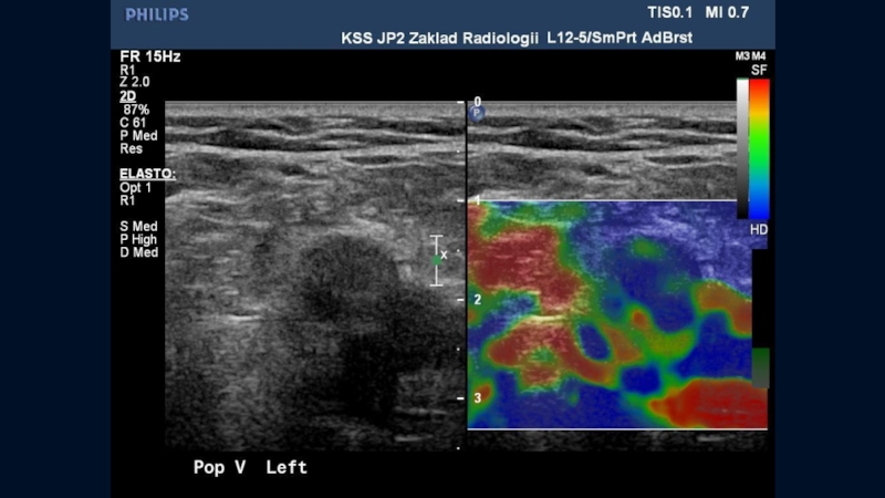
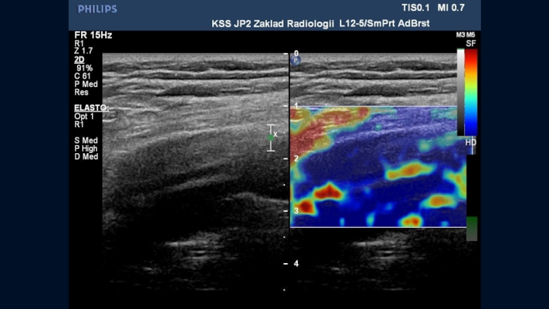

Слайд 2What is Elastography?

Elastography is an imaging technique to measure the stiffness

of tissues

Images are acquired before and after soft compression of tissues and the deformation is evaluated

Initially elastography used manual compression and was only qualitative, now some methods appears to apply a non operator dependant compression


Слайд 3Elastography was developed first in the US field

Three step approach:

1.

Organs mechanically stressed by either external or internal forces.

2. Measurement of tissues movement induced

3. Qualitative or quantitative evaluation of tissue elastic properties from the measured displacement of tissues.

Слайд 4Several Approaches

Manual compression by operator using the transducer (static

elastography - strain elastography)

Organ compression by heartbeat or vascular pulsations

Push pulse waves compression

Supersonic shear waves


Слайд 5Sonoelastography
Strain elastography (manual mechanical compression of transducer) is a ultrasonic (US)

technique invented by Hitachi Medical and Tsukuba University in Japan
Dynamic Elastography ( Shear Wave Elastography) introduced by SuperSonic Imagine in France

Слайд 6Strain elastography

technically simpler option is available in multiple devices, many

manufacturers of ultrasound equipment
main application in the study of pathologies of superficial structures - breast, thyroid, lymph nodes, prostate,

…but they are also successful attempts to study the structure of venous thrombosis

Слайд 7Axial and lateral deformations
after an axial constraint

STRAIN (STATIC) ELASTOGRAPHY
B-mode
Elastography

mode

Слайд 8Application of vein elastography

What is the age of the thrombus?

Whether there

are features of recanalization?

Слайд 9Mostly green coloring of fresh thrombosis
It is possible to capture the

changes from a soft thrombus in the acute phase to a hard thrombus in the chronic phase

Слайд 10First signs of recanalization, identified as areas of relatively high elasticity

of the
clot structure

Слайд 11Further extension of recanalization channel


Слайд 12 an extension of recanalization channel


Слайд 35Forehead the thrombus


Слайд 37POPLITEAL VEIN


Слайд 39Conclusions

USEG usage for dynamic monitoring of acute venous thrombosis can help

in identifying
of initial signs of thrombus recanalization

Changes in clot structure can define probable prognosis of recanalization.

Elastography would be useful for evaluating the tissue characteristics of venous thrombi, enabling selection of the treatment method and forecast of complications

Further review is desired

Слайд 40REFERENCES

1.USEFULNESS OF ULTRASONOGRAPHIC ELASTOGRAPHY FOR EVALUATION OF VENOUS THROMBOEMBOLISM
K. Shimizu,T. Tomaru,

Cardiology Center,Toho University Sakura Hospital, Sakura
Circulation Journal Vol 73, Suppl 1, 2009
2.AGING OF VENOUS THROMBOSIS - COMPARISON BETWEEN HISTOLOGICAL AND TISSUE
ELASTICITY IMAGING K.Uno, A. Tonomura, M. Yamakawa, T. Ishizu, Y. Seo, S. Honma.
T. Shiina, K. Aonuma, Circulation Journal Vol 72, Suppl 1, 2008
3.ULTRASOUND ELASTICITY IMAGING CAN DETECT HIGH RISK PATIENTS WITH DEEP VEIN THROMBOSIS WHO RESULTS IN PULMONARY EMBOLISM
H.Kato, M.Nishino, M. Hara, T. Yoshimura, D.Nakamura, Y. Lee, S. Nakatani, A. Hashimoto, K. Yamagami, Y. Egami, R. Shutta, H. Yamaguchi, J.Tanouchi, Y. Yamada
Division of Cardiology, Osaka Rosai Hospital, Sakai, Circulation Journal Vol 73, Suppl 1, 2009
4.AGING OF VENOUS THROMBOSIS - COMPARISON BETWEEN HISTOLOGICAL AND TISSUE
ELASTICITY IMAGING
K. Uno, A. Tonomura, M. Yamakawa, T. Ishizu, Y. Seo, S. Honma, T. Shiina, K. Aonuma, Circulation Journal Vol 72, Suppl 1, 2008
5.ULTRASONIC ELASTOGRAPHY IMAGING OF DEEP VENOUS THROMBOSIS
K. Shimizu, T. Tomaru, K. Nakamura, K. Hirano, H. Noike, M. Takahashi, T. Sakurai. Department of Cardiovascularcenter, Sakura Hospital,Toho University School of Medicine, Chiba, Circulation Journal Vol 71 Suppl 1 PJ-709, 2007
6.APPLICATION OF ULTRASONIC ELASTOGRAPHY FOR EVALUATION OF DEEP VENOUS THROMBOSIS
K. Shimizu, K. Nakamura, T. Hirohashi, et al.,Circulation Journal Vol 70 Suppl 1 PE-623, 2006
7.EXPERIENCE OF USING ULTRASOUND ELASTOGRAPHY FOR DYNAMIC MONITORING OF RECANALISATION OF VENOUS THROMBOSIS OF LOWER EXTREMITIES IN ADULTS AND CHILDREN A. R. Zubarev, N. Krivosheeva, A. Demidova, I. Rychkova, E. A. Zubareva; ECR 2012
8.SONOGRAPHISCHE KLASSIFIKATION DER TIEFEN VENOSEN THROMBOSE S.Siebers, B.Geier, M.Vogt, U.Scheipers, A.Mumme, H.Ermert, Ultraschall in der Medizin, No. 25. Sept.2004, p.S35

Слайд 41Thank you for the attention


Обратная связь

Если не удалось найти и скачать презентацию, Вы можете заказать его на нашем сайте. Мы постараемся найти нужный Вам материал и отправим по электронной почте. Не стесняйтесь обращаться к нам, если у вас возникли вопросы или пожелания:

Email: Нажмите что бы посмотреть 

Что такое ThePresentation.ru?

Это сайт презентаций, докладов, проектов, шаблонов в формате PowerPoint. Мы помогаем школьникам, студентам, учителям, преподавателям хранить и обмениваться учебными материалами с другими пользователями.


Для правообладателей

Яндекс.Метрика