- Главная
- Разное
- Дизайн
- Бизнес и предпринимательство
- Аналитика
- Образование
- Развлечения
- Красота и здоровье
- Финансы
- Государство
- Путешествия
- Спорт
- Недвижимость
- Армия
- Графика
- Культурология
- Еда и кулинария
- Лингвистика
- Английский язык
- Астрономия
- Алгебра
- Биология
- География
- Детские презентации
- Информатика
- История
- Литература
- Маркетинг
- Математика
- Медицина
- Менеджмент
- Музыка
- МХК
- Немецкий язык
- ОБЖ
- Обществознание
- Окружающий мир
- Педагогика
- Русский язык
- Технология
- Физика
- Философия
- Химия
- Шаблоны, картинки для презентаций
- Экология
- Экономика
- Юриспруденция
Multiple Myeloma презентация
Содержание
- 1. Multiple Myeloma
- 2. Multiple Myeloma Definition: B-cell malignancy characterised
- 3. Multiple Myeloma = M-CRAB Monoclonal protein Calcium Renal failure Anemia Bone pain with lytic lesions
- 4. Disorders Associated with M- Protein
- 5. Multiple Myeloma Clinical forms: multiple myeloma
- 6. Multiple Myeloma Clinical manifestations are related to
- 7. Multiple Myeloma Clinical symptoms: bone pain,
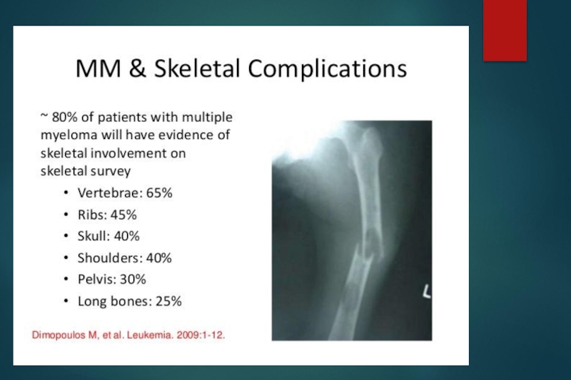
- 8. Lytic Bone Lesion
- 10. MM: PET Scan
- 11. Multiple Myeloma Laboratory tests: ESR > 100
- 13. Diagnostic Criteria for Multiple Myeloma Major criteria
- 14. Multiple Myeloma All 3 criteria must
- 15. Smoldering Multiple Myeloma SMM, Asymptomatic
- 16. Monoclonal Gammopathy of Undetermined Significance (MGUS)
- 17. Monoclonal gammopathy of undetermined significance ( MGUS)
- 18. POEMS Syndrome Osteosclerotic myeloma Polyneuropathy Organomegaly Endocrinopathy Monoclonal protein Skin changes
- 19. MM: Evaluation CBC and differential,peripheral blood smear
- 20. MM Evaluation Serum viscosity should be measured
- 21. Staging for MM International staging system (ISS) Stage I — B2M
- 22. MM Survival by ISS
- 23. Cytogenenetics, Interphase FISH Poor prognosis (median survival
- 27. MM: RISK STRATIFICATION FISH for detection of
- 28. Multiple Myeloma Poor prognosis factors cytogenetic
- 29. MM: Indications for Treatment Anemia (hemoglobin 11.5
- 30. Treatment of Multiple Myeloma Patients fit< 65
- 31. Treatment of Multiple Myeloma Conventional chemotherapy Melphlan
- 32. Treatment of Multiple Myeloma Autologous transplantation Fit
- 33. Treatment of Multiple Myeloma New methods Reduced
- 34. Treatment of Multiple Myeloma Supportive treatment biphosphonates, calcitonin recombinant erythropoietin immunoglobulins plasmapheresis radiation therapy
- 35. Monoclonal gammopathy of undetermined significance ( MGUS)
- 36. The end

Слайд 2Multiple Myeloma
Definition:
B-cell malignancy characterised by abnormal proliferation of plasma cells
able to produce a monoclonal immunoglobulin ( M protein )
Incidence:
3 - 9 cases per 100000 population / year
more frequent in elderly
modest male predominance
Incidence:
3 - 9 cases per 100000 population / year
more frequent in elderly
modest male predominance

Слайд 3Multiple Myeloma = M-CRAB
Monoclonal protein
Calcium
Renal failure
Anemia
Bone pain with lytic lesions

Слайд 4 Disorders Associated with M- Protein
Neoplastic cell proliferation
multiple myeloma
solitary plasmacytoma
Waldenstrom
macroglobulinemia, CLL
heavy chain disease
primary amyloidosis AL
Undetermined significance
monoclonal gammopathy of undetermined significance (MGUS)
Transient M protein
viral infection
post-valve replacement
Solid Malignacy
bowel cancer, breast cancer
Immune dysregulation
AIDS, old age
Chronic inflamation
heavy chain disease
primary amyloidosis AL
Undetermined significance
monoclonal gammopathy of undetermined significance (MGUS)
Transient M protein
viral infection
post-valve replacement
Solid Malignacy
bowel cancer, breast cancer
Immune dysregulation
AIDS, old age
Chronic inflamation

Слайд 5Multiple Myeloma
Clinical forms:
multiple myeloma
solitary plasmacytoma
plasma cell leukaemia
M protein:
-
is seen in 99% of cases in serum and/or urine
IgG > 50%, IgA 20-25%, IgE or IgD 1-3%
light chain 20%
- 1% of cases are nonsecretory
IgG > 50%, IgA 20-25%, IgE or IgD 1-3%
light chain 20%
- 1% of cases are nonsecretory

Слайд 6Multiple Myeloma
Clinical manifestations are related to malignant
behaviour of plasma cells
and abnormalities produced
by M protein
plasma cell proliferation:
multiple osteolytic bone lesions
hypercalcemia
bone marrow suppression ( pancytopenia )
monoclonal M protein
decreased level of normal immunoglobulins
hyper viscosity, RENAL FAILURE, amyloidosis
by M protein
plasma cell proliferation:
multiple osteolytic bone lesions
hypercalcemia
bone marrow suppression ( pancytopenia )
monoclonal M protein
decreased level of normal immunoglobulins
hyper viscosity, RENAL FAILURE, amyloidosis

Слайд 7Multiple Myeloma
Clinical symptoms:
bone pain, pathologic fractures
weakness and fatigue
serious infection
renal
failure
bleeding diathesis (hyper viscosity)
bleeding diathesis (hyper viscosity)
")


Слайд 11Multiple Myeloma
Laboratory tests:
ESR > 100
anaemia, thrombocytopenia
Rouleau in peripheral blood smears
marrow plasmacytosis
> 10 -15%
hyperproteinaemia
hypercalcemia
proteinuria
renal failure
hyperproteinaemia
hypercalcemia
proteinuria
renal failure


Слайд 13Diagnostic Criteria for Multiple Myeloma
Major criteria
I. Plasmacytoma on tissue
biopsy
II. Bone marrow plasma cell > 30%
III. Monoclonal M spike on electrophoresis IgG > 3,5g/dl,
IgA > 2g/dl, light chain > 1g/dl in 24h urine sample
Minor criteria
a. Bone marrow plasma cells 10-30%
b. M spike but less than above
c. Lytic bone lesions
d. Normal IgM < 50mg, IgA < 100mg, IgG < 600mg/dl
II. Bone marrow plasma cell > 30%
III. Monoclonal M spike on electrophoresis IgG > 3,5g/dl,
IgA > 2g/dl, light chain > 1g/dl in 24h urine sample
Minor criteria
a. Bone marrow plasma cells 10-30%
b. M spike but less than above
c. Lytic bone lesions
d. Normal IgM < 50mg, IgA < 100mg, IgG < 600mg/dl
I + b; I + c; I + d
II + b; II + c; II + d
III + a; III + c; III + d
a + b +c
a + b+ d

Слайд 14Multiple Myeloma
All 3 criteria must be met (except unsecretory):
Presence of
a serum or urinary monoclonal protein
Presence of clonal plasma cells in the bone marrow or a plasmacytoma
Presence of end organ damage felt related to the plasma cell dyscrasia, such as:
Increased calcium concentration
Lytic bone lesions
Anemia
Renal failure
Presence of clonal plasma cells in the bone marrow or a plasmacytoma
Presence of end organ damage felt related to the plasma cell dyscrasia, such as:
Increased calcium concentration
Lytic bone lesions
Anemia
Renal failure
:Presence of a serum or urinary")
Слайд 15 Smoldering Multiple Myeloma
SMM, Asymptomatic
Both criteria must be met:
Serum monoclonal
protein ≥3 g/dL and/or bone marrow plasma cells ≥10 percent
No end organ damage related to plasma cell dyscrasia
No end organ damage related to plasma cell dyscrasia

Слайд 16Monoclonal Gammopathy of Undetermined Significance (MGUS)
All 3 criteria must be met:
Serum
monoclonal protein <3 g/dL
Bone marrow plasma cells <10 percent
No end organ damage related to plasma cell dyscrasia or a related B cell lymphoproliferative disorder
Bone marrow plasma cells <10 percent
No end organ damage related to plasma cell dyscrasia or a related B cell lymphoproliferative disorder
 All 3 criteria must be met:Serum monoclonal protein")
Слайд 17Monoclonal gammopathy of undetermined significance ( MGUS)
M protein
3% of people >
70 years
15% of people > 90 years
MGUS is diagnosed in 67% of patients with an M protein
10% of patients with MGUS develop multiple myeloma, 1% per year
15% of people > 90 years
MGUS is diagnosed in 67% of patients with an M protein
10% of patients with MGUS develop multiple myeloma, 1% per year
M protein3% of people > 70 years15% of people")
Слайд 18POEMS Syndrome
Osteosclerotic myeloma
Polyneuropathy
Organomegaly
Endocrinopathy
Monoclonal protein
Skin changes

Слайд 19MM: Evaluation
CBC and differential,peripheral blood smear
Chemistry: serum calcium, creatinine, albumin, LDH
, beta-2 microglobulin, and C-reactive protein
Serum protein electrophoresis (SPEP) + IF
Quantification of immunoglobulins
Urinalysis and a 24-hour urine collection for electrophoresis (UPEP) + IF
Serum free monoclonal light chain (FLC)
Serum protein electrophoresis (SPEP) + IF
Quantification of immunoglobulins
Urinalysis and a 24-hour urine collection for electrophoresis (UPEP) + IF
Serum free monoclonal light chain (FLC)

Слайд 20MM Evaluation
Serum viscosity should be measured if the M-protein concentration is
high
Bone marrow aspiration and biopsy with immunophenotyping, conventional cytogenetics, and fluorescence in situ hybridization (FISH)
Metastatic bone survey with plain radiographs including the humeri and femoral bones should be performed in all patients.
MRI, CT, or PET/CT
Bone marrow aspiration and biopsy with immunophenotyping, conventional cytogenetics, and fluorescence in situ hybridization (FISH)
Metastatic bone survey with plain radiographs including the humeri and femoral bones should be performed in all patients.
MRI, CT, or PET/CT

Слайд 21Staging for MM
International staging system (ISS)
Stage I — B2M
and serum albumin ≥3.5 g/dL
Stage II — neither stage I nor stage III
Stage III — B2M ≥5.5 mg/L
Median overall survival for patients with ISS stages I, II, and III are 62, 44, and 29 months
Stage II — neither stage I nor stage III
Stage III — B2M ≥5.5 mg/L
Median overall survival for patients with ISS stages I, II, and III are 62, 44, and 29 months
 Stage I — B2M")

Слайд 23Cytogenenetics, Interphase FISH
Poor prognosis (median survival 25 months): t(4;14)(p16;q32), t(14;16)(q32;q23), and
-17p13
Intermediate prognosis (median survival 42 months): -13q14
Good prognosis (median survival 50 months): all others
: t(4;14)(p16;q32), t(14;16)(q32;q23), and -17p13Intermediate prognosis (median")

Слайд 27MM: RISK STRATIFICATION
FISH for detection of t(4;14), t(14;16), and del17p13
Conventional cytogenetics
(karyotyping) for detection of del 13 or hypodiploidy
The presence of any of the above markers defines high risk myeloma, which encompasses the 25 percent of MM patients who have a median survival of approximately two years or less despite standard treatment
The presence of any of the above markers defines high risk myeloma, which encompasses the 25 percent of MM patients who have a median survival of approximately two years or less despite standard treatment
, t(14;16), and del17p13Conventional cytogenetics (karyotyping) for detection of")
Слайд 28 Multiple Myeloma
Poor prognosis factors
cytogenetic abnormalities
High β-2 microglobulin
Advanced stage
Hypercalcemia
Renal
failure
Plasma cell leukaemia
Plasma cell leukaemia

Слайд 29MM: Indications for Treatment
Anemia (hemoglobin
normal)
Hypercalcemia (serum calcium >11.5 mg/dL)
Renal insufficiency (serum creatinine>2 mg/dL)
Lytic bone lesions or severe osteopenia
Extramedullary plasmacytoma
Hypercalcemia (serum calcium >11.5 mg/dL)
Renal insufficiency (serum creatinine>2 mg/dL)
Lytic bone lesions or severe osteopenia
Extramedullary plasmacytoma
Renal insufficiency (serum creatinine>2 mg/dL)Lytic bone lesions or severe")
Слайд 30Treatment of Multiple Myeloma
Patients fit< 65 years
induction with combination
of IMIDS, cyclophosphamide, dexamethasone and velcade
High dose chemo with autologous stem cell transplantation
Patients > 65 years
conventional chemotherapy, new drugs
High dose chemo with autologous stem cell transplantation
Patients > 65 years
conventional chemotherapy, new drugs

Слайд 31Treatment of Multiple Myeloma
Conventional chemotherapy
Melphlan + Prednisone
M2 ( Vincristine, Melphalan, Cyclophosphamid,
BCNU, Prednisone)
VAD (Vincristin, Adriamycin, Dexamethasone)
Response rate 50-60% patients (CR very low)
Long term survival 5-10% patients
VAD (Vincristin, Adriamycin, Dexamethasone)
Response rate 50-60% patients (CR very low)
Long term survival 5-10% patients
VAD (Vincristin, Adriamycin,")
Слайд 32Treatment of Multiple Myeloma
Autologous transplantation
Fit patients < 65
treatment related mortality 5-10%
response
rate 80%
long term survival 40-50%
allogeneic stem cell transplantation
patients < 45-50 years with HLA-identical donor
Poor prognostic factors
treatment related mortality 40-50%
long term survival 20-30%
long term survival 40-50%
allogeneic stem cell transplantation
patients < 45-50 years with HLA-identical donor
Poor prognostic factors
treatment related mortality 40-50%
long term survival 20-30%

Слайд 33Treatment of Multiple Myeloma
New methods
Reduced intensity allogeneic transplantation
Thalidomide, Revlimid, Pomalidomide
Proteasome inhibitors
– bortezomib, carfilsomibe
New drugs – anti IL-6, HDAC inhibitors, anti CD38 (DARATUMOMAB)
New drugs – anti IL-6, HDAC inhibitors, anti CD38 (DARATUMOMAB)

Слайд 34Treatment of Multiple Myeloma
Supportive treatment
biphosphonates, calcitonin
recombinant erythropoietin
immunoglobulins
plasmapheresis
radiation therapy

Слайд 35Monoclonal gammopathy of undetermined significance ( MGUS)
M protein presence, stable
levels of M protein: IgG < 3,5g IgA < 2g LC<1g/day
normal immunoglobulins - normal levels
marrow plasmacytosis < 5%
complete blood count - normal
no lytic bone lesions
no signs of disease
 M protein presence, stable levels of M protein:")