- Главная
- Разное
- Дизайн
- Бизнес и предпринимательство
- Аналитика
- Образование
- Развлечения
- Красота и здоровье
- Финансы
- Государство
- Путешествия
- Спорт
- Недвижимость
- Армия
- Графика
- Культурология
- Еда и кулинария
- Лингвистика
- Английский язык
- Астрономия
- Алгебра
- Биология
- География
- Детские презентации
- Информатика
- История
- Литература
- Маркетинг
- Математика
- Медицина
- Менеджмент
- Музыка
- МХК
- Немецкий язык
- ОБЖ
- Обществознание
- Окружающий мир
- Педагогика
- Русский язык
- Технология
- Физика
- Философия
- Химия
- Шаблоны, картинки для презентаций
- Экология
- Экономика
- Юриспруденция
Urinary system презентация
Содержание
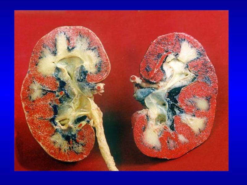
- 1. Urinary system
- 2. А: 1 - канальцы пронефроса; 2 -
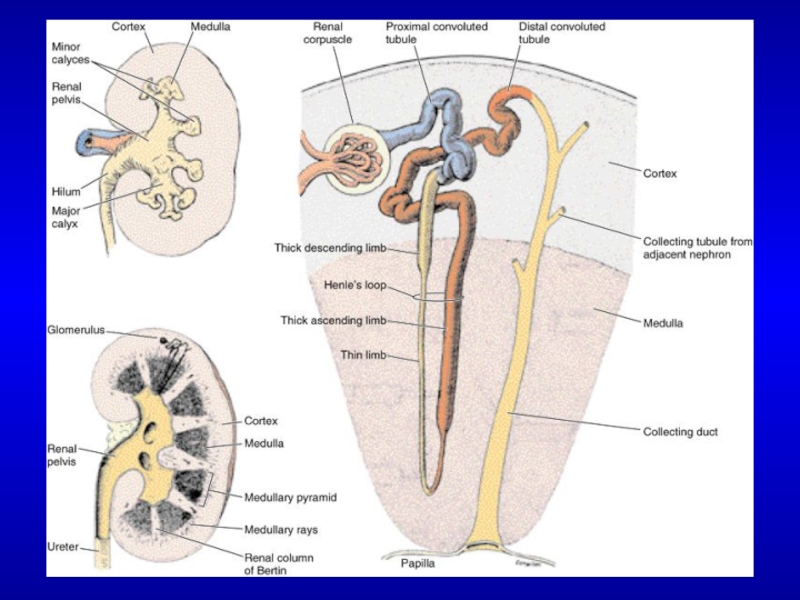
- 6. RENAL CORTEX
- 7. Photomicrograph of renal cortex. A macula
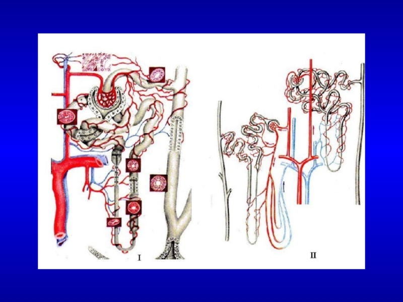
- 8. The renal corpuscle. The upper part
- 9. Photomicrograph of an afferent arteriole entering a
- 10. Schematic representation of a glomerular capillary with
- 12. Mesangial cell located between capillaries enveloped by the basement membrane.
- 14. Renal cortex section showing a proximal convoluted
- 15. 1-descending thick limb of the Henle loop
- 16. Collecting tubule
- 18. THE URINARY BLADDER Transitional epithelium Submucosa layer Muscle layer
- 19. Transitional epithelium
- 20. Compare the structure of the transitional epithelium

Слайд 2А: 1 - канальцы пронефроса; 2 - проток пронефроса; 3 -
канальцы мезонефроса; 4 - клоака;
Б: 1 — канальцы пронефроса (дегенерирующие); 2 — канальцы мезонефроса с нефростомами; 3 - канальцы мезонефроса без нефростомов; 4 — проток метанефроса; 5 - клоака;
В: 1 - канальцы пронефроса; 2 - канальцы мезонефроса без нефростомов; 4 - аллантоис; 5 - проток мезонефроса; 6 - проюк метанефроса; 7 - клоака;
Г: 1 - мюллеровы протоки; 2 — семенник; 3 — канальцы мезонефроса; 4 - аллантоис; 5 - проток метанефроса; 6 - клоака; 7 - канальцы метанефроса; 8 - яичник; 9 — дегенерирующие канальцы и проток мезонефроса; 10 - оофорон и пароофорон (по Пэттену в модификации).


Слайд 7Photomicrograph of renal cortex.
A macula densa is clearly seen (arrow)
at the vascular pole of a renal corpuscle. Picrosirius-hematoxylin (PSH) stain. Medium magnification.
 at the vascular pole")
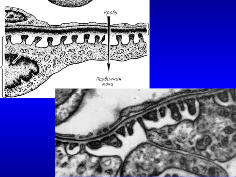
Слайд 8The renal corpuscle.
The upper part of the drawing shows the vascular
pole, with afferent and efferent arterioles and the macula densa. Note the juxtaglomerular cells in the wall of the afferent arteriole. Podocyte processes cover the outer surfaces of the glomerular capillaries; the part of the podocyte containing the nucleus protrudes into the urinary space. Note the flattened cells of the parietal layer of Bowman’s capsule. The lower part of the drawing shows the urinary pole and the proximal convoluted tubule.

Слайд 9Photomicrograph of an afferent arteriole entering a renal corpuscle. The wall
of this arteriole shows the renin-producing juxtaglomerular (JG) cells (broken line). At the upper right is a distal convoluted tubule (DCT) with many elongated mitochondria. PT stain. High magnification.

Слайд 10Schematic representation of a glomerular capillary with the visceral layer of
Bowman’s capsule (formed of podocytes). In this capillary, endothelial cells are fenestrated, but the basal lamina on which they rest is continuous. At left is a podocyte shown in partial section. As viewed from the outside, the part of the podocyte that contains the nucleus protrudes into the urinary space. Each podocyte has many primary processes, from which arise an even greater number of secondary processes that are in contact with the basal lamina.


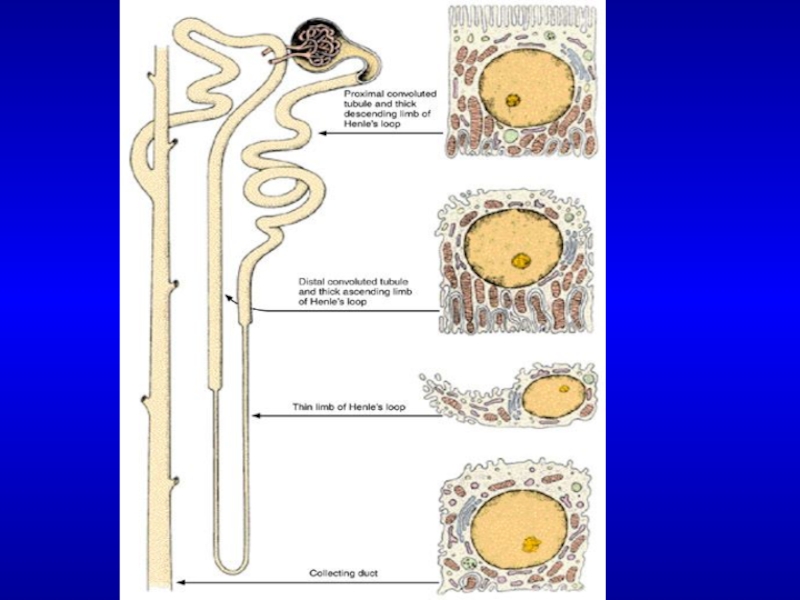
Слайд 14Renal cortex section showing a proximal convoluted tubule (PCT) with its
large cuboidal cells presenting a brush border formed by numerous microvilli. Distal convoluted tubules (DCT) are also present.
 with its large cuboidal cells presenting")
Слайд 151-descending thick limb of the Henle loop
2-ascending thick limb of the
Henle loop
3-blood vessels
3-blood vessels


Слайд 20Compare the structure of the transitional epithelium when the urinary bladder
is empty (A) or full (B). When the bladder is full, the capacity of epithelial cells to slide upon one another reduces the thickness of the epithelium. As a result, the interior surface of the bladder increases. In B, note the thin strands of collagen fibers separating bundles of smooth muscle cells. PSH stain. Medium magnification.
 or")