Performed by:Ordabekov A
Group 16-2
Faculty:General medicine
Checked by:Bizhanova A.
- Главная
- Разное
- Дизайн
- Бизнес и предпринимательство
- Аналитика
- Образование
- Развлечения
- Красота и здоровье
- Финансы
- Государство
- Путешествия
- Спорт
- Недвижимость
- Армия
- Графика
- Культурология
- Еда и кулинария
- Лингвистика
- Английский язык
- Астрономия
- Алгебра
- Биология
- География
- Детские презентации
- Информатика
- История
- Литература
- Маркетинг
- Математика
- Медицина
- Менеджмент
- Музыка
- МХК
- Немецкий язык
- ОБЖ
- Обществознание
- Окружающий мир
- Педагогика
- Русский язык
- Технология
- Физика
- Философия
- Химия
- Шаблоны, картинки для презентаций
- Экология
- Экономика
- Юриспруденция
The heart презентация
Содержание
- 1. The heart
- 2. The heart is a muscular organ in
- 3. In humans, other mammals, and
- 4. Left Heart The left heart has
- 5. Right heart The right heart consists of
- 6. Diseases Cardiovascular diseases, which include diseases of
- 7. Cardiomyopathy is a noticeable deterioration
- 8. Pericardial disease Diseases may also affect the
- 9. Diagnosis Heart disease is diagnosed by the
- 10. Treatment Cardiac arrhythmias Main articles: Heart arrhythmia,
Слайд 1 Kazakh National Medical University named after S.D.
Asfendiyarov
Department of foreign languages
Report
Theme: The Heart disease
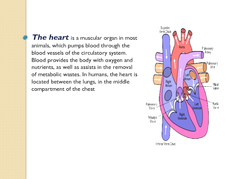
Слайд 2The heart is a muscular organ in most animals, which pumps
blood through the blood vessels of the circulatory system. Blood provides the body with oxygen and nutrients, as well as assists in the removal of metabolic wastes. In humans, the heart is located between the lungs, in the middle compartment of the chest
Слайд 3
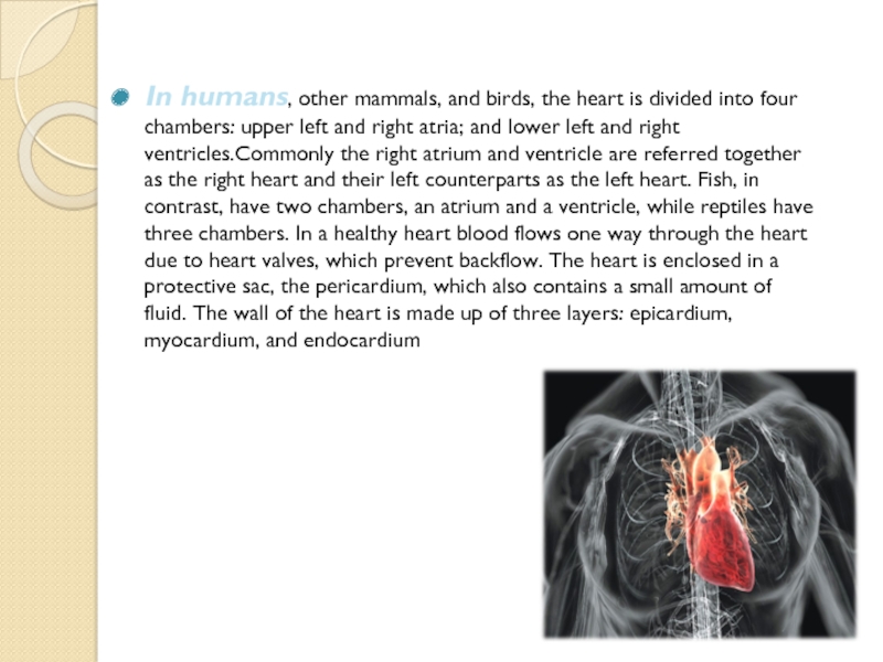
In humans, other mammals, and birds, the heart is divided
into four chambers: upper left and right atria; and lower left and right ventricles.Commonly the right atrium and ventricle are referred together as the right heart and their left counterparts as the left heart. Fish, in contrast, have two chambers, an atrium and a ventricle, while reptiles have three chambers. In a healthy heart blood flows one way through the heart due to heart valves, which prevent backflow. The heart is enclosed in a protective sac, the pericardium, which also contains a small amount of fluid. The wall of the heart is made up of three layers: epicardium, myocardium, and endocardium
Слайд 4Left Heart
The left heart has two chambers: the left atrium, and
the left ventricle, separated by the mitral valve.
The left atrium receives oxygenated blood back from the lungs via one of the four pulmonary veins. The left atrium has an outpouching called the left atrial appendage. Like the right atrium, the left atrium is lined by pectinate muscles. The left atrium is connected to the left ventricle by the mitral valve.
The left ventricle is much thicker as compared with the right, due to the greater force needed to pump blood to the entire body. Like the right ventricle, the left also has trabeculae carneae, but there is no moderator band. The left ventricle pumps blood to the body through the aortic valve and into the aorta. Two small openings above the aortic valve carry blood to the heart itself, the left main coronary artery and the right coronary artery
Слайд 5Right heart
The right heart consists of two chambers, the right atrium
and the right ventricle, separated by a valve, the tricuspid valve.
The right atrium receives blood almost continuously from the body's two major veins, the superior and inferior venae cavae. A small amount of blood from the coronary circulation also drains into the right atrium via the coronary sinus, which is immediately above and to the middle of the opening of the inferior vena cava.In the wall of the right atrium is an oval-shaped depression known as the fossa ovalis, which is a remnant of an opening in the fetal heart known as the foramen ovale. Most of the internal surface of the right atrium is smooth, the depression of the fossa ovalis is medial, and the anterior surface has prominent ridges of pectinate muscles, which are also present in the right atrial appendage
The right atrium receives blood almost continuously from the body's two major veins, the superior and inferior venae cavae. A small amount of blood from the coronary circulation also drains into the right atrium via the coronary sinus, which is immediately above and to the middle of the opening of the inferior vena cava.In the wall of the right atrium is an oval-shaped depression known as the fossa ovalis, which is a remnant of an opening in the fetal heart known as the foramen ovale. Most of the internal surface of the right atrium is smooth, the depression of the fossa ovalis is medial, and the anterior surface has prominent ridges of pectinate muscles, which are also present in the right atrial appendage
Слайд 6Diseases
Cardiovascular diseases, which include diseases of the heart, are the leading
cause of death worldwide. The majority of cardiovascular disease is noncommunicable and related to lifestyle and other factors, becoming more prevalent with ageing.Heart disease is a major cause of death, accounting for an average of 30% of all deaths in 2008, globally This rate varies from a lower 28% to a high 40% in high-income countries Doctors that specialise in the heart are called cardiologists. Many other medical professionals are involved in treating diseases of the heart, including doctors such as general practitioners, cardiothoracic surgeons and intensivists, and allied health practitioners including physiotherapists and dieticians.
Слайд 7 Cardiomyopathy is a noticeable deterioration of the heart muscle's
ability to contract, which can lead to heart failure. The causes of many types of cardiomyopathy are poorly understood; some identified causes include alcohol, toxins, systemic disease such as sarcoidosis, and congenital conditions such as HOCM. The types of cardiomyopathy are described according to how they affect heart muscle. Cardiomyopathy can cause the heart to become enlarged (hypertrophic cardiomyopathy), constrict the outflow tracts of the heart (restrictive cardiomyopathy), or cause the heart to dilate and impact the efficiency of its beating (dilated cardiomyopathy). HOCM is often undiagnosed and can cause sudden death in young athletes.
Слайд 8Pericardial disease
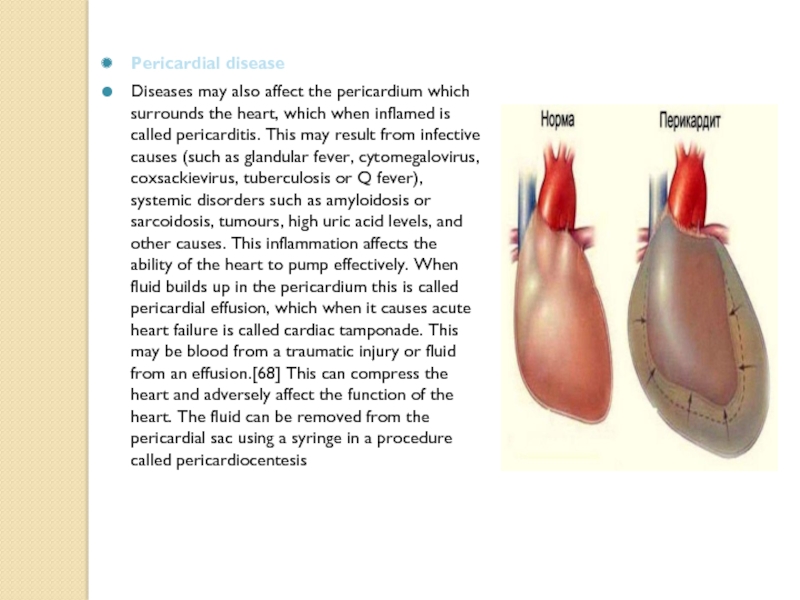
Diseases may also affect the pericardium which surrounds the heart,
which when inflamed is called pericarditis. This may result from infective causes (such as glandular fever, cytomegalovirus, coxsackievirus, tuberculosis or Q fever), systemic disorders such as amyloidosis or sarcoidosis, tumours, high uric acid levels, and other causes. This inflammation affects the ability of the heart to pump effectively. When fluid builds up in the pericardium this is called pericardial effusion, which when it causes acute heart failure is called cardiac tamponade. This may be blood from a traumatic injury or fluid from an effusion.[68] This can compress the heart and adversely affect the function of the heart. The fluid can be removed from the pericardial sac using a syringe in a procedure called pericardiocentesis
Слайд 9Diagnosis
Heart disease is diagnosed by the taking of a medical history,
a cardiac examination, and further investigations, including blood tests, echocardiograms, ECGs and imaging. Other invasive procedures such as cardiac catheterisation can also play a role.
Слайд 10Treatment
Cardiac arrhythmias
Main articles: Heart arrhythmia, Radiofrequency ablation, and Artificial cardiac pacemaker
Abnormal
heart rhythms (arrhythmias) can be treated using antiarrhythmic drugs. These may work by manipulating the flow of electrolytes across the cell membrane (such as calcium channel blockers, sodium channel blockers, amiodarone, or digoxin), or modify the autonomic nervous system's effect on the heart (beta blockers and atropine). In some arrhythmias such as atrial fibrillation which increase the risk of stroke, this risk can be reduced using anticoagulants such as warfarin or novel oral anticoagualants
