- Главная
- Разное
- Дизайн
- Бизнес и предпринимательство
- Аналитика
- Образование
- Развлечения
- Красота и здоровье
- Финансы
- Государство
- Путешествия
- Спорт
- Недвижимость
- Армия
- Графика
- Культурология
- Еда и кулинария
- Лингвистика
- Английский язык
- Астрономия
- Алгебра
- Биология
- География
- Детские презентации
- Информатика
- История
- Литература
- Маркетинг
- Математика
- Медицина
- Менеджмент
- Музыка
- МХК
- Немецкий язык
- ОБЖ
- Обществознание
- Окружающий мир
- Педагогика
- Русский язык
- Технология
- Физика
- Философия
- Химия
- Шаблоны, картинки для презентаций
- Экология
- Экономика
- Юриспруденция
Radiology and imaging of the mammаry gland презентация
Содержание
- 1. Radiology and imaging of the mammаry gland
- 2. Normal anatomy Normal Structures Normal breast is
- 3. Parenchyma The lobules are glandular units and
- 4. . The breast lobes are intertwined and
- 7. Connective tissue Trabecular structures, which are condensations
- 8. Fat The breast is composed of a
- 9. Lymph Nodes Lymph nodes are
- 10. Veins Veins are seen traversing the
- 11. Arteries Arteries appear as slightly
- 12. Skin Skin lines are normally thin
- 13. Normal variants The normal anatomical variants of
- 15. The dense breast Diffuse increase in the
- 16. The increased density of the parenchyma seen
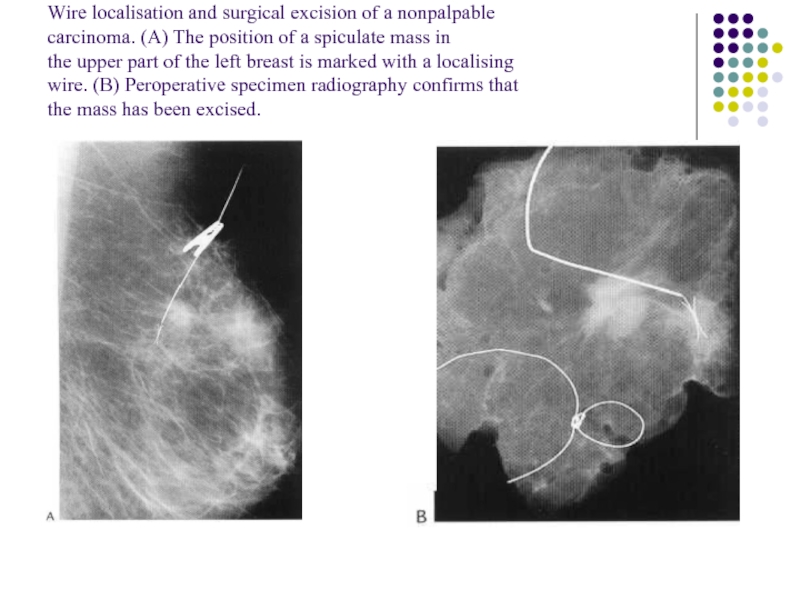
- 17. Mammography The film-screen mammogram is created with
- 18. The routine examination consists of two views
- 19. The C-C view can be considered
- 20. A flat, plastic compression paddle is then
- 23. Compression may be uncomfortable, and may even
- 24. Compression Firm compression is essential for
- 25. The effects of compression are: (i) reduced
- 26. Mammography projections and normal appearances The standard
- 27. More breast tissue is demonstrated on the
- 28. The standard craniocaudal film is obtained with
- 29. Supplementary views For demonstration of tissue in
- 30. Magnification views Magnification views are obtained
- 31. Localized compression views Localized compression views
- 33. Screening Mammography The standard mammogram (along with
- 34. Diagnostic Mammography The diagnostic mammogram begins with
- 35. Indications for diagnostic mammography are:
- 36. Indications of mammography • Screening asymptomatic women
- 37. • Investigation of symptomatic women aged 35
- 38. Patient Preparation For the mammogram, two-piece clothing
- 39. Mammography is generally limited to ambulatory, cooperative
- 40. Computer-Aided Detection Computer-aided detection (CAD) utilizes complex
- 41. _ Ultrasonography Technique High-quality images of the
- 42. The side being examined is raised
- 43. The indications for ultrasonography are: (1) a
- 44. Ultrasonography is a highly reliable technique for
- 45. A limitation A limitation of ultrasonography
- 46. Normal breast ultrasound: 1 = skin; 2
- 47. Magnetic Resonance Imaging The role of
- 48. (5) screening of high-risk patients (6) evaluating
- 50. Axial T1 -weighted (A) and T2 -weighted
- 51. _ Ductography Ductography, or galactography, uses mammographic imaging with contrast injection into the breast ducts.
- 52. The indication The indication
- 53. The patient lies in supine position while
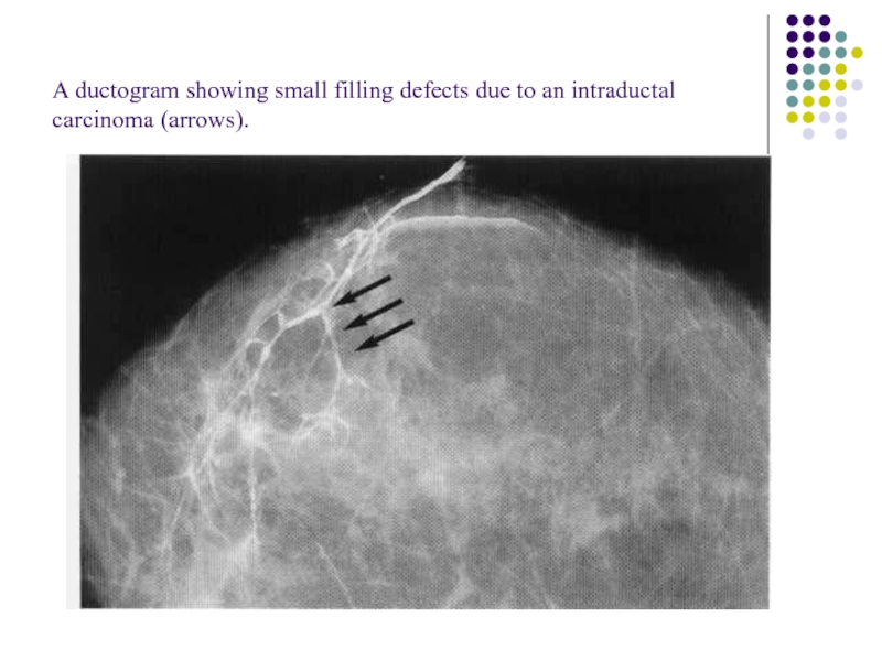
- 54. A ductogram showing small filling defects due to an intraductal carcinoma (arrows).
- 55. Image-Guided Needle Aspiration and Biopsy The first
- 56. Needle biopsy is used in this case
- 57. 1 4G needle and automated biopsy device used for ultrasound and stereotactic core breast biopsy.
- 58. Stereotactic-guided fine needle aspiration. The check pair
- 59. Stereotactic core biopsy. Stereo film pair showing
- 60. Image-Guided Needle Localization When a nonpalpable breast
- 61. Once the needle is in the appropriate
- 62. Wire localisation and surgical excision of a
- 63. Patient Preparation Patients for whom stereotactic biopsy
- 64. Approach to the Palpable Lump When a
- 65. Second, is the lump new or enlarged?
- 66. Third, how big is the lump? Tiny
- 67. Fourth, how old is the patient? If
- 68. If the patient is below 35 years
- 69. For women between the ages of 30
- 70. Bi - rads assessment
- 71. Circumscribed mass A circumscribed mass is analysed
- 72. Radiolucent lesions Lipoma Oil cyst Galactocele.
- 73. Mixed density lesions adenolipoma hamartoma galactocele hematoma lymph node
- 74. Radiopaque (soft-tissue density) lesions Benign lesions *
- 75. Malignant lesions * Mucinous carcinoma * Medullary
- 76. Calcifications Arterial: curvilinear, parallel line calcifications along
- 77. Cyst: curvilinear calcification may occur in the
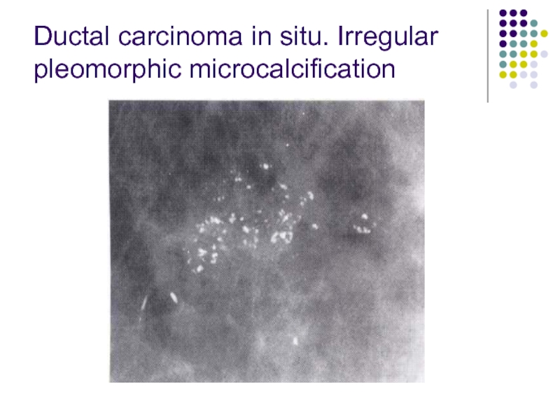
- 78. Ductal carcinoma in situ. Irregular pleomorphic microcalcification
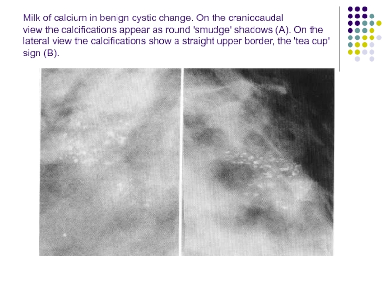
- 79. Milk of calcium in benign cystic change.
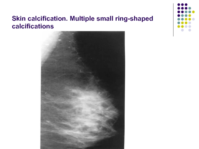
- 80. Skin calcification. Multiple small ring-shaped calcifications
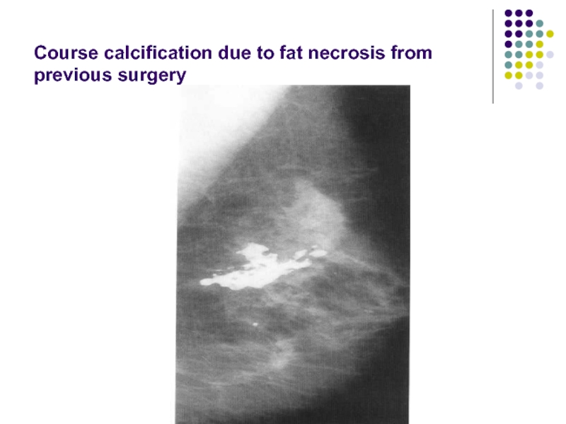
- 81. Course calcification due to fat necrosis from previous surgery
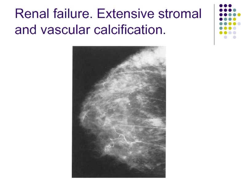
- 82. Renal failure. Extensive stromal and vascular calcification.
- 84. Spiculate mass A spiculate mass is the
- 85. 2. Large or superficially positioned tumors may
- 86. The typical ultrasound features are Most spiculate
- 87. the presence of these signs, however,
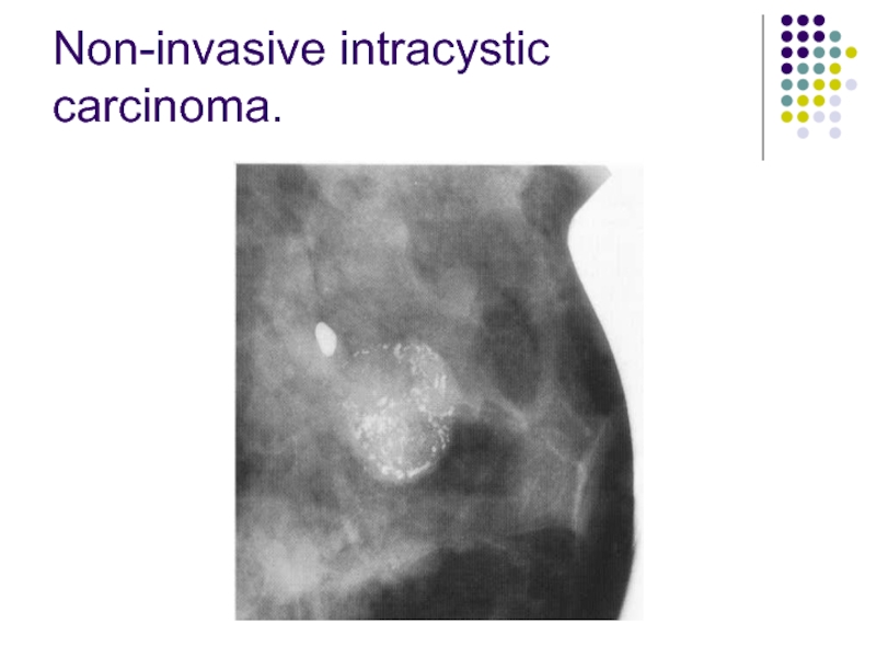
- 93. Non-invasive intracystic carcinoma.
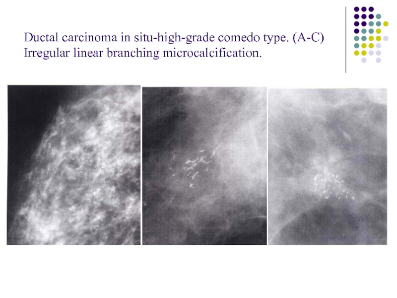
- 94. Ductal carcinoma in situ-high-grade comedo type. (A-C) Irregular linear branching microcalcification.
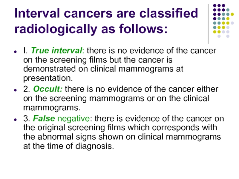
- 95. Interval cancers are classified radiologically as follows:
- 96. 4. Minimal sign: there are subtle features
- 97. Fibroadenoma Fibroadenoma are characteristically
- 98. The typical ultrasound appearance of a fibroadenoma
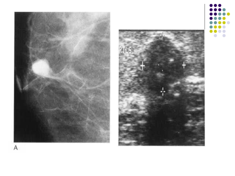
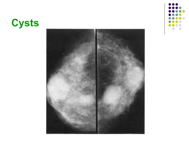
- 101. Cyst Cyst are the most common cause
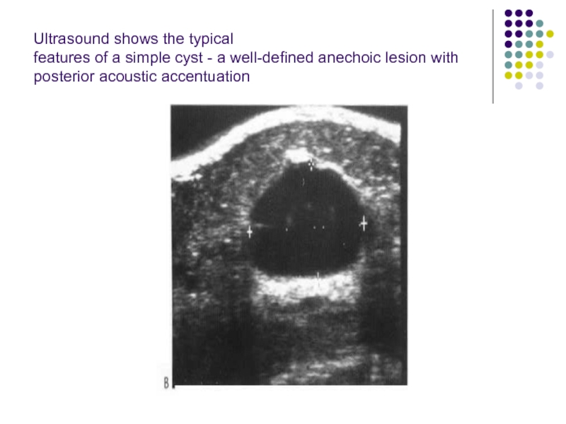
- 102. Cysts can be readily diagnosed with ultrasound.
- 103. When these features are present, a cyst
- 104. Cysts
- 105. Ultrasound shows the typical features of a
Слайд 2Normal anatomy
Normal Structures
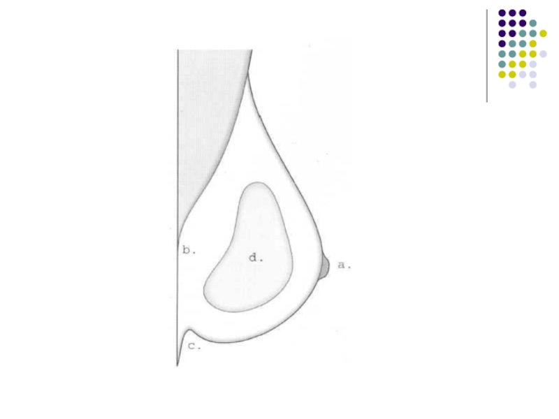
Normal breast is composed:
mainly of parenchyma (lobules and ducts)
fat
Lobules are drained by ducts. There are about 15 to 20 lobes in the breast. The lobar ducts converge upon the nipple.
Слайд 3Parenchyma
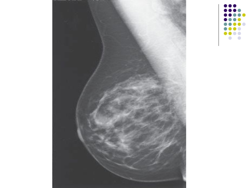
The lobules are glandular units and are seen as ill-defined, splotchy
Слайд 4. The breast lobes are intertwined and are therefore not discretely
The amount and distribution of glandular tissue are highly variable. Younger women tend to have more glandular tissue than do older women.
Слайд 7Connective tissue
Trabecular structures, which are condensations of connective tissue, appear as
Слайд 8Fat
The breast is composed of a large amount of fat, which
Слайд 10Veins
Veins are seen traversing the breast as uniform, linear opacities, about
Слайд 11Arteries
Arteries appear as slightly thinner, uniform, linear densities and are best
Слайд 12Skin
Skin lines are normally thin and are not easily seen without
Слайд 13Normal variants
The normal anatomical variants of the brest result from the
Слайд 15The dense breast
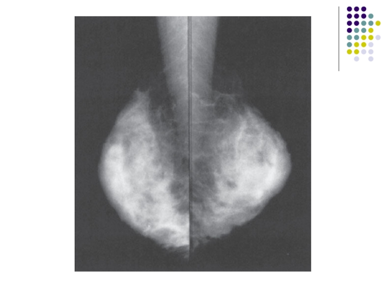
Diffuse increase in the density of the breast tissue
by oedema (see the `Oedematous breast' below)
by an increase in the glandular tissue
or fibrous tissue
This is commonly seen in benign breast change, and may be accompanied by evidence of cysts, and in women who are taking hormone replacement therapy (HRT) for menopausal symptoms.
Слайд 16The increased density of the parenchyma seen as a result of
Слайд 17Mammography
The film-screen mammogram is created with x-rays, radiographic film, and intensifying
The digital mammogram is created using a similar system, but replacing the film and screen with a digital detector.
Слайд 18The routine examination consists of two views of each breast:
the craniocaudal
the mediolateraloblique (MLO) view, with a total of four films.
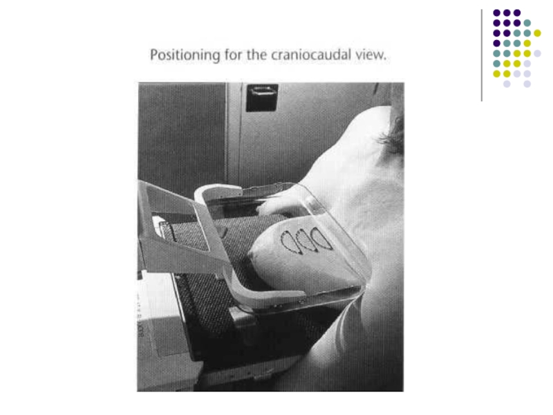
Слайд 19 The C-C view can be considered the “top-down” view, and
The patient undresses from the waist up and stands for the examination, leaning slightly against the mammography unit.
The technologist must mobilize, elevate, and pull the breast to place as much breast tissue as possible on the surface of the film cassette holder.
Слайд 20A flat, plastic compression paddle is then gently but firmly lowered
This compression achieves both immobilizations during exposure and dispersion of breast tissue shadows over a larger area, thereby permitting better visual separation of imaged structures
Слайд 23Compression may be uncomfortable, and may even be painful in a
However, most patients accept this level of discomfort for the few seconds required for each exposure, particularly if they understand the need for compression and know what to expect during the examination.
Mammography has proved to be more cost-effective, while maintaining resolution high enough to demonstrate early malignant lesions, than any other breast imaging technique.
Слайд 24Compression
Firm compression is essential for high-quality mammograms and is applied
Слайд 25The effects of compression are:
(i) reduced dose;
(ii) reduced scatter-improved contrast;
(iii) reduced
(iv) reduced movement unsharpness;
(v) reduced range of breast thickness;
(vi) reduced tissue overlap improved resolution.
Слайд 26Mammography projections and normal appearances
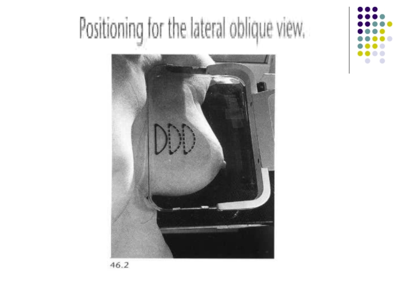
The standard examination for women undergoing either
The lateral oblique view is usually obtained with the tube angled at 45° to the horizontal, but tube angulation from 30° to 60° may be needed depending on the build of the woman.
Слайд 27More breast tissue is demonstrated on the lateral oblique projection than
Careful positioning is essential for satisfactory demonstration of the breast.
Слайд 28The standard craniocaudal film is obtained with a vertical X-ray beam
Слайд 29Supplementary views
For demonstration of tissue in the most posterolateral part of
Слайд 30Magnification views
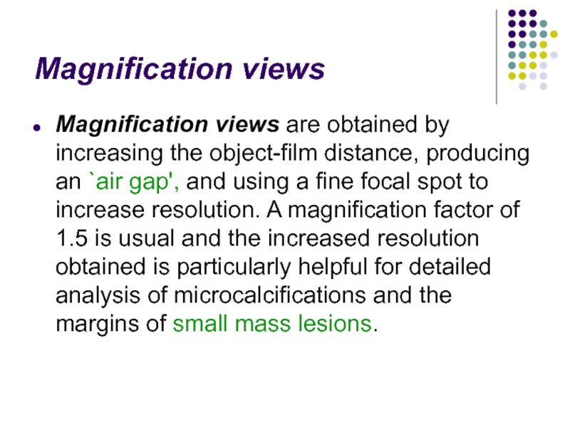
Magnification views are obtained by increasing the object-film distance,
Слайд 31Localized compression views
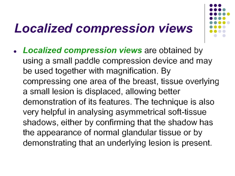
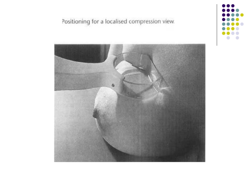
Localized compression views are obtained by using a
Слайд 33Screening Mammography
The standard mammogram (along with appropriate history taking) makes up
Such patients should be referred for diagnostic mammography.
Слайд 34Diagnostic Mammography
The diagnostic mammogram begins with the two-view standard mammogram. Additional
Слайд 35 Indications for diagnostic mammography are:
(1) a palpable mass or
or nipple discharge that is clear or bloody)
(2) a radiographic abnormality on a screening mammogram.
Additionally, patients with a personal history of breast cancer may be considered in the diagnostic category.
Слайд 36Indications of mammography
• Screening asymptomatic women aged 50 years and over
•
-women who have one or more first degree relatives who have been diagnosed with premenopausal breast cancer
-women with histologic risk factors found at previous surgery, e.g. atypical ductal hyperplasia
Слайд 37• Investigation of symptomatic women aged 35 years and over with
• Surveillance of the breast following local excision of breast carcinoma
• Evaluation of a breast lump in women following augmentation mammoplasty
• Investigation of a suspicious breast lump in a man
Слайд 38Patient Preparation
For the mammogram, two-piece clothing is most convenient as the
Слайд 39Mammography is generally limited to ambulatory, cooperative patients because of the
Слайд 40Computer-Aided Detection
Computer-aided detection (CAD) utilizes complex algorithms to analyze the data
calcifications
masses
architecture distortion
Слайд 41_ Ultrasonography
Technique
High-quality images of the normal and abnormal breast can be
Слайд 42 The side being examined is raised and the arm placed
Слайд 43The indications for ultrasonography are:
(1) a mammographically detected mass, the nature
(2) a palpable mass that is not seen on mammography
(3) a palpable mass in a patient below the age recommended for routine mammography
(4) guidance for intervention.
Слайд 44Ultrasonography is a highly reliable technique for differentiating cystic from solid
Слайд 45A limitation
A limitation of ultrasonography is that it is very
Also, it images only a small part of the breast at any one moment. Therefore, an overall inclusive survey is not possible in one image, and lesions may easily be missed.
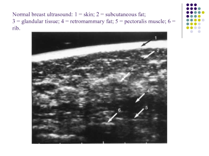
Слайд 46Normal breast ultrasound: 1 = skin; 2 = subcutaneous fat; 3 =
Слайд 47Magnetic Resonance Imaging
The role of MRI in mammography continues to
(1) staging of and surgical planning for breast tumors
(2) searching for a primary tumor in patients who present with cancerous axillary lymph nodes
(3) evaluating tumor response to neoadjuvant chemotherapy
(4) differentiating tumor recurrence from posttreatment changes in patients with previous breast-conserving surgery and radiation
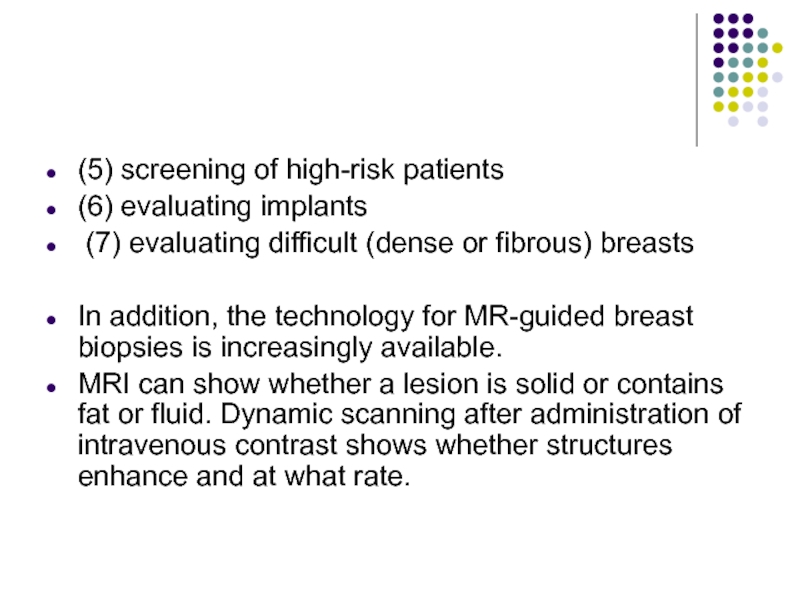
Слайд 48(5) screening of high-risk patients
(6) evaluating implants
(7) evaluating
In addition, the technology for MR-guided breast biopsies is increasingly available.
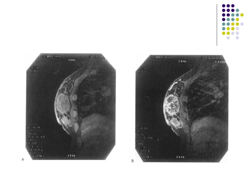
MRI can show whether a lesion is solid or contains fat or fluid. Dynamic scanning after administration of intravenous contrast shows whether structures enhance and at what rate.
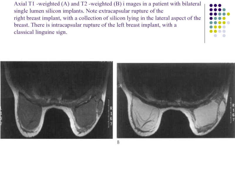
Слайд 50Axial T1 -weighted (A) and T2 -weighted (B) i mages in
Слайд 51_ Ductography
Ductography, or galactography, uses mammographic imaging with contrast injection into
Слайд 52 The indication
The indication for use is a profuse,
If these conditions are not present, the ductogram is likely to be of little help. The purpose is to reveal the location of the ductal system involved.
The cause of the discharge is frequently not identified.
Слайд 53The patient lies in supine position while the discharging duct is
Слайд 55Image-Guided Needle Aspiration and Biopsy
The first indication is aspiration of cystic
The second indication concerns solid lesions.
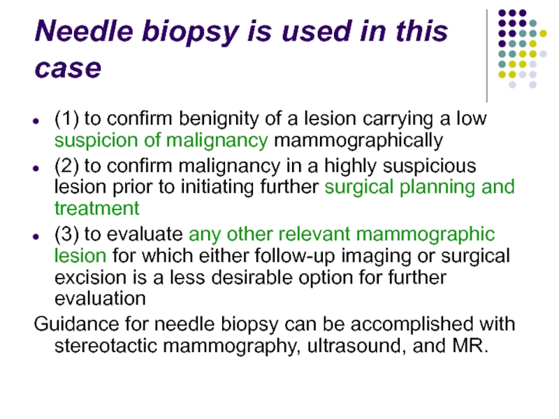
Слайд 56Needle biopsy is used in this case
(1) to confirm benignity
(2) to confirm malignancy in a highly suspicious lesion prior to initiating further surgical planning and treatment
(3) to evaluate any other relevant mammographic lesion for which either follow-up imaging or surgical excision is a less desirable option for further evaluation
Guidance for needle biopsy can be accomplished with stereotactic mammography, ultrasound, and MR.
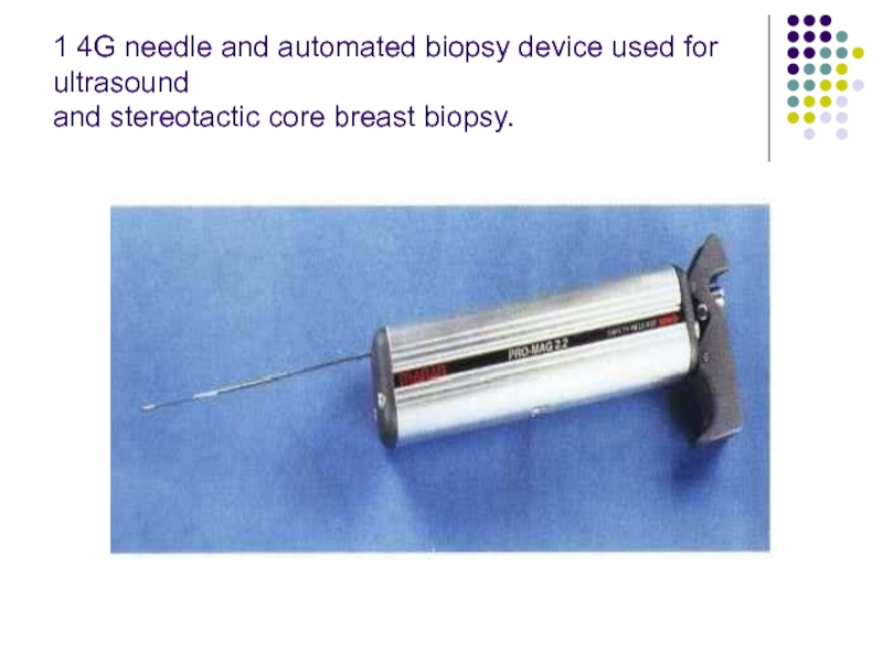
Слайд 571 4G needle and automated biopsy device used for ultrasound and stereotactic
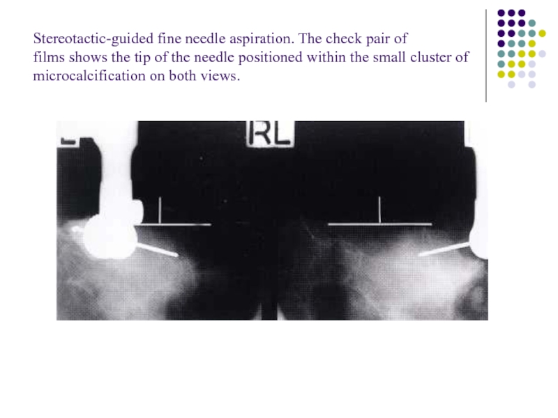
Слайд 58Stereotactic-guided fine needle aspiration. The check pair of films shows the tip
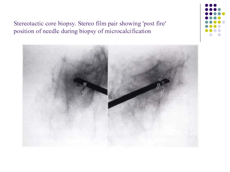
Слайд 59Stereotactic core biopsy. Stereo film pair showing 'post fire' position of needle
Слайд 60Image-Guided Needle Localization
When a nonpalpable breast lesion must be excised, imaging
Слайд 61Once the needle is in the appropriate position, a hook wire
Слайд 62Wire localisation and surgical excision of a nonpalpable carcinoma. (A) The position
Слайд 63Patient Preparation
Patients for whom stereotactic biopsy is being considered should be
Слайд 64Approach to the Palpable Lump
When a breast lump is found, several
First, given that lumpy breasts are a normal variant, when is a lump significant?
Experts in CBE advise palpation with the flat surface of two to three fingers, and not with the fingertips. With this technique, nonsignificant lumps will disperse into background breast density, but a significant lump will stand out as a dominant mass.
Слайд 65Second, is the lump new or enlarged? A new lump is
Слайд 66Third, how big is the lump? Tiny pea-sized or smaller lumps,
Слайд 67Fourth, how old is the patient? If the patient is less
For patients over the age of 35 years, breast imaging begins with a diagnostic mammogram at the time a lump is deemed to be significant. The mammogram provides a view of the lump, as well as of the remainder of the involved breast and the opposite breast, where associated findings may aid in diagnosis and treatment planning.
Слайд 68If the patient is below 35 years of age, a significant
Слайд 69For women between the ages of 30 and 40 years, judgment
If the primary care physician is uncertain of the significance of the findings of CBE, evaluation by a breast specialist may be helpful prior to requesting radiologic tests.
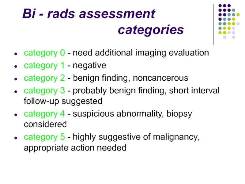
Слайд 70 Bi - rads assessment
category 0 - need additional imaging evaluation
category 1 - negative
category 2 - benign finding, noncancerous
category 3 - probably benign finding, short interval follow-up suggested
category 4 - suspicious abnormality, biopsy considered
category 5 - highly suggestive of malignancy, appropriate action needed
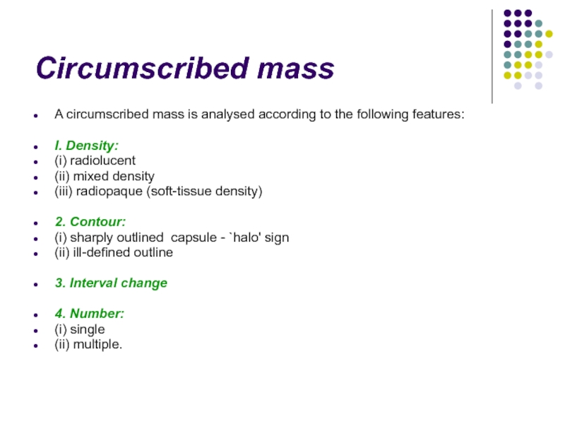
Слайд 71Circumscribed mass
A circumscribed mass is analysed according to the following features:
I.
(i) radiolucent
(ii) mixed density
(iii) radiopaque (soft-tissue density)
2. Contour:
(i) sharply outlined capsule - `halo' sign
(ii) ill-defined outline
3. Interval change
4. Number:
(i) single
(ii) multiple.
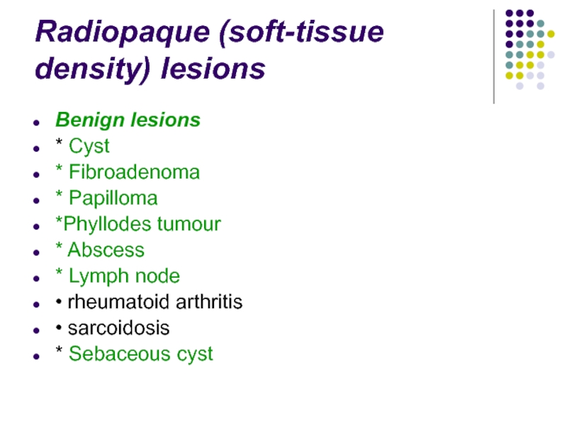
Слайд 74Radiopaque (soft-tissue density) lesions
Benign lesions
* Cyst
* Fibroadenoma
* Papilloma
*Phyllodes tumour
* Abscess
* Lymph
• rheumatoid arthritis
• sarcoidosis
* Sebaceous cyst
Слайд 75Malignant lesions
* Mucinous carcinoma
* Medullary carcinoma
* Papillary carcinoma
* Invasive ductal carcinoma
*
* Metastasis
• melanoma
• lung
• ovary
* Lymphoma
* Sarcoma
* Pathological lymph node
• breast cancer
• Phyllodes tumour
• lymphoma
• metastasis
Recurrent breast cancer
Слайд 76Calcifications
Arterial: curvilinear, parallel line calcifications along the course of a blood
Skin calcification: multiple small ring-shaped calcifications.
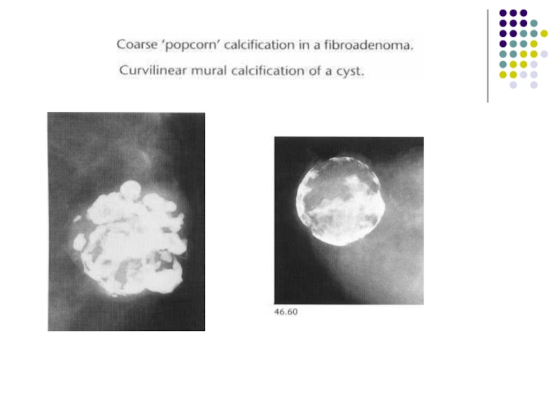
Fibroadenoma: coarse `popcorn' type calcification associated with a soft-tissue mass. Less commonly the calcifications may he fine, irregular or curvilinear `eggshell' type related to the periphery of the lesion.
Слайд 77Cyst: curvilinear calcification may occur in the wall of a cyst.
Carcinoma:
Слайд 79Milk of calcium in benign cystic change. On the craniocaudal view the
Слайд 84Spiculate mass
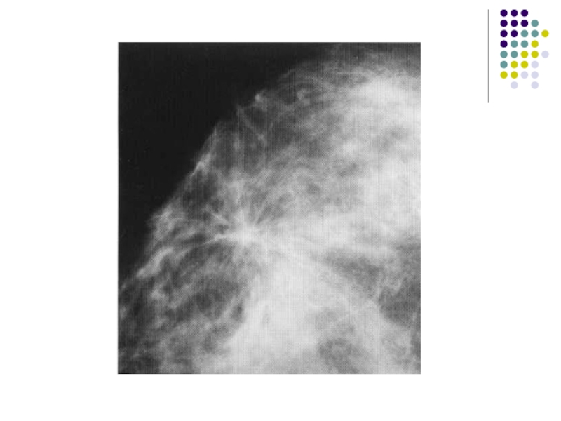
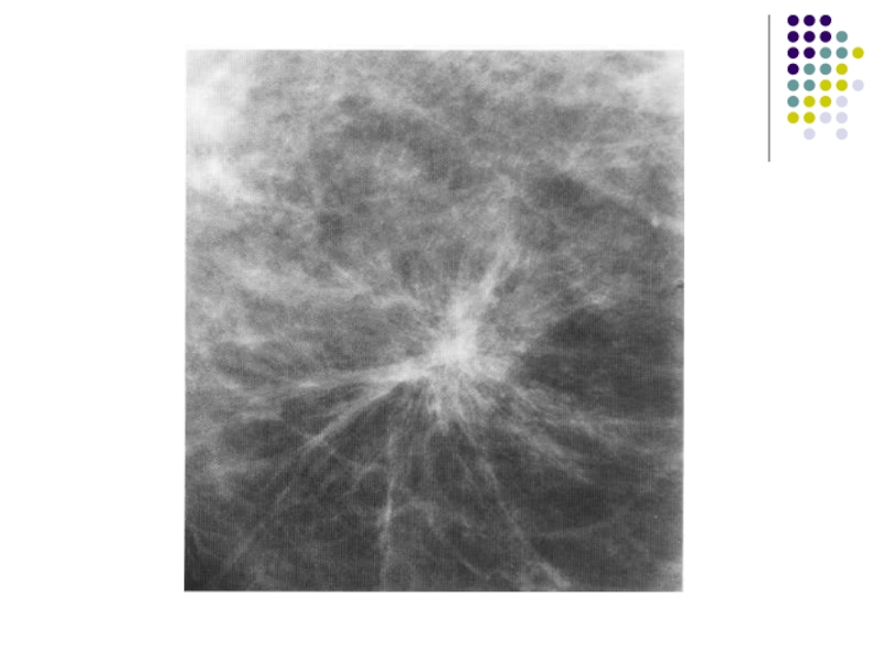
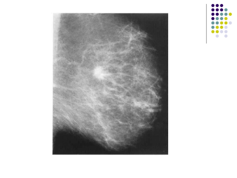
A spiculate mass is the commonest mammographic appearance of invasive
1. It consists of a central soft-tissue tumor mass from the surface of which spicules extend into the surrounding breast tissue. There is often associated distortion of the surrounding breast tissue with straightening of the trabeculae due to retraction.
Слайд 852. Large or superficially positioned tumors may be associated with localized
3. Deeply positioned tumors may be associated with tethering of the pectoralis muscle.
4. Irregular microcalcifications due to associated ductal carcinoma in situ may be found within the tumour or in the surrounding breast tissue, sometimes extending to the nipple.
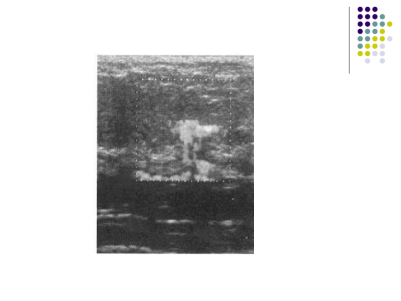
Слайд 86The typical ultrasound features are
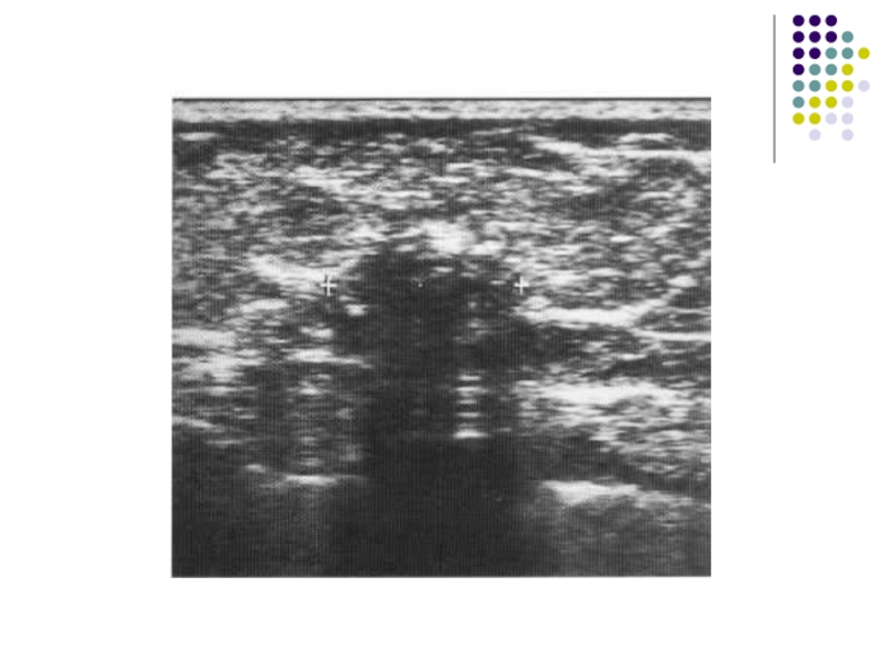
Most spiculate carcinomas of I cm diameter
of an echo-poor mass, with poorly defined margins and posterior acoustic shadowing
distortion of the surrounding breast tissue may be visible and a rim of increased reflectivity around the tumour mass may be seen
Слайд 87 the presence of these signs, however, is variable: acoustic shadowing
similar suspicious ultrasound appearances may be caused by a sclerosing fibroadenoma or benign complex sclerosing lesion
Слайд 94Ductal carcinoma in situ-high-grade comedo type. (A-C) Irregular linear branching microcalcification.
Слайд 95Interval cancers are classified radiologically as follows:
I. True interval: there is
2. Occult: there is no evidence of the cancer either on the screening mammograms or on the clinical mammograms.
3. False negative: there is evidence of the cancer on the original screening films which corresponds with the abnormal signs shown on clinical mammograms at the time of diagnosis.
Слайд 964. Minimal sign: there are subtle features on the screening mammograms
5. Unclassified: mammography was not performed at the time of diagnosis and therefore the presence of mammographic signs of malignancy on the previous screening films cannot he verified.
Слайд 97 Fibroadenoma
Fibroadenoma are characteristically
sharply outlined
low soft tissue density
they are usually solitary but may be multiple with increasing age, they may undergo
fibroadenoma can, however, show very fine calcifications with some pleomorphism which can raise the suspicion of malignancy
fibroadenoma do not arise de novo in women aged 40 years or more but may grow in menopausal women who are taking HRT
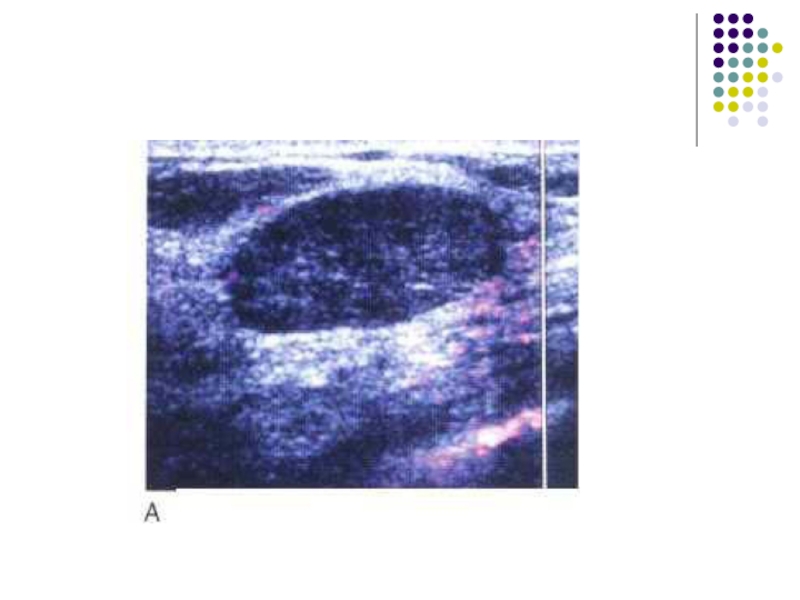
Слайд 98The typical ultrasound appearance of a fibroadenoma is
a well circumscribed round
the ultrasound findings alone therefore cannot be used to confirm the diagnosis of a circumscribed solid lesion found on mammography
Слайд 101Cyst
Cyst are the most common cause of a discrete breast mass.
they
they are common between the ages of 20 and 50 years, with a peak incidence between 40 and 50 years
simple cysts are not associated with an increased risk of malignancy and have no malignant potential
On mammography they are seen as well-defined, round or oval masses. Sometimes a characteristic halo is visible on mammography
Слайд 102Cysts can be readily diagnosed with ultrasound.
They have:
well-defined margins
are
show an absence of internal echoes indicating the presence of fluid
the area of breast tissue behind a cyst appears bright on ultrasound (posterior enhancement) due to improved transmission on the ultrasound beam through the cyst fluid When these features are present, a cyst can be diagnosed with certainty. Aspiration is easily performed under ultrasound guidance to alleviate symptoms or when there is diagnostic uncertainty. Cytology on cyst fluid is not routinely performed unless there are atypical imaging features or the aspirate is bloodstained
Слайд 103When these features are present, a cyst can be diagnosed with
Cytology on cyst fluid is not routinely performed unless there are atypical imaging features or the aspirate is bloodstained
