- Главная
- Разное
- Дизайн
- Бизнес и предпринимательство
- Аналитика
- Образование
- Развлечения
- Красота и здоровье
- Финансы
- Государство
- Путешествия
- Спорт
- Недвижимость
- Армия
- Графика
- Культурология
- Еда и кулинария
- Лингвистика
- Английский язык
- Астрономия
- Алгебра
- Биология
- География
- Детские презентации
- Информатика
- История
- Литература
- Маркетинг
- Математика
- Медицина
- Менеджмент
- Музыка
- МХК
- Немецкий язык
- ОБЖ
- Обществознание
- Окружающий мир
- Педагогика
- Русский язык
- Технология
- Физика
- Философия
- Химия
- Шаблоны, картинки для презентаций
- Экология
- Экономика
- Юриспруденция
Pheochromocytomas презентация
Содержание
- 1. Pheochromocytomas
- 2. Pheochromocytomas rare, catecholamine-secreting, vascular, neuroendocrine tumors
- 3. Pheochromocytoma localization
- 4. Epidemiology rare cause of secondary hypertension
- 5. Tumor characteristics ~ 95% of catecholamine-secreting tumors
- 6. Clinical presentation The “classic triad”: episodic headache,
- 7. PHEO may bee asymptomatic incidental imaging discovery (incidentaloma) genetic survey autopsy
- 8. Familial pheochromocytoma MEN 2 syndrome 95%
- 9. Familial pheochromocytoma MEN 2A (Sipple's syndrome)
- 10. Familial pheochromocytoma MEN 2B (mucosal neuroma syndrome)
- 11. Familial pheochromocytoma Neurofibromatosis type 1
- 12. Familial pheochromocytoma von Hippel–Lindau disease (VHL)
- 13. Familial pheochromocytoma Familial paraganglioma syndromes Paraganglioma syndrome
- 14. Genetic vs. sporadic PHEO
- 15. When to suspect PHEO? Hyperadrenergic spells Resistant
- 16. Catecholemine metabolism Metanephrine Normetanephrine Norepinephrine Vanillylmandelic acid COMT COMT MAO MAO
- 17. Pheochromocytoma diagnosis 24-hour urine collection for fractionated
- 18. Pheochromocytoma diagnosis Plasma fractionated metanephrines metanephrine
- 20. Pheochromocytoma diagnosis 24-hour urinary vanillylmandelic acid (VMA)
- 21. Pheochromocytoma imaging CT or MRI of the
- 22. Pheochromocytoma imaging a- FDG-PET b- abdominal CT c- DOTATATE-Scan
- 23. Pheochromocytoma treatment all patients should undergo a

Слайд 2Pheochromocytomas
rare, catecholamine-secreting,
vascular, neuroendocrine tumors
arising from chromaffin cells of the
adrenal
medulla ~80%
extra-adrenal pheochromocytoma
or paraganglioma (PGL) ~15–20%
extra-adrenal pheochromocytoma
or paraganglioma (PGL) ~15–20%


Слайд 4Epidemiology
rare cause of secondary hypertension
less than 0.2% of patients with
HTN
incidence is approximately 0.8/100,000 p-y
0.05% in the autopsy (report from China)
occur at any age, most common in 40-50 y
male and female equally
incidence is approximately 0.8/100,000 p-y
0.05% in the autopsy (report from China)
occur at any age, most common in 40-50 y
male and female equally

Слайд 5Tumor characteristics
~ 95% of catecholamine-secreting tumors are in the abdomen
85-90% of
which are intraadrenal (PHEO)
10-15% of catecholamine-secreting tumors are extra-adrenal (paraganglioma(
5-10% multiple PHEO
~ 10% malignant PHEO: local invasion into surrounding tissues and organs (kidney, liver) or distant metastases
10-15% of catecholamine-secreting tumors are extra-adrenal (paraganglioma(
5-10% multiple PHEO
~ 10% malignant PHEO: local invasion into surrounding tissues and organs (kidney, liver) or distant metastases
10-15%")
Слайд 6Clinical presentation
The “classic triad”: episodic headache, sweating, and tachycardia – rarely
seen
Blood pressure: paroxysmal hypertension (50%); persistent hypertension (30%) or normal BP(15%)
Other symptoms: palpitations, tremor, pallor, dyspnea, weakness, syncope, panic attack, orthostatic hypotension, weight loss, polyuria, constipation, hyperglycemia, cardiomyopathy, pulmonary edema
Paroxysmal elevations in BP, tachycardia, or arrhythmia during diagnostic procedures, surgery, induction of anesthesia, with certain foods or drugs

 genetic surveyautopsy")
Слайд 8Familial pheochromocytoma
MEN 2 syndrome
95% autosomal dominant RET proto-oncogene mutation
prevalence ~1/
35,000 individuals
~ 50% of patients with MEN 2 syndrome develop PHEO in the adrenal glands
rarely malignant
younger age (30-40 years)
~ 50% of patients with MEN 2 syndrome develop PHEO in the adrenal glands
rarely malignant
younger age (30-40 years)

Слайд 9Familial pheochromocytoma
MEN 2A (Sipple's syndrome)
medullary thyroid cancer (MTC) in all
patients, PHEO in 50%, primary hyperparathyroidism in 20%, and cutaneous lichen amyloidosis in 5%
 medullary thyroid cancer (MTC) in all patients, PHEO in 50%,")
Слайд 10Familial pheochromocytoma
MEN 2B (mucosal neuroma syndrome)
MTC in all patients, PHEO
in 50%, mucocutaneous neuromas,
skeletal deformities, marfanoid
habitus and intestinal
ganglioneuromas (Hirschsprung's
disease)
skeletal deformities, marfanoid
habitus and intestinal
ganglioneuromas (Hirschsprung's
disease)
 MTC in all patients, PHEO in 50%, mucocutaneous neuromas,")
Слайд 11Familial pheochromocytoma
Neurofibromatosis type 1
prevalence ~ 1/ 3,000 individuals
neurofibromas, multiple café-au-lait
spots, axillary and inguinal freckling,
iris hamartomas (Lisch nodules),
macrocephaly and cognitive deficits
~ 2% solitary, benign adrenal PHEO
iris hamartomas (Lisch nodules),
macrocephaly and cognitive deficits
~ 2% solitary, benign adrenal PHEO

Слайд 12Familial pheochromocytoma
von Hippel–Lindau disease (VHL)
prevalence ~2–3/ 100,000 persons
hemangioblastoma (cerebellum, spinal
cord or brainstem), retinal angioma, clear cell renal carcinoma, pancreatic tumors, endolymphatic sac tumors of the middle ear
bilateral or malignant PHEO, paraganglioma in the mediastinum, abdomen and pelvis
bilateral or malignant PHEO, paraganglioma in the mediastinum, abdomen and pelvis
prevalence ~2–3/ 100,000 persons hemangioblastoma (cerebellum, spinal cord or brainstem), retinal")
Слайд 13Familial pheochromocytoma
Familial paraganglioma syndromes
Paraganglioma syndrome type 1-4
usually nonfunctional parasympathetic paragangliomas
at skull base and neck
sometimes adrenal pheochromocytoma
type 4 may be malignant PHEO
sometimes adrenal pheochromocytoma
type 4 may be malignant PHEO


Слайд 15When to suspect PHEO?
Hyperadrenergic spells
Resistant hypertension
A familial syndrome that predisposes to
PHEO (MEN2, NF1, VHL(
A family history of pheochromocytoma
An incidentally discovered adrenal mass
Hypertension and new onset or atypical DM
Pressor response to anesthesia, surgery, or angiography
Onset of hypertension at a young age (<20 years)
A family history of pheochromocytoma
An incidentally discovered adrenal mass
Hypertension and new onset or atypical DM
Pressor response to anesthesia, surgery, or angiography
Onset of hypertension at a young age (<20 years)

Слайд 16Catecholemine metabolism
Metanephrine
Normetanephrine
Norepinephrine
Vanillylmandelic acid
COMT
COMT
MAO
MAO

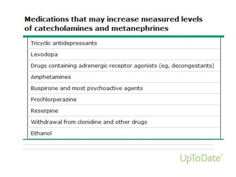
Слайд 17Pheochromocytoma diagnosis
24-hour urine collection for fractionated metanephrines and catecholamines
Norepinephrine >170 mcg/24
h
Epinephrine >35 mcg/24 h
Dopamine >700 mcg/24 h
Normetanephrine >900 mcg/24 h
Metanephrine >400 mcg/24 h
may be false-positive
have to be used if clinical probability is low
Epinephrine >35 mcg/24 h
Dopamine >700 mcg/24 h
Normetanephrine >900 mcg/24 h
Metanephrine >400 mcg/24 h
may be false-positive
have to be used if clinical probability is low
measurement of urinary creatinine to verify an adequate collection

Слайд 18Pheochromocytoma diagnosis
Plasma fractionated metanephrines
metanephrine
nmol\l (fast), <0.9 nmo\l (non-fast)
high predictive value of a negative test
high rate of false-positive tests
have to be used if clinical probability is high
high predictive value of a negative test
high rate of false-positive tests
have to be used if clinical probability is high

Слайд 20Pheochromocytoma diagnosis
24-hour urinary vanillylmandelic acid (VMA) excretion
poor diagnostic
sensitivity and specificity
Chromogranine A in serum
increased in 80% of patients with PHEO
not specific for PHEO and may be seen with other neuroendocrine tumors (carcinoid), and in a variety of other conditions (atrophic gastritis, cirrhosis, CRF, PPI treatment …)
Chromogranine A in serum
increased in 80% of patients with PHEO
not specific for PHEO and may be seen with other neuroendocrine tumors (carcinoid), and in a variety of other conditions (atrophic gastritis, cirrhosis, CRF, PPI treatment …)
 excretion poor diagnostic sensitivity and specificityChromogranine A in")
Слайд 21Pheochromocytoma imaging
CT or MRI of the abdomen and pelvis
Pheo Imaging characteristics
•
Usually large size (>3 sm)
May be bilateral
• Cystic and hemorrhagic changes
• Increased mass vascularity
Increased attenuation on non-enhanced CT (>20HU)
Additional imaging: MIBG, FDG-PET, DOTATATE-Scan
Biopsy of suspected Pheo should be avoided!
May be bilateral
• Cystic and hemorrhagic changes
• Increased mass vascularity
Increased attenuation on non-enhanced CT (>20HU)
Additional imaging: MIBG, FDG-PET, DOTATATE-Scan
Biopsy of suspected Pheo should be avoided!


Слайд 23Pheochromocytoma treatment
all patients should undergo a resection of the Pheo (laparascopic
or open adrenalectomy)
preoperative medical therapy
hypertension and tachycardia control:
target BP 120/80 mmHg
combined α-adrenergic blockade (Phenoxybenzamine,
Prazocine, Doxazocine) and β-blockade (Deralin)
volume expansion (high sodium diet, IV 0.9% NS)
prevention of the hypertensive crisis during surgery (Nitroprusside, Phentolamine, Nicardipine)
preoperative medical")