- Главная
- Разное
- Дизайн
- Бизнес и предпринимательство
- Аналитика
- Образование
- Развлечения
- Красота и здоровье
- Финансы
- Государство
- Путешествия
- Спорт
- Недвижимость
- Армия
- Графика
- Культурология
- Еда и кулинария
- Лингвистика
- Английский язык
- Астрономия
- Алгебра
- Биология
- География
- Детские презентации
- Информатика
- История
- Литература
- Маркетинг
- Математика
- Медицина
- Менеджмент
- Музыка
- МХК
- Немецкий язык
- ОБЖ
- Обществознание
- Окружающий мир
- Педагогика
- Русский язык
- Технология
- Физика
- Философия
- Химия
- Шаблоны, картинки для презентаций
- Экология
- Экономика
- Юриспруденция
Peptic ulcer disease презентация
Содержание
- 1. Peptic ulcer disease
- 2. Plan of the lecture 1. Definition
- 3. Peptic ulcer disease (PUD) -
- 4. PUD morbidity is 1 case for 1000
- 5. Etiology of PUD The most significant factor
- 6. Predisposing factors HP contamination Early formula
- 7. Environment factors Can change ratio of
- 8. HP strains of the first type
- 9. Pathogenesis Hereditary predisposition in
- 10. Shiaya balance- ratio of main protective and
- 11. Classic clinics of typical pain syndrome
- 12. Clinics Pain syndrome Fasting pain appearance
- 13. PUD peculiarities in children Classic clinics can
- 14. Differences among stomach and duodenum ulcer disease
- 16. Most helpful diagnostic examining Endoscopy . X-ray
- 17. PUD classification Severity (first defined, mild-recurrence once
- 18. PUD complications BLEEDING – most frequent (80%)
- 19. Diagnostics approach algorithm in the case of
- 20. Perforation (8 %) – sudden knife-like
- 21. Differential diagnosis Must be performed with acute
- 22. Endocrine. Very rare development in diabetes,
- 23. Clinics of symptomatic ulcers Diagnostic difficulties
- 24. Treatment goals To reduce PUD symptoms and
- 25. PUD treatment
- 26. Medication treatment of PUD PUD is obligatory
- 27. HELICOBACTER PYLORI eradication provides regression
- 28. Regulations for antihelicobacter therapy If usage of
- 29. Main medications activity locuse
- 30. Antisecretory medications Selective M-cholinolytics (pirenzepim,
- 31. Antisecretory therapy 1. Н2-histamine receptor blockers
- 32. 2. Peripheral M- choline receptors blockers (gastrocepin,
- 33. Cytoprotectors 1. Film-forming medications(decrease backward diffusion of
- 34. 2. PROSTOGLANDINS – increase bicarbonates and
- 35. If accompanied dysmotility is present (duodeno-gastral
- 36. Bleeding treatment Urgent hospitalization to provide endoscopic
- 37. Duration of hospitalization in the case
- 38. Efficacy criteria of therapy is clinic
- 39. Dispensary Doctor’s examination must be
- 40. During complete remission period diet №
- 41. Questions Prevention of peptic ulcer

Слайд 2Plan of the lecture
1. Definition of peptic ulcer disease
2.
Etiologic factors
3. Classification
4. Clinical presentation of peptic ulcer disease
5. Treatment
6. The differential diagnosis of peptic ulcer disease
3. Classification
4. Clinical presentation of peptic ulcer disease
5. Treatment
6. The differential diagnosis of peptic ulcer disease

Слайд 3Peptic ulcer disease (PUD) -
- is polygene inherited chronic
recurrent disease, manifested by formation of ulcer in stomach or duodenum that can be progressive or develop complications
Code after World wide disease classification (WDC) -10:
К 25 – stomach ulcer
К 26 – duodenum ulcer
Code after World wide disease classification (WDC) -10:
К 25 – stomach ulcer
К 26 – duodenum ulcer
 - - is polygene inherited chronic recurrent disease, manifested by formation")
Слайд 4PUD morbidity is 1 case for 1000 healthy children. Before puberty
PUD morbidity is the same in boys and girls, later it’s more frequent in males because of protective influence of female sexual hormones.
In PUD structure in children PUD of duodenum is more frequent and compound 81% of all cases, 13% are due to stomach PUD and 6% are combination of duodenum and stomach affection.
In PUD structure in children PUD of duodenum is more frequent and compound 81% of all cases, 13% are due to stomach PUD and 6% are combination of duodenum and stomach affection.

Слайд 5Etiology of PUD
The most significant factor of PUD formation is hereditary
predisposition (family load is 60 - 80 %, and as for aggressive features of stomach juice in one of the parents it’s defined in all 100% of cases)

Слайд 6Predisposing factors
HP contamination
Early formula feeding (it can induce activation of
gastrin produced cells and histamine produced cells formation in mucous membrane of stomach antrum)
Alimentary inaccuracy
Prolonged consumption of some drugs ( salicylic acid, glucocorticoids, cytostatics etc.))
Peculiarities of family habits – life style, family type of feeding, family relationship
Hypodynamia or physical over loadings
Chronic infection focuses
Intestine parasites
Neuro -psychic over loading
Smoking and drug abuse
Food allergy

Слайд 7Environment factors
Can change ratio of some regulatory system compartments, actively
influence to peptic acid factor, change protective properties of mucous barrier.
Prolonged acidity in pyloric and duodenal region induce methaplasia of epithelium in this compartment and predispose to HP invasion. HP can impair epithelium and suppress protective mucous membrane properties , initiate auto-aggressive reactions.
Prolonged acidity in pyloric and duodenal region induce methaplasia of epithelium in this compartment and predispose to HP invasion. HP can impair epithelium and suppress protective mucous membrane properties , initiate auto-aggressive reactions.

Слайд 8
HP strains of the first type has the highest cytolytic activity
so this strain is 4 times more in virulence as compared to another strains. In 90% of affected patients this strain is defined.

Слайд 9Pathogenesis
Hereditary predisposition in PUD has such features:
Hereditary determined
peculiarities of mucous membranes structure – elevated quantity of gastrin produced and histamine produced cells, hyperplasia of fundal glands with increased quantity of main and acidic cells.
Increased acidic- peptic aggression due to hereditary increased secretion of pepsinogene A( responsible gene is situated in 11 chromosome) and also quality of these pepsinogene with dominating of A type that induce synthesis of PG3 type.
Decreased resistance of mucous membranes due to suppression of mucin and bicarbonates production.
Peculiarities of motor stomach function- decreased obturative reflex that prevent acidic antrum content to pass into duodenum before its alkalizing in antrum.

Слайд 10Shiaya balance- ratio of main protective and aggressive factors that define possibility
of ulcer formation
Protective factors
Mucous-bicarbonate barrier
Proper circulation
Epithelium regeneration
Immune defence
Prostaglandins
Antro-duodenal acidic brake
Ulcer absence
Ulcer
Aggressive factors
Acids and pepsin excess production
Motor impairment
Drugs
Helicobacter pylori
Gastrin excess production
Fundic mucus hyperplasia
Lesion of gastro-duodenal mucous membrane
Neuendocrine regulation
Genetic factors

Слайд 11
Classic clinics of typical pain syndrome in PUD was described at
the beginning of 20 century by Monigan.

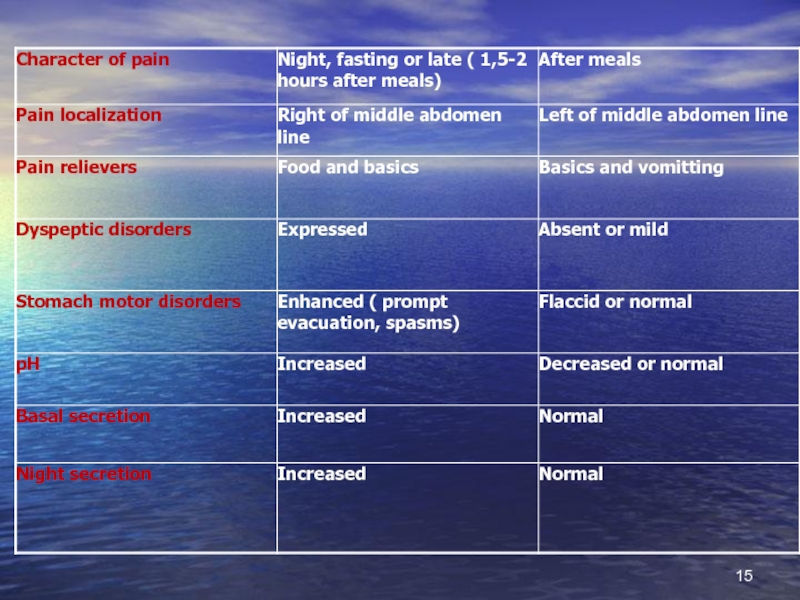
Слайд 12Clinics
Pain syndrome
Fasting pain appearance or 1,5-2 hours after feeding (
Moinigan rythm)
Nocturnal pain predominance
Intensity ranges from slight to severe unbearable
Localized in epigastrium . If accompanied GERD is present it can irradiate retrosternum space.
DYSPEPTIC SYNDROME
Heartburn ( usually together with GERD)
Acidic regurgitation
Vomiting with relieving pain
ABDOMEN PALPATION
Painfulness in epigastrium, sometimes local in pyloric-duodenal region
VAGOTONY SYMPTOMS (in teenagers):
Cold, moist palms
Hyperhydrosis
Acrocyanosis
Decreasing of BP
Pulse lability
SEASONAL FALL –SPRING EXACERBATIONS ARE TYPICAL FOR PUD
Nocturnal pain predominanceIntensity")
Слайд 13PUD peculiarities in children
Classic clinics can be seen less than in
50% patients
In 15% children complaints are absent ( silent ulcer)
In 3% patient first presentation of disease can be complicated ( by bleeding, perforation)
More younger the child more atypical clinics is seen
In 15% children complaints are absent ( silent ulcer)
In 3% patient first presentation of disease can be complicated ( by bleeding, perforation)
More younger the child more atypical clinics is seen


Слайд 16Most helpful diagnostic examining
Endoscopy .
X-ray (not obedient for non-complicated cases).
Examining of
secretory function (increasing of basal and stimulating secretion fractions)- is helpful to define functional disorders but not ulcer itself .
Helicobacter pyloric contamination.
Helicobacter pyloric contamination.
.Examining of secretory function (increasing of")
Слайд 17PUD classification
Severity (first defined, mild-recurrence once per year and less ,
moderate – relapse 2 times per year, severe – recurrence more than 2 times per year and complicatiuons).
Phase – exacerbation, partial remission, remission.
Clinic-endoscopic stage – fresh ulcer, scarring defect, scar, scar-ulcer deformity.
Ulcer localization – Stomach (cardiac, subcardial portion, little, big curvature, pyloric region; duodenum (bulbus, post bulbar region)
Gastritis character (superficial, atrophic, and localization of it) gastroduodenitis (active, erosions, hyperplastic, associated with H.P.)
Functional characteristics (with decreased acidic production, preserved or increased ).
Complications– penetration in pancreas, hepatoduodenal legamentum, gall bladder, liver, colon), acute bleeding , perforation, stenosis (compensated, subcompensated, decompensated, reflux-esophagitis)

Слайд 18PUD complications
BLEEDING – most frequent (80%) complication.
Clinics: emesis, melena, symptoms of
acute blood loss .
Coffe-like vomiting (Hb under influence of HCl turn into hematin with dark-brown color)
Melena – is black stool can be seen after the loss of more than 60 ml of blood ( ferrum sulfate realizes under the influence of digestive enzymes)
Symptoms of blood loss appear in the case of big blood loss weakness, nausea, paleness, tachycardia, cold, clammy sweat, BP decreasing, dizziness, vertigo, conscience loss sometimes
Bleeding can be hidden . In stool you can find hidden blood (positive Gregersen reaction)
 complication.Clinics: emesis, melena, symptoms of acute blood loss .Coffe-like")
Слайд 19Diagnostics approach algorithm in the case of PUD bleeding
Taking history and
patient inspection
Blood group and Rh defining
Endoscopy and X-ray of stomach and duodenum if necessary
Ultrasound diagnostics of abdomen
Blood group and Rh defining
Endoscopy and X-ray of stomach and duodenum if necessary
Ultrasound diagnostics of abdomen

Слайд 20
Perforation (8 %) – sudden knife-like pain in epigastrium, nausea, defans
of anterior abdomen wall , vomiting without condition improvement
Penetration (1,5 %) – spreading of ulcer into surrounding tissues. It can be defined by X-ray examining by changing of the near organs functioning
Pyloro- duodenal stenosis (11 %). Formed steadily . Accompanied by sensation of stomach overfilling, nausea, regurgitation, burning, vomiting with relief of condition. The splash sound in epigastrium. By X-ray stomach dilation with retardation of its emptying.
 – sudden knife-like pain in epigastrium, nausea, defans of anterior abdomen wall")
Слайд 21Differential diagnosis
Must be performed with acute symptomatic ulcers.
STRESS -ulcers They
can appear in burnings, trauma, freezing. Clinics is scanty. The first presentation can be bleeding, more rae –perforation.
Due to medicine influences Appear after consuming the medications that can disturb barrier properties of mucus (non-steroid and steroid drugs, cytostatics, etc). They are presented by asymptomatic course. Bleeding can be the first manifestation,
Hepatogenic. Can appear if inactivation of gastrin and histamine is impaired in liver. Clinics is vague and atypical, course is torpid, badly corrected by treatment.
Pancreogenic. Appear in the case of decreased production of bicarbonates and increased production of kinins. Pain syndrome is manifested and induced by food consuming. Course is constant.

Слайд 22
Endocrine. Very rare development in diabetes, hypothyroidism. Course of this ulcer
disease id similar to severe course of PUD.
Zollinger-Ellison syndrome – gastrin produced neoplasms (gastrinoma) . IT’s usually localized in antral part of stomach or in pancreas, in 16% of cases it can be malignant. It’s resistant to PUD therapy. Screening test is elevation of gastrin in fasting condition in serum.
Allergic ulceration more frequently can be developed in the case of food allergy.
In chronic renal failure due to impairment of gastrin degradation in kidneys and as a result disturb of protective barrier in stomach
In diffuse connective tissue disorders due to impairment of microcirculation .

Слайд 23
Clinics of symptomatic ulcers
Diagnostic difficulties
Absence of typical pain syndrome and dyspeptic
symptoms
Absence of seasonal periodic exacerbation
High risk of life threatening conditions ( bleeding, perforation)
Endoscopic data
Ulcers can be singular or multiple
Ulcer diameter usually is not more than 1 cm .
Shape of ulcer defect is oval or round, so called “punched ulcers”
Bottom of ulcer defect is plant crater-like.
Around the defect there is bright red crown, but inflammatory ring is absent.
Main localization is stomach
Prompt epithelization.

Слайд 24Treatment goals
To reduce PUD symptoms and provide reparation of ulcer defect
Eradicate
contamination of H.P. of mucus.
Not only get the healing of defect but restitute functional capacity of mucous membrane.
Prevent development of exacerbations and complications.
Not only get the healing of defect but restitute functional capacity of mucous membrane.
Prevent development of exacerbations and complications.

Слайд 25PUD treatment
PUD treatment is directed to
suppress aggression factors like acidic –peptic factor and contamination of mucous membrane by HELICOBACTER PYLORI .
Main principles :
Reject of smoking, alcohol taking.
Stop to get non-steroid and steroid medications, if it can’t be stopped to decrease dosages.
Rational feeding. It means frequent intake 5-6 times per day with excluding of spicy products. Diet N 1-b in the case of exacerbation signs.
Medication treatment.
HELICOBACTER PYLORI eradication
Suppressing of acidity and peptic factors production
Correct motor evacuative function .
Stimulation of reparative processes.

Слайд 26Medication treatment of PUD
PUD is obligatory indication for H.P. eradicative therapy
in any stage of disease
Treatment include first and second line of eradicative therapy.
First line is performed after diagnosis of PUD in any period (exacerbation or remission) and complications.
Control of its efficiency is performed a month later the treatment not earlier predominantly by noninvasive methods: breathing test (carbonic C13 or Helic-test) or test of H.P defining in stool.
If test is positive for H.P. second line therapy is proposed. If test is negative therapy is stopped.

Слайд 27 HELICOBACTER PYLORI eradication provides regression of inflammatory and dystrophic
changes and restitutes protective properties of stomach mucous membranes.
Antihelicobacter pylori medications – methronidazole, Clarythromycine, Amoxycylline, colloid Bismuthi subcytrate.
Approximal eradicative schemes:
Omeprazol + Clarythromycine + Methronidazole
Omeprazol + Amoxycilline + Clarythromycine
Omeprazole + colloid Bismuthi subcytrate +Methronidazole + Amoxycilline
COMBINED ANTIBACTERIAL MEDICATIONS.
Gastrostat (colloid Bismuthi subcytratis + Tetracycline + Methronidazole)
Gastropak – (colloid Bismuthi subcytratis + Amoxicilline + Methronidazole)
Pylorid (ranitidin + colloid Bismuthi subcytratis )
Helicocide (Amoxicilline+ Methronidazole)
Therapy according to schemes is continued for 7 days, later they live only antisecretory therapy.

Слайд 28Regulations for antihelicobacter therapy
If usage of the eradication scheme doesn’t provide
complete H.pylori eradication you needn’t to repeat it once more. It means that H.P . get resistance to one of the components in this scheme.
If usage of one scheme later another scheme don’t provide complete H.P. eradication you need to check susceptibility of Helicobacter pylori to all the spectrum of prescribed antibiotics.
Appearance of H.pylori in patient earlier than one year after eradication means recurrence of infection but not reinfection. You need to choose more effective treatment scheme.
Decreasing of antibiotic quantity in scheme leads to H. pylori resistance formation. After finishing of 7 day combined eradicative treatment you can prolong it for 4-5 days in the case o duodenal ulcer and 7-8 weeks in stomach one with usage of one antisecretory drug.

Слайд 29Main medications activity locuse
Parietal cell
Н+/K+-АТP ase
protone pomp
Histamine
Gastrin
Acetylcholine
ulcer
H.pylori
Mucouse-bicarbonate barrier
Н2-blockers
Proglumid
Atropin
Gastrocepin
Prostaglandins
Omeprazole
HCl
Antacids
Antibacterial drugs
Sucralfate
De-nol

Слайд 30Antisecretory medications
Selective M-cholinolytics (pirenzepim, gastrocepin)
Н2-histamine receptor blockers (ranitidin, famotidin)
Protone
pomp inhibitors – blockers of Н+/К+АТP –ase in parietal cells (omeprazole)
 Н2-histamine receptor blockers (ranitidin, famotidin)Protone pomp inhibitors – blockers")
Слайд 31Antisecretory therapy
1. Н2-histamine receptor blockers
Selectively block secretion of HCl
Decrease volume
of gastric juice
Decrease the level of pepsin
Cimetedine group – 1 generation (Cimetedin, Tagamet, Histodyl, Cimehexal, Neutronorm, rimamet)
Ranitidin group – 2 and 3 generation (Ranitidin, Ranisan, Zantak, Ulkodin, Zoran, Histak, Ranigast, Ranitab, Ranitard, Ranitin)
Famotidin group – 2 and 3 generation (Lecidyl, Gastrocydin, Quamatel, Famocyd, Ulfamid, Famodin)
Nizatidin group (Axid)
Roxatidin group (Roxan)

Слайд 322. Peripheral M- choline receptors blockers
(gastrocepin, pyrenzepin, gastrozem, gastril, pyren)
Suppress HCL
and pepsin production
Increase protective properties of ventricular mucus
3. Н+/К+-АТP ase blockers ( protone pomp inhibitors)
(omeprazol, omez, omeprol, omezak, ornatol, losek)
Inhibit HCl production
Increase protective properties of ventricular mucus
3. Н+/К+-АТP ase blockers ( protone pomp inhibitors)
(omeprazol, omez, omeprol, omezak, ornatol, losek)
Inhibit HCl production
Suppress HCL and pepsin productionIncrease protective")
Слайд 33Cytoprotectors
1. Film-forming medications(decrease backward diffusion of Hydrogen ion):
Colloid Bismuthi subcytrate, De-nol
(Tribimol, Ventrixol). Increase prostaglandin production, adsorb pepsin, has antihelicobacter activity.
Sukralfat (Venter, antepsyn). Connect Aluminium with sulfate polysaccharide, in acidic surroundings get adhesive properties. On the surface of erosions and ulcers perform complex compound with protein –helate and create mechanic protective barrier.
Film-forming medications are basic remedies in peptic ulcers with normal secretory function.
:Colloid Bismuthi subcytrate, De-nol (Tribimol, Ventrixol). Increase prostaglandin")
Слайд 34
2. PROSTOGLANDINS – increase bicarbonates and mucus production, increase protective layer
thickness, improve microcirculation. It’s mesoprostol (Arboprostyl, Enprostyl)
It must be taken before meals and before sleeping. Course is 4 weeks.
It must be taken before meals and before sleeping. Course is 4 weeks.

Слайд 35
If accompanied dysmotility is present (duodeno-gastral reflux, gastro-esophageal reflux) DOPA-receptor blockers
(cerucal, motilium) 1mg/kg TID or cizaprid (Coordinax, Propulsid) 0,4-0,5 mg/kg /day can be used.
Spasmolytics (no-spa, Papaverin, Platiohyllin, Buskopan)
Spasmolytics (no-spa, Papaverin, Platiohyllin, Buskopan)
 DOPA-receptor blockers (cerucal, motilium) 1mg/kg TID")
Слайд 36Bleeding treatment
Urgent hospitalization to provide endoscopic treatment (diathermo coagulation, laser coagulation).
Intravenous
infusion of haemostatic medications (Vicasol, Calcium, Androxol)
Oral intake of 5 % Sol. Of Aminocapronic acid with Thrombin and Androxol
Prescribing of Н2 –histamine blockers IM (Quamatel, 2mg/kg/day IV)
If bleeding is significant transfusion of plasma or blood (only of the same group)
Oral intake of 5 % Sol. Of Aminocapronic acid with Thrombin and Androxol
Prescribing of Н2 –histamine blockers IM (Quamatel, 2mg/kg/day IV)
If bleeding is significant transfusion of plasma or blood (only of the same group)
.Intravenous infusion of haemostatic medications")
Слайд 37
Duration of hospitalization in the case of Duodenum PUD is 28
days, in Stomach PUD – 30-35 days, in the case of severe course it can be 6-8 weeks.
After ulcer healing (phase of incomplete remission) treatment can be prolonged at ambulatory regimen. In the phase of remission sanatorium treatment is desirable.
After ulcer healing (phase of incomplete remission) treatment can be prolonged at ambulatory regimen. In the phase of remission sanatorium treatment is desirable.

Слайд 38
Efficacy criteria of therapy is clinic and endoscopic remission, exacerbation symptoms
absence, healing of ulcer defect and absence of inflammatory signs while endoscopic examining. Observation must be provided for 5 years. It can be finished if remission is stable for 5 years.

Слайд 39Dispensary
Doctor’s examination must be performed 2-4 times per year depending
on severity of disease.
If exacerbations are absent FGDS must be performed once per year. It will be done in the case of therapy inefficiency “on demand” during exacerbation period.
Stomach secretion must be examined by pH-metry once per year.
Stool analysis for hidden blood must be performed twice per year.
If exacerbations are absent FGDS must be performed once per year. It will be done in the case of therapy inefficiency “on demand” during exacerbation period.
Stomach secretion must be examined by pH-metry once per year.
Stool analysis for hidden blood must be performed twice per year.

Слайд 40
During complete remission period diet № 1 is taken for 4-6
mo.
Child is freed from physical training in the main group.
During dispensary period two times per year ( usually on fall and spring period) prophylactic treatment courses for 3-4 weeks are performed.
Sanatorium treatment can be recommended only in period of complete remission or period of recovery if signs of gastro-duodenitis are absent. If bleeding has been present sanatorium is permitted not earlier than 6 mo after gaining full remission ( such sanatorium will be preferable like Truskavets, Morshin, Berezovsky mineral waters, Ray-Yelenovka etc.)
If child has no exacerbations for 5 years and he is in complete remission he is stopped to undergo dispensary examination.

Слайд 41
Questions
Prevention of peptic ulcer disease
Frequency and prognosis
Clinical symptoms of peptic ulcer
disease
Additional (instrumental) methods of invastigations
Prevention of complications of peptic ulcer disease
Principles of treatment of peptic ulcer disease
Additional (instrumental) methods of invastigations
Prevention of complications of peptic ulcer disease
Principles of treatment of peptic ulcer disease
")