- Главная
- Разное
- Дизайн
- Бизнес и предпринимательство
- Аналитика
- Образование
- Развлечения
- Красота и здоровье
- Финансы
- Государство
- Путешествия
- Спорт
- Недвижимость
- Армия
- Графика
- Культурология
- Еда и кулинария
- Лингвистика
- Английский язык
- Астрономия
- Алгебра
- Биология
- География
- Детские презентации
- Информатика
- История
- Литература
- Маркетинг
- Математика
- Медицина
- Менеджмент
- Музыка
- МХК
- Немецкий язык
- ОБЖ
- Обществознание
- Окружающий мир
- Педагогика
- Русский язык
- Технология
- Физика
- Философия
- Химия
- Шаблоны, картинки для презентаций
- Экология
- Экономика
- Юриспруденция
Obstetric procedures презентация
Содержание
- 1. Obstetric procedures
- 2. Content: Procedures for fetal prenatal diagnosis: Ultrasound.
- 3. Ultrasound: Types: Tran abdominal. Transvagina Guidelines for
- 4. Guide for second and third trimesters: Fetal
- 5. Amniocentesis: Def: it is a technique for
- 6. Gestation age: from 11
- 7. Chorionic villus sampling: Dif: small samples of
- 8. Indications (CVS): Maternal age : 35 yo
- 9. Fetal blood sampling (cordocentesis) Indication: Assessment and
- 10. Complications: (cordocentesis )
- 11. Operative vaginal delivery: Forceps: Parts:
- 12. Cont. Forceps: Classification: Outlet forceps. Low forceps.
- 13. Pre-requisites for forceps application: Head engaged. Vertex
- 14. Complication of forceps: Lacerations and episiotomy. Urinary
- 15. Vacuum Extraction (Ventouse): Types: Metal cup vacuum.
- 16. Cont. complication of ventouse: 7. Macrosomia. 8.
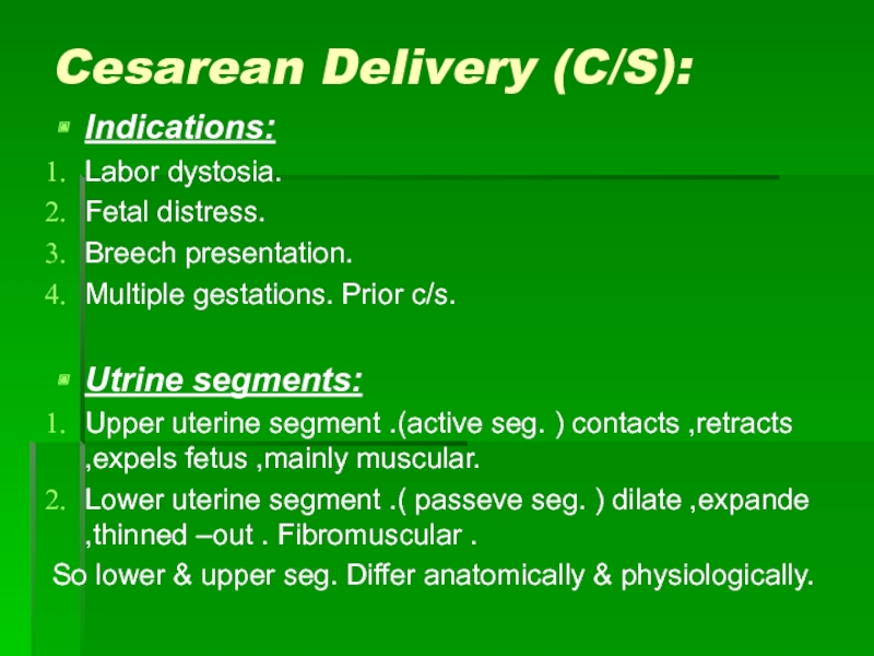
- 17. Cesarean Delivery (C/S): Indications: Labor dystosia. Fetal
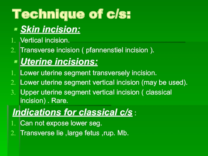
- 18. Technique of c/s: Skin incision: Vertical incision.
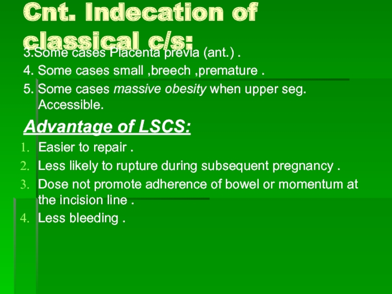
- 19. Cnt. Indecation of classical c/s: 3.Some cases
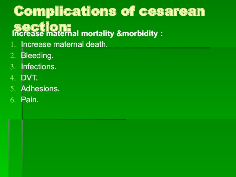
- 20. Complications of cesarean section: Increase maternal mortality
- 21. Vaginal birth after prior cesarean: Benefit: Short
- 22. Cont. VBAC: LSCS. Institute ER C/S with
- 23. External cephalic version :
- 24. Cont. ECV : AGOC recommendation ECV shoud
- 25. Cont. Contraindications ECV : 3. No reassuring
- 26. Cont. ECV: Preprocedure requisites: U/S , Bladder
Слайд 2Content:
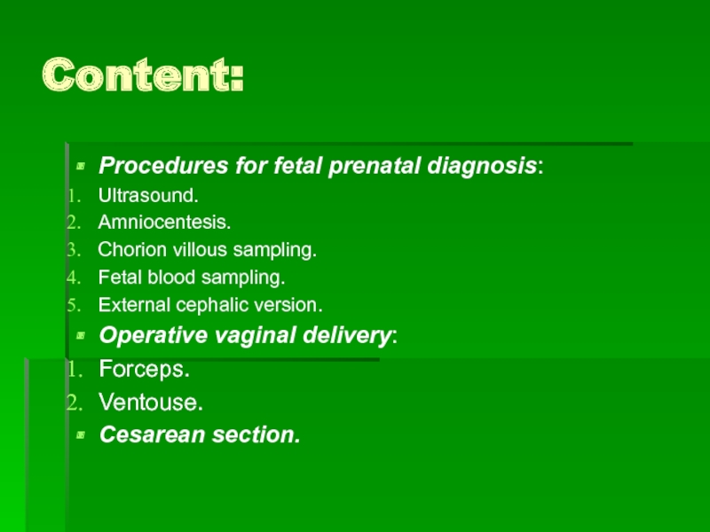
Procedures for fetal prenatal diagnosis:
Ultrasound.
Amniocentesis.
Chorion villous sampling.
Fetal blood sampling.
External cephalic version.
Operative
vaginal delivery:
Forceps.
Ventouse.
Cesarean section.
Forceps.
Ventouse.
Cesarean section.
Слайд 3Ultrasound:
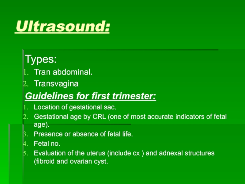
Types:
Tran abdominal.
Transvagina
Guidelines for first trimester:
Location of gestational sac.
Gestational age by CRL
(one of most accurate indicators of fetal age).
Presence or absence of fetal life.
Fetal no.
Evaluation of the uterus (include cx ) and adnexal structures (fibroid and ovarian cyst.
Presence or absence of fetal life.
Fetal no.
Evaluation of the uterus (include cx ) and adnexal structures (fibroid and ovarian cyst.
Слайд 4Guide for second and third trimesters:
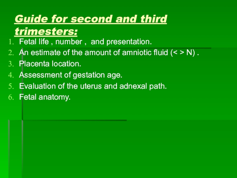
Fetal life , number , and
presentation.
An estimate of the amount of amniotic fluid (< > N) .
Placenta location.
Assessment of gestation age.
Evaluation of the uterus and adnexal path.
Fetal anatomy.
An estimate of the amount of amniotic fluid (< > N) .
Placenta location.
Assessment of gestation age.
Evaluation of the uterus and adnexal path.
Fetal anatomy.
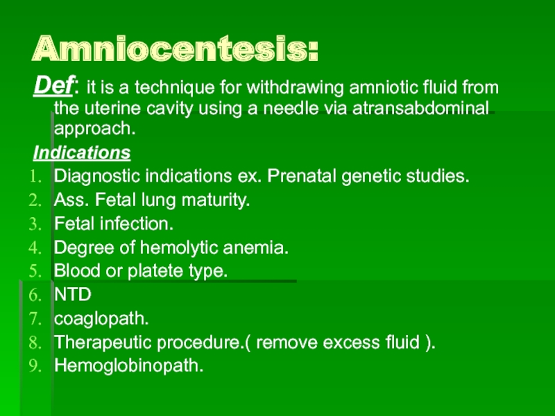
Слайд 5Amniocentesis:
Def: it is a technique for withdrawing amniotic fluid from the
uterine cavity using a needle via atransabdominal approach.
Indications
Diagnostic indications ex. Prenatal genetic studies.
Ass. Fetal lung maturity.
Fetal infection.
Degree of hemolytic anemia.
Blood or platete type.
NTD
coaglopath.
Therapeutic procedure.( remove excess fluid ).
Hemoglobinopath.
Indications
Diagnostic indications ex. Prenatal genetic studies.
Ass. Fetal lung maturity.
Fetal infection.
Degree of hemolytic anemia.
Blood or platete type.
NTD
coaglopath.
Therapeutic procedure.( remove excess fluid ).
Hemoglobinopath.
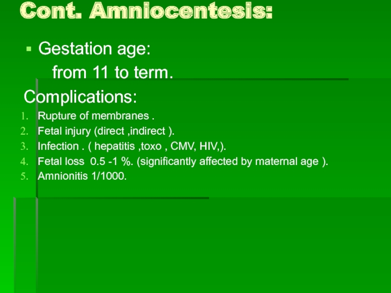
Слайд 6Gestation age:
from 11 to term.
Complications:
Rupture of membranes .
Fetal
injury (direct ,indirect ).
Infection . ( hepatitis ,toxo , CMV, HIV,).
Fetal loss 0.5 -1 %. (significantly affected by maternal age ).
Amnionitis 1/1000.
Infection . ( hepatitis ,toxo , CMV, HIV,).
Fetal loss 0.5 -1 %. (significantly affected by maternal age ).
Amnionitis 1/1000.
Cont. Amniocentesis:
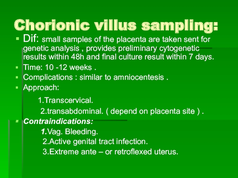
Слайд 7Chorionic villus sampling:
Dif: small samples of the placenta are taken sent
for genetic analysis , provides preliminary cytogenetic results within 48h and final culture result within 7 days.
Time: 10 -12 weeks .
Complications : similar to amniocentesis .
Approach:
1.Transcervical.
2.transabdominal. ( depend on placenta site ) .
Contraindications:
1.Vag. Bleeding.
2.Active genital tract infection.
3.Extreme ante – or retroflexed uterus.
Time: 10 -12 weeks .
Complications : similar to amniocentesis .
Approach:
1.Transcervical.
2.transabdominal. ( depend on placenta site ) .
Contraindications:
1.Vag. Bleeding.
2.Active genital tract infection.
3.Extreme ante – or retroflexed uterus.
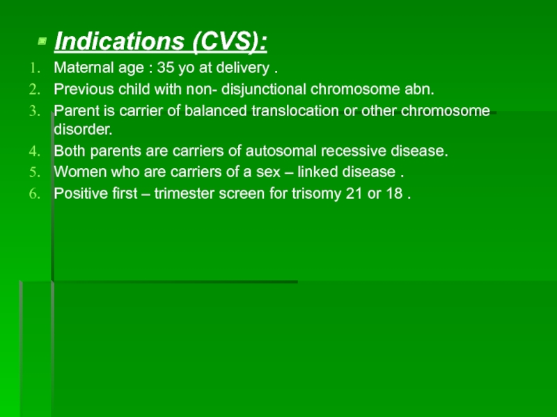
Слайд 8Indications (CVS):
Maternal age : 35 yo at delivery .
Previous child with
non- disjunctional chromosome abn.
Parent is carrier of balanced translocation or other chromosome disorder.
Both parents are carriers of autosomal recessive disease.
Women who are carriers of a sex – linked disease .
Positive first – trimester screen for trisomy 21 or 18 .
Parent is carrier of balanced translocation or other chromosome disorder.
Both parents are carriers of autosomal recessive disease.
Women who are carriers of a sex – linked disease .
Positive first – trimester screen for trisomy 21 or 18 .
Слайд 9Fetal blood sampling (cordocentesis)
Indication:
Assessment and treatment of confirmed red cell or
platelet alloimmunization.
Analysis of non – immune hydrope.
Karyotyping of fetal blood and congenital infection.
Analysis of metabolic & hematological status.
Procedure:
The operator punctures the umbilical vein , usually at or near its placental origin.
Analysis of non – immune hydrope.
Karyotyping of fetal blood and congenital infection.
Analysis of metabolic & hematological status.
Procedure:
The operator punctures the umbilical vein , usually at or near its placental origin.
Слайд 10Complications: (cordocentesis )
As amniocentesis.
Cord
hematoma.
Cord vessel bleeding.
Fetal – maternal hemorrhage.
Fetal bradycardia.
Fetal death.
Cord vessel bleeding.
Fetal – maternal hemorrhage.
Fetal bradycardia.
Fetal death.
Слайд 11Operative vaginal delivery:
Forceps:
Parts:
Blade.( cephalic ,pelvic curve )
Shank.
Lock. (sliding
lock , English lock )
Handle.
Function of forceps:
Traction.
Rotation.
Both.
Handle.
Function of forceps:
Traction.
Rotation.
Both.
Слайд 12Cont. Forceps:
Classification:
Outlet forceps.
Low forceps.
Midpelvic forceps.
High forceps.
Indications for forceps:
Heart disease , pulmonary
inj. Or compromise.
Intrapartum infection.
Exhaustion.
Prolonged second stage of labour.
Certain neurological condition.
Non- reassuring CTG.
Prolapsed cord , premature separation of placenta.
Intrapartum infection.
Exhaustion.
Prolonged second stage of labour.
Certain neurological condition.
Non- reassuring CTG.
Prolapsed cord , premature separation of placenta.
Слайд 13Pre-requisites for forceps application:
Head engaged.
Vertex presentation.
Known position.
Fully dilated cx.
Mb ruptured.
Role out
CPD.
Local or regional anesthesia.
Bladder should be empty.
Expert operator.
Local or regional anesthesia.
Bladder should be empty.
Expert operator.
Слайд 14Complication of forceps:
Lacerations and episiotomy.
Urinary dysfunction .
Rectal & anal dysfunction.
Febrile morbidity.
Low
Apgar score.
Cephalohematoma.
Caput.
Facial nerve injury.
Fetal truma ( Erb palsy ,fratured clavicle ).
Retinal hemorrhage.
Cephalohematoma.
Caput.
Facial nerve injury.
Fetal truma ( Erb palsy ,fratured clavicle ).
Retinal hemorrhage.
Слайд 15Vacuum Extraction (Ventouse):
Types:
Metal cup vacuum.
Soft cup vacuum (silastic cup ).
Indications &prerequisites:
as forceps.
Contraindications:
Inexperience.
Inability to ass. Fetal position.
High station.
Suspicion of CPD.
Non vertex presentations.
Fetal coagulopathy.
Contraindications:
Inexperience.
Inability to ass. Fetal position.
High station.
Suspicion of CPD.
Non vertex presentations.
Fetal coagulopathy.
Слайд 16Cont. complication of ventouse:
7. Macrosomia.
8. Recent scalp blood sampling.
Vacuum extraction is
reserved for fetus 34 weeks or older.
Complications:
Scalp lacerations & bruising.
Subgaleal hematoma.
Cephalohematomas.
Intracranial hemorrhage.
Neonatal jaundice.
Subconjuctival hemorrhage.
Clavicular fracture.
Complications:
Scalp lacerations & bruising.
Subgaleal hematoma.
Cephalohematomas.
Intracranial hemorrhage.
Neonatal jaundice.
Subconjuctival hemorrhage.
Clavicular fracture.
Слайд 17Cesarean Delivery (C/S):
Indications:
Labor dystosia.
Fetal distress.
Breech presentation.
Multiple gestations. Prior c/s.
Utrine segments:
Upper uterine
segment .(active seg. ) contacts ,retracts ,expels fetus ,mainly muscular.
Lower uterine segment .( passeve seg. ) dilate ,expande ,thinned –out . Fibromuscular .
So lower & upper seg. Differ anatomically & physiologically.
Lower uterine segment .( passeve seg. ) dilate ,expande ,thinned –out . Fibromuscular .
So lower & upper seg. Differ anatomically & physiologically.
Слайд 18Technique of c/s:
Skin incision:
Vertical incision.
Transverse incision ( pfannenstiel incision ).
Uterine incisions:
Lower
uterine segment transversely incision.
Lower uterine segment vertical incision (may be used).
Upper uterine segment vertical incision ( classical incision) . Rare.
Indications for classical c/s :
Can not expose lower seg.
Transverse lie ,large fetus ,rup. Mb.
Lower uterine segment vertical incision (may be used).
Upper uterine segment vertical incision ( classical incision) . Rare.
Indications for classical c/s :
Can not expose lower seg.
Transverse lie ,large fetus ,rup. Mb.
Слайд 19Cnt. Indecation of classical c/s:
3.Some cases Placenta previa (ant.) .
4. Some
cases small ,breech ,premature .
5. Some cases massive obesity when upper seg. Accessible.
Advantage of LSCS:
Easier to repair .
Less likely to rupture during subsequent pregnancy .
Dose not promote adherence of bowel or momentum at the incision line .
Less bleeding .
5. Some cases massive obesity when upper seg. Accessible.
Advantage of LSCS:
Easier to repair .
Less likely to rupture during subsequent pregnancy .
Dose not promote adherence of bowel or momentum at the incision line .
Less bleeding .
Слайд 20Complications of cesarean section:
Increase maternal mortality &morbidity :
Increase maternal death.
Bleeding.
Infections.
DVT.
Adhesions.
Pain.
Слайд 21Vaginal birth after prior cesarean:
Benefit:
Short stay at hospital.
Less blood loss .
Fewer
transfusion.
Fewer infections.
Fewer DVT.
Risk of VBAC:
Uterine rupture.
Hysterectomy.
Increase morbidity .(b.tran.).
Fetal death or damage.
Fewer infections.
Fewer DVT.
Risk of VBAC:
Uterine rupture.
Hysterectomy.
Increase morbidity .(b.tran.).
Fetal death or damage.
Слайд 22Cont. VBAC:
LSCS.
Institute ER C/S with anest &physician immd. Available.
NO contraindications for
vag. Delivery.
No other uterine scare.
No other uterine scare.
Слайд 23External cephalic version :
( ECV )
Definition: It
is a procedure by which an obstetrician turns the baby from the breech to the cephalic position by manipulating the baby through the maternal abdomen .
The procedure increases the chance of cephalic presentation at onset of labor and decreases the rate of cesarean delivery.
The procedure increases the chance of cephalic presentation at onset of labor and decreases the rate of cesarean delivery.
Слайд 24Cont. ECV :
AGOC recommendation ECV shoud be available and offered to
women with breech presentation at term .
Risk : Discomfort , ERC/S , Transient bradycardia is less common ,placental abruption , premature labor .
Cost : cost-effective .
Contraindications :
Indications for c/s .
Rupture membranes .
Risk : Discomfort , ERC/S , Transient bradycardia is less common ,placental abruption , premature labor .
Cost : cost-effective .
Contraindications :
Indications for c/s .
Rupture membranes .
Слайд 25Cont. Contraindications ECV :
3. No reassuring CTG.
4. Hyper extended fetal head.
5. Significant fetal or uterine anomaly .
6. Abruptio placentae .
Relative contraindications :
Previous c/s .
Maternal hypertension, obesity .
IUGR.
Слайд 26Cont. ECV:
Preprocedure requisites: U/S , Bladder empty .
Timing : completed 36
weeks of gestation .
Women who RH – negative receive anti-D immunoglobulin .
If the procedure is un successful or the baby reverts to breech , a retrial of version to be considered .
Women who RH – negative receive anti-D immunoglobulin .
If the procedure is un successful or the baby reverts to breech , a retrial of version to be considered .
