- Главная
- Разное
- Дизайн
- Бизнес и предпринимательство
- Аналитика
- Образование
- Развлечения
- Красота и здоровье
- Финансы
- Государство
- Путешествия
- Спорт
- Недвижимость
- Армия
- Графика
- Культурология
- Еда и кулинария
- Лингвистика
- Английский язык
- Астрономия
- Алгебра
- Биология
- География
- Детские презентации
- Информатика
- История
- Литература
- Маркетинг
- Математика
- Медицина
- Менеджмент
- Музыка
- МХК
- Немецкий язык
- ОБЖ
- Обществознание
- Окружающий мир
- Педагогика
- Русский язык
- Технология
- Физика
- Философия
- Химия
- Шаблоны, картинки для презентаций
- Экология
- Экономика
- Юриспруденция
Meningococcal infection презентация
Содержание
- 1. Meningococcal infection
- 2. Etiology the causative agent is meningococcus
- 3. Epidemiology the sources of infection are
- 4. Pathogenesis and Pathology The portal of
- 5. Purulent meningitis develops due to the ingress
- 6. Acute swelling and edema of the brain
- 7. Classification Location form: Nasopharyngitis; Carriers. Generalized
- 8. Nasopharyngitis headache, painful swallowing, subfebrile temperature
- 9. Meningitis The onset of the disease
- 10. Meningeal symptoms hyperesthesia of the skin
- 11. Spinal fluid increased pressure turbid
- 12. Blood leukocytosis (up to 20-40-109/1)
- 13. Meningococcemia The onset is acute and
- 15. Hypertoxic (fulminating) form A sudden turbulent
- 16. Waterhouse-Friderichsen syndrome Multiple petechiae and hemorrhage
- 17. Features peculiar to meningitis in infants
- 18. Complications Pneumonia, Purulent otitis Hydrocephalus
- 19. Diagnosis the clinical symptomatology and its
- 20. Differential diagnosis Tuberculosis meningitis starts gradually
- 21. Differential diagnosis Acute serous meningitis differs
- 22. Meningeal form of poliomyelitis The cerebrospinal
- 23. Other purulent meningitis (staphylococcus, pneumococcus, Afanasyev-Pfeiffer bacillus,
- 24. Meningococcemia of thrombopenic purpura and hemorrhagic vasculitis
- 25. Prognosis Mortality from epidemic meningitis was
- 26. Etiotropic treatment Penicillin was first given
- 27. Toxicosis can be controlled by administration of
- 28. Prophylaxis The following in an epidemic
- 29. Acute Epidemic Poliomyelitis
- 30. Etiology the causative agent of polyomyelitis (Poliovirus
- 31. Epidemiology Sources of infection - patients with
- 32. Pathogenesis The most probable portal of entry
- 33. Clinical Manifestations The incubation period of poliomyelitis
- 34. Preparalytic stage The disease starts acutely with
- 35. Paralytic stage The temperature falls at the
- 36. Signs of damage of the peripheral neuron
- 37. Stage of residual phenomena The stage
- 38. Clinical forms of poliomyelitis paralytic poliomyelitis:
- 39. Paralytic poliomyelitis The spinal form is characterized
- 40. Aparalytic poliomyelitis The visceral (or abortive) form
- 41. Diagnosis Rapid investigation suspected cases
- 42. Viral Isolation isolate
- 43. Treatment NO curative treatment Supportive care:
- 44. Prophylaxis Isolation of poliomyelitis patient and suspected

Слайд 2Etiology
the causative agent
is meningococcus
(Neisseria meningitidis).
this microorganism
has the
grows on media containing human protein (blood serum)
very unstable and perishes rapidly outside the organism
several serotypes of meningococ (A, B, C, D, Z, X, and Y) have been discovered
. this microorganism has the form of a diplococcus,")
Слайд 3Epidemiology
the sources of infection are patient and carriers
meningococcus expel
Infection is transmitted by the aerial-droplet route
The susceptibility of man to meningococcal infection is slight: the susceptibility index does not exceed 0.5 %
The meningococcal infection is characterized by periodic rises of the incidence every 10-15 year or longer

Слайд 4Pathogenesis and Pathology
The portal of the infection entry is the
The carrier state develops frequently, while nasopharyngitis and generalized form (in 0.5-1 % of cases) occurs significantly less frequently
The important role in mingococcemia belongs to marked intoxication with the endotoxin released during decomposition of the microbial bodies - microcirculation is thus affected to provoke thrombosis and extravasates
Necrosis in the adrenal glands with diffuse hemorrhages and decomposition of the glandular tissue - fulminating forms (Waterhause-Friderichsen syndrome )

Слайд 5Purulent meningitis develops due to the ingress of the meningococcus into
Pathogenesis and Pathology
Purulent exudates is particularly abundant in the base, and on the surface of the frontal and parietal lobes of the brain - "purulent cap"

Слайд 6Acute swelling and edema of the brain can cause protrusion of
Pathogenesis and Pathology

Слайд 7Classification
Location form:
Nasopharyngitis;
Carriers.
Generalized form
Meningitis;
Mingococcemia;
Fulminating form;
Meningitis+ mingococcemia.
Atypical form:
Iridocyclochorioiditis;
Pneumonia
Endocarditic.

Слайд 8Nasopharyngitis
headache, painful swallowing, subfebrile temperature
hyperemia of the nasopharyngeal mucosa
rhinitis with scanty discharge, and difficult nasal breathing

Слайд 9Meningitis
The onset of the disease is usually violent, and a
The patient's posture is lying on his side with head tossed back and legs flexed to the abdomen

Слайд 10Meningeal symptoms
hyperesthesia of the skin and increased sensitivity to light
stiffness of the occipital muscles
Kernig's
Brudzinsky's
Mental disturbances are also frequent (lethargy, drowsiness, etc.).
In young children clonik and tonic convulsions are not infrequent

Слайд 11Spinal fluid
increased pressure
turbid and purulent
neutrophilosis (from several hundreds
considerable protein content (up to 1-2 g/l)
sugar content is lowered

Слайд 12Blood
leukocytosis (up to 20-40-109/1)
neutrophilosis with a shift to the
aneosinophilia
the ESR is considerably increased
 neutrophilosis with a shift to the left aneosinophilia the ESR")
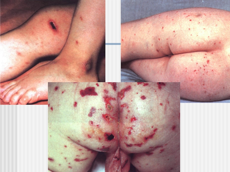
Слайд 13Meningococcemia
The onset is acute and violent, with intermittent fever
The
size; they are
hard on palpation
and are often
elevated
Meningococcal
are found in blood
smears taken
from the periphery
of the lesions

Слайд 15Hypertoxic (fulminating) form
A sudden turbulent onset
Severe toxemia (uncontrollable vomiting,
Meningeal symptoms are sharply pronounced
Death usually ensues within 12 to 24 hours after the onset
Swelling of the brain and protrusion of the cerebellar tonsils into the great foramen is one of the frequent causes of death
 form A sudden turbulent onset Severe toxemia (uncontrollable vomiting, convulsions, mental confusion, cardiovascular")
Слайд 16Waterhouse-Friderichsen syndrome
Multiple petechiae and hemorrhage into the skin
The arterial
progressively
The pulse is rapid and hard
Cyanosis, vomiting
(often with blood) and convulsions
The patient dies in 16-30
hours after the onset
of the disease unless an urgent
and effective therapy is given

Слайд 17Features peculiar to meningitis in infants
The disease is accompanied with
Frequent dyspeptic disturbances
Infants cry loudly
Meningeal symptoms and red dermographism are often mild or absent
Even with modern methods of treatment, mortality remains high

Слайд 18Complications
Pneumonia,
Purulent otitis
Hydrocephalus
The symptoms of which appeared already at
Paralysis, paresis
Asthenic syndrome, headache
Various functional disorders

Слайд 19Diagnosis
the clinical symptomatology and its course: acute onset and rapid
The most important diagnostic aid is lumbar puncture and examination of the cerebrospinal fluid
The diagnosis is undiscutable when meningococcus is detected by bacterioscopy or is found in a cerebrospinal fluid culture

Слайд 20Differential diagnosis
Tuberculosis meningitis
starts gradually and is accompanied with moderate pyrexia
anamnesis and the results of tuberculin tests
the X-ray of the lungs
cerebrospinal fluid is slightly opalescent; cell count is moderately increased due to an increase in the lymphocyte number; sugar and CL content is lowered; protein is elevate

Слайд 21Differential diagnosis
Acute serous meningitis
differs in the cerebrospinal fluid findings :

Слайд 22Meningeal form of poliomyelitis
The cerebrospinal fluid is transparent
A slight
Lymphocytes predominate among the cells
Differential diagnosis

Слайд 23Other purulent meningitis (staphylococcus, pneumococcus, Afanasyev-Pfeiffer bacillus, streptococcus )
develops secondarily to
gram-positive cocci and diplococci are found in the cerebrospinal fluid
Differential diagnosis
develops secondarily to purulent otitis, pneumonia, sepsis")
Слайд 24Meningococcemia of thrombopenic purpura and hemorrhagic vasculitis
meningococcemia is characterized by
Accurate diagnosis is established bacteriologically
Differential diagnosis

Слайд 25Prognosis
Mortality from epidemic meningitis was very high (30 to 40
The worst outcome in meningitis is prognoses in cases with the Waterhouse-Frederickson syndrome and the hypertoxic clinical form
 The")
Слайд 26Etiotropic treatment
Penicillin was first given dose of 300 000-400 000
Levomycetin sodium succinate can be given (100 mg/kg a day), ampicillin (150-200 mg/ kg a day), cephalosporins, oxacillin or methicillin are also recommended
Stopped antibiotic therapy need after sanayshin liquor: citosis is less then 100 cell of lymphocytes!

Слайд 27Toxicosis can be controlled by administration of large amounts of liquids
Dehydration therapy should be especially intensive in the presence of brain swelling
Corticosteroids should be given simultaneously 5-10-15 mg/kg with septic shock
Pathogenetic treatment

Слайд 28Prophylaxis
The following in an epidemic focus
The patient is hospitalized
Contacts and carriers should be treated with rifampicini for 3 days as a prophylactic measure, the standard dose being given 3 times a day
Terminal disinfection is carried out after isolation of the patient
Polysaccharide meningococcal vaccines have been recently developed in some countries


Слайд 30Etiology
the causative agent of polyomyelitis (Poliovirus hominis)
a very small virus
contains RNA
is
Three types of poliovirus (I, II, III) are known
a very small viruscontains RNAis very stable in the")
Слайд 31Epidemiology
Sources of infection - patients with clinically manifest poliomyelitis, persons suffering
The infectivity of patients is greatest during the acute stage. Most are free of the virus in 15 to 20 days after an attack
The mechanism of infection - of fecal mode of transmission
Susceptibility to poliomyelitis is low (75 to 90 % )

Слайд 32Pathogenesis
The most probable portal of entry of the infection - the
The poliomyelitis virus is isolated, as a rule, from lesions of the nervous system
The most pronounced pathological changes are in the ventral horns of the gray matter of the cervical and lumbar enlargements of the spinal cord
The nerve cells undergo dystrophic necrotic changes, and perish

Слайд 33Clinical Manifestations
The incubation period of poliomyelitis averages from 5 to 14
Four stages are distinguished in the course of the disease:
a) initial (preparalytic),
b) paralytic,
c) restitution,
d) the stage of residual phenomena

Слайд 34Preparalytic stage
The disease starts acutely with a marked rise of temperature
Catarrh
General and local hyperhidrosis
Symptoms of irritation on the nervous system : headache, vomiting, adynamia, lassitude, drowsiness or insomnia, sometimes delirium, tremor, muscular jerking, and convulsions
This stage usually lasts from 2 to 5 days

Слайд 35Paralytic stage
The temperature falls at the end of the initial stage,
Paralysis usually suddenly; may wake up paralyses in the morning ("morning paralysis")
Careful examination will have revealed hypotonia, muscular weakness, and loss of reflexes

Слайд 36Signs of damage of the peripheral neuron characterize
the paresis and paralysis
absence of tendon reflexes,
cutaneous reflexes may also disappear,
muscular appear one or two weeks after the onset of paralysis

Слайд 37Stage of residual phenomena
The stage of residual phenomena is characterized

Слайд 38Clinical forms of poliomyelitis
paralytic poliomyelitis:
a) spinal,
b) bulbar,
c) pontine,
d) encephalitic
aparalytic poliomyelitis:
visceral (or abortive)
meningeal
 spinal, b) bulbar, c) pontine, d) encephaliticaparalytic poliomyelitis:visceral (or")
Слайд 39Paralytic poliomyelitis
The spinal form is characterized by flaccid paralysis of the
The bulbar form, which is fraught with the greatest danger, is accompanied with swallowing, speech, and respiratory disturbances
The pontine form is expressed in implication of the nucleus of the facial nerve with paresis of the facial muscles
The encephalitic form is characterized by general cerebral phenomena and symptoms of focal lesions in the brain

Слайд 40Aparalytic poliomyelitis
The visceral (or abortive) form shows symptoms of the initial
In the meningeal form there are the same signs as in the visceral, with meningeal symptoms in addition. Findings in the cerebrospinal fluid - elevation of cell count (lymphocytes) and a normal or slightly elevated protein content
 form shows symptoms of the initial stage of poliomyelitis. There")
Слайд 41Diagnosis
Rapid investigation suspected cases
critical to identifying
Clinical case definition
Acute onset of a flaccid paralysis of one or more limbs with decreased or absent tendon reflexes in the affected limbs, without other apparent cause, and without sensory or cognitive loss.

Слайд 42Viral Isolation
isolate wild polio virus from stool
do genetic “finger printing” of virus to see wild type and where from
Serology
neutralizing antibodies: early and may be high
by the time the patient is hospitalized
may not see 4 fold rise in titer
Laboratory Diagnosis

Слайд 43Treatment
NO curative treatment
Supportive care:
aseptic meningitis- fluids, acetomenоphen,
rest until fever
paralysis- pain medications, +/-ventilator,
manage muscle spasms, treat 2o infection,
longer term –physiotherapy & occupational therapy

Слайд 44Prophylaxis
Isolation of poliomyelitis patient and suspected cases - hospitalization in special
After the patient is isolated (for 21 days from the onset of the disease) final disinfections is performed in his swelling
Contacts are observed for 20 days after isolation of the patient
Active immunization - with pertussis-diphtheria-tetanus vaccine beginning from 3 months of age 3 times with 30 days