Franc Hudspeth, PhD, NCC, RPh, RPT-S, ACS
Mississippi LPC & Board Qualified Supervisor
Director of the Institute for Play Therapy &
Assistant Professor of Counselor Education,
Henderson State University
Editor, International Journal of Play Therapy
hudspee@hsu.edu
Alabama Association for Play Therapy
8:30 am-4:30 pm
- Главная
- Разное
- Дизайн
- Бизнес и предпринимательство
- Аналитика
- Образование
- Развлечения
- Красота и здоровье
- Финансы
- Государство
- Путешествия
- Спорт
- Недвижимость
- Армия
- Графика
- Культурология
- Еда и кулинария
- Лингвистика
- Английский язык
- Астрономия
- Алгебра
- Биология
- География
- Детские презентации
- Информатика
- История
- Литература
- Маркетинг
- Математика
- Медицина
- Менеджмент
- Музыка
- МХК
- Немецкий язык
- ОБЖ
- Обществознание
- Окружающий мир
- Педагогика
- Русский язык
- Технология
- Физика
- Философия
- Химия
- Шаблоны, картинки для презентаций
- Экология
- Экономика
- Юриспруденция
Medicated Children and Adolescents in Play Therapy. Therapists about the Intersection of Neurobiology and Psychopharmacology презентация
Содержание
- 1. Medicated Children and Adolescents in Play Therapy. Therapists about the Intersection of Neurobiology and Psychopharmacology
- 2. Goals for Today Following the workshop, participants
- 3. Brain Complexities
- 4. Nervous System
- 5. Nervous System (cont) Sympathetic NS Arouses (fight-or-flight)
- 6. Endocrine System The Endocrine System is the
- 7. The Basic Brain Self-regulation, problem solving, goal
- 8. Brainstem
- 9. The Limbic System is a doughnut-shaped system
- 10. The “little brain” attached to the rear
- 11. Amygdala The Amygdala [ah-MIG-dah-la] consists of two
- 12. Hypothalamus The Hypothalamus lies below (hypo) the
- 13. The Cerebral Cortex The intricate fabric of
- 14. Functions of the Cortex The Motor Cortex
- 15. Brain Growth AGE
- 16. Brain Changes At birth, most neurons
- 17. Brain Changes (cont) Overproduction of neurons and
- 18. Brain Changes (cont) Anatomical studies of brain
- 19. Brain Changes (cont) Myelin & Age
- 20. Brain Changes - Critical Events (Toga & Mazziotta, 2000)
- 21. Brain Changes and Important Developments Brain areas
- 22. Impacting Brain Development Genes form neurons, connections
- 23. Brain Areas and Anatomical Development Brainstem (0-1)--Regulation
- 24. Brain Areas and Anatomical Development Brain stem
- 25. Normal Development and Regulation Consider: The
- 26. “DIR” Model (Greenspan & Wieder, 1997; Willis,
- 27. Functional Emotional Developmental Levels (Greenspan & Wieder,
- 28. Individual Differences Sensory Processing systems Cortical
- 29. Relational Context in Early Childhood Parent –
- 30. Relational Context in Early Childhood Socio-Emotional Co-Regulation
- 31. Adaptive Functioning (Shore, 2001, 2009)
- 32. The Right Brain
- 33. Order of
- 34. The Ventral
- 35. What’s Functional? 3 Types of
- 36. Neurobiology and Attachment Secure Attachment- a person
- 37. Attachment Neurobiology Process
- 38. Polyvagal Theory The more primitive branch elicits
- 39. Polyvagal Theory The vagus nerve is a
- 40. Polyvagal Theory Dorsal branch unmylenated primal survival
- 41. Okay, So Let’s Consider Dysfunction and Dysregulation?
- 42. Abnormal Development and Dysregulation Consider: The
- 43. Attachment Trauma/Disturbances Impairments in the development of
- 44. Right Brain Development: Affect Regulation (Schore, 2001)
- 45. Traumatic Brain Injury Childhood illnesses (high fevers,
- 46. The Neurochemical Origins of Disruptive Behaviors Those
- 47. Disruptive Behaviors, Neurotransmitters, and Brain Regions Emotional
- 48. Disruptive Behaviors, Neurotransmitters, and Brain Regions (cont)
- 49. Another Point We Now Have a Big Problem!
- 50. The ACE Study (Anda et al., 2005;
- 51. CDC (1998-2010)
- 52. Stress, the Brain, & the Body Stress
- 54. Early Childhood Disturbances from Trauma and Risk
- 55. The Continuum Attachment Disturbance ADHD, Bipolar
- 56. What’s The Point? We Now Have
- 57. Neurotransmitters Categorized into three major groups:
- 58. Neurotransmitters & Function Acetylcholine - voluntary movement
- 59. Neurotransmitter (Excitation vs. Inhibition) EXCITATORY Acetylcholine Aspartate
- 60. Dopamine (DA) Dopamine is transmitted via three
- 61. Serotonin (5-HT) The principal centers for serotonergic
- 62. Norepinephrine (NE) Many regions of the brain
- 63. Gamma-aminobutyric acid (GABA) GABA is the main
- 64. Glutamate In the normal brain the prominent
- 65. Acetylcholine (Ach) There are three Acetylcholine pathways
- 66. Transmission
- 67. Research, Use, & Age >6 months –diazepam
- 68. Research, Use, & Age (cont) >7yrs- fluoxetine
- 70. Gamma-aminobutyric acid (GABA) GABA is the main
- 71. Antianxiety Agents GABA receptors Valium (diazepam) Ativan (lorazepam) Klonopin (clonazepam) Xanax (alprazolam)
- 72. Antianxiety Agents (cont) Valium/Ativan/Klonopin/Xanax Clumsiness Sleepiness Dizziness Irritability Unsteadiness Confusion Problems with memory
- 73. Serotonin (5-HT) The principal centers for serotonergic
- 74. Antianxiety Agents (cont) 5HT Receptors Buspar (buspirone)
- 75. Antianxiety Agents (cont) 5HT Buspar Confusion, Dizziness,
- 76. Norepinephrine (NE) Many regions of the brain
- 77. Serotonin (5-HT) The principal centers for serotonergic
- 78. Antidepressants TCA (NE and/or 5HT reuptake presynaptic)
- 79. Antidepressants (cont) TCA Elavil/Tofranil/Pamelor Fatigue Drowsiness/Insomnia Mild Tremors Nightmares Restlessness Confusion
- 80. Serotonin (5-HT) The principal centers for serotonergic
- 81. Antidepressants (cont) SSRI (selective seratonin reuptake inhibitors)
- 82. Antidepressants (cont) SSRI Celexa/Prozac/Paxil/Zoloft/Lexapro/Viibryd Agitation Nervousness Fatigue Sleep Problems Vertigo Sexual Side Effects
- 83. Antidepressants (cont) MAOI (monoamine oxidase inhibitors) Nardil (phenelzine) Parnate (tranylcypromine) Marplan (isocarbozide)
- 84. Antidepressants (cont) MAOI Nardil/Parnate/Marplan Dizziness Headache Sleep Problems
- 85. Norepinephrine (NE) Many regions of the brain
- 86. Serotonin (5-HT) The principal centers for serotonergic
- 87. Antidepressants (cont) MISC (MOA unclear) Desyrel
- 88. Antidepressants (cont) MISC Desyrel/Wellbutrin/Effexor/Serzone/Cymbalta/ Pristiq/Remeron
- 89. , Intake Gathering Information Initial Treatment Plan
- 90. Gathering Information The Initial Play Therapy Session
- 91. Intake Past medications: List, in chronological order,
- 92. Intake Current medications: List, in chronological order,
- 93. Medication/Behavioral/Cognitive/Emotional/Developmental Time Line
- 94. The Initial Treatment Plan How will you
- 96. Addressing Medication Side Effects in the Treatment
- 97. Left and Right Brain LEFT BRAIN FUNCTIONS
- 98. Working with Lethargy in Play Therapy
- 99. Working with Lethargy in Play Therapy (cont)
- 100. Dopamine (DA) Dopamine is transmitted via three
- 101. Antipsychotics Phenothiazine Derv. (DA receptor antagonist)
- 102. Antipsychotics (cont) Phenothiazine derv. Thorazine/Stelazine/Mellaril Akathisia Akinesia Sleepiness Cognitive Blunting Stiffness
- 103. Antipsychotics (cont) Phenylbutylpiperadine derv. Haldol (haloperidol) Orap (pimozide)
- 104. Antipsychotics (cont) Phenylbutylpiperadine derv. Haldol/Orap Akathisia Akinesia Blurred Vision Sleepiness Cognitive Blunting
- 105. Dopamine (DA) Dopamine is transmitted via three
- 106. Serotonin (5-HT) The principal centers for serotonergic
- 107. Glutamate In the normal brain the prominent
- 108. Acetylcholine (Ach) There are three Acetylcholine pathways
- 110. Antipsychotics (cont) Dibenzapine derv. Loxitane (loxapine)
- 111. Antipsychotics (cont) Dibenzapine derv. Loxitane/Zyprexa/Seroquel
- 112. Antipsychotics (cont) Dihydroindolones Geodone (ziprasidone) Moban (molindone)
- 113. Antipsychotics (cont) Dihydroindolones Geodone/Moban Sleepiness Confusion Quinolinone
- 114. Working With Cognitive Cloudiness in Play Therapy
- 115. Working With Cognitive Cloudiness in Play Therapy
- 116. Working With Cognitive Cloudiness in Play Therapy
- 117. Working With Emotional Blunting in Play Therapy Rhythm Music Dance Bibliotherapy
- 118. Working With Emotional Blunting in Play Therapy
- 119. Working With Emotional Blunting in Play Therapy
- 120. Working with Coordination Difficulties in Play Therapy
- 121. Gross Motor Skills Involve the following in
- 122. Gross Motor Skills (cont) Involve the following
- 123. Fine Motor Skills Involve the following in
- 124. Fine Motor Skills (cont) Crafts
- 125. Other Things Consult or get to know an Occupational Therapist
- 126. Dopamine (DA) Dopamine is transmitted via three
- 127. CNS Stimulants Analeptic Provigil (modafinil)
- 128. CNS Stimulants (cont) Analeptic Provigil Irritability
- 129. CNS Stimulants (cont) Non-Amphetamines Ritalin/Concerta/Metadate/Methylin (methylphenidate) Cylert (pemoline) Focalin (dexmethylphenidate) Daytrana (methylphenidate)---Patch
- 130. CNS Stimulants (cont) Non-Amphetamines Ritalin/Concerta/Daytrana/Metadate/Methylin Sleep Problems
- 131. Norepinephrine (NE) Many regions of the brain
- 132. MISC ADHD Medications Strattera (atomoxetine) potent inhibitor of presynaptic NE transporter
- 133. MISC ADHD Medications (cont) Strattera Fatigue Sleep Disturbance
- 134. Working with Agitation/Aggression in Play Therapy
- 135. Working with Agitation/Aggresion in Play Therapy (cont)
- 136. Gamma-aminobutyric acid (GABA) GABA is the main
- 137. Sedative/Hypnotics (GABA) Newer Ambien (zolpidem) ProSom
- 138. Sedative/Hypnotics (cont) GABA Ambien/Prosom/Lunesta/Sonata/Halcion/Restoril Fatigue Clumsiness
- 139. Sedative/Hypnotics (cont) Melatonin Rozerem (ramelteon) Fatigue Clumsiness
- 140. Gamma-aminobutyric acid (GABA) GABA is the main
- 141. Anticonvulsants/Psychiatric Uses Tegretol/Carbatrol (carbamazepine) Trileptal (oxcarbazepine) Neurontin
- 142. Anticonvulsants/Psychiatric Uses (cont) Tegretol/Carbatrol Dizziness, Drowsiness, Blurred
- 143. Acetylcholine (Ach) There are three Acetylcholine pathways
- 144. Antiparkinsons/Psychiatric Uses Cogentin (bentropine) Artane (trihexyphenidyl) No major negative effects
- 145. MISC MISC MISC/Psychiatric Uses Benadryl (diphenhyramine)—with older
- 146. MISC MISC MISC Psychiatric Uses (cont) Benadryl Sedation, Cognitive Impairments
- 147. Medication Antihypertensives
- 148. Norepinephrine (NE) Many regions of the brain
- 149. MISC MISC MISC/Psychiatric Uses Inderal (propranolol)---IED, PTSD
- 150. Antihypetensives Inderal (propranolol) Drowsiness, Hypotension Catapres
- 151. Items We Should All Have: They Accomplish
- 152. Games We Should All Have: They Accomplish
- 153. Conclusion Remember: The goal is to go
- 154. References Aarts, E., van Holstein, M., &
- 155. References (cont) Centers for Disease Control and
- 156. References (cont) Gogtay, N., Giedd, J.
- 157. References (cont) Ingersoll, R. E., Bauer,
- 158. References (cont) Nestler, E. J., Hyman, S.
- 159. References (cont) Schore, A. N. (2005). Right-brain
- 160. References (cont) Toga, A. W., & Mazziotta,
- 161. Recommended videos: Medicating Kids—Frontline
Слайд 1Medicated Children and Adolescents in Play Therapy: Teaching Play Therapists about
the Intersection of Neurobiology and Psychopharmacology

Слайд 2Goals for Today
Following the workshop, participants will be able to:
Discuss basic
neurobiology, neurotransmitters, and brain functioning.
Identify different medications and their mechanisms of action.
Discuss the interaction of neurobiology, medication, and Play Therapy.
Identify how beneficial effects of medication may facilitate Play Therapy.
Utilize Play Therapy techniques to compensate for the side effects of medications.
Develop an individualized Play Therapy plan for each medicated child.


Слайд 5Nervous System (cont)
Sympathetic NS
Arouses
(fight-or-flight)
Parasympathetic
NS
Calms
(rest and digest)
Sympathetic NSArouses(fight-or-flight)ParasympatheticNS Calms(rest and digest)")
Слайд 6Endocrine System
The Endocrine System is the body’s slow chemical communication system.
Communication is carried out through hormones synthesized by a set of glands.

Слайд 7The Basic Brain
Self-regulation, problem solving, goal setting, & social cognition
Vision and
perception
Sensory motor perception, &
spatial abilities
Hearing, language,
memory, & social emotional function

Слайд 8Brainstem
The Thalamus [THAL-uh-muss] is the brain’s sensory switchboard, located on top
of the brainstem. It directs messages to the sensory areas in the cortex and transmits replies to the cerebellum and medulla.
Reticular Formation is a nerve network in the brainstem that plays an important role in controlling arousal.

Слайд 9The Limbic System is a doughnut-shaped system of neural structures at
the border of the brainstem and cerebrum, associated with emotions such as fear, aggression and drives for food and sex. It includes the hippocampus, amygdala, and hypothalamus.
The Limbic System

Слайд 10The “little brain” attached to the rear of the brainstem. It
helps coordinate voluntary movements and balance.
Cerebellum

Слайд 11Amygdala
The Amygdala [ah-MIG-dah-la] consists of two lima bean-sized neural clusters linked
to the emotions of fear and anger.

Слайд 12Hypothalamus
The Hypothalamus lies below (hypo) the thalamus. It directs several maintenance
activities like eating, drinking, body temperature, and control of emotions. It helps govern the endocrine system via the pituitary gland.
 the thalamus. It directs several maintenance activities like eating, drinking,")
Слайд 13The Cerebral Cortex
The intricate fabric of interconnected neural cells that covers
the cerebral hemispheres. It is the body’s ultimate control and information processing center.

Слайд 14Functions of the Cortex
The Motor Cortex is the area at the
rear of the frontal lobes that control voluntary movements. The Sensory Cortex (parietal cortex) receives information from skin surface and sense organs.

Слайд 15Brain Growth
AGE BRAIN WEIGHT (GRAMS)
20 WEEKS
GESTATION 100
BIRTH 400
18 MONTHS 800
3 YEARS OLD 1100
ADULT 1300 - 1400
BIRTH 400
18 MONTHS 800
3 YEARS OLD 1100
ADULT 1300 - 1400
20 WEEKS GESTATION 100BIRTH 40018 MONTHS 8003 YEARS OLD 1100ADULT")
Слайд 16Brain Changes
At birth, most neurons the brain will have are
present (approx. 100 billion neurons)
By age 2 years, brain is 80% of adult size
What keeps growing?
Other brain cells (glia)
New neuron connections
approx. 1000 trillion connections by age 3 yrs.
By age 2 years, brain is 80% of adult size
What keeps growing?
Other brain cells (glia)
New neuron connections
approx. 1000 trillion connections by age 3 yrs.

Слайд 17Brain Changes (cont)
Overproduction of neurons and connections among neurons
Selective reduction of
neurons and connections among neurons
Waves of intense branching and connecting followed by reduction in neurons
Before birth through 3-years-old
Again at 11- or 12-years-old
Waves of intense branching and connecting followed by reduction in neurons
Before birth through 3-years-old
Again at 11- or 12-years-old
Overproduction of neurons and connections among neuronsSelective reduction of neurons and connections among")
Слайд 18Brain Changes (cont)
Anatomical studies of brain development show
Occipital lobes
show earliest pruning
Frontal and Temporal lobes show growth of neural connections longer than other areas of the brain…through 3 years old
Frontal and Temporal lobes show pruning of connections longer than other areas of the brain
Greatest change between 2 years and 5 years
Frontal and Temporal lobes show growth of neural connections longer than other areas of the brain…through 3 years old
Frontal and Temporal lobes show pruning of connections longer than other areas of the brain
Greatest change between 2 years and 5 years
Anatomical studies of brain development show Occipital lobes show earliest pruningFrontal and Temporal")
Слайд 19Brain Changes (cont)
Myelin & Age Changes
Speed of connection and conductivity
Begins
at birth, rapidly increases to 2-years old
Continues to increase more slowly through 30-years-
old
Continues to increase more slowly through 30-years-
old
 Myelin & Age ChangesSpeed of connection and conductivityBegins at birth, rapidly increases")
")
Слайд 21Brain Changes and Important Developments
Brain areas with longest periods of organization
related to…
self-regulation,
problem-solving,
language/communication
Social bonding
Most vigorous growth, pruning, connecting, and activity occurs between 1-1/2 years through 3 or 4 years old.
May be one of the most important periods for developing self-regulation, problem-solving, social-emotional, and language/communication behaviors.

Слайд 22Impacting Brain Development
Genes form neurons, connections among major brain regions.
Environment and
experience refines the connections; enhancing some connections while eliminating others.
Brain development is “activity-dependent”
Every experience excites some neural circuits and
leaves others alone.
Neural circuits used over and over strengthen,
those that are not used are dropped resulting in
“pruning”.
Medication ?????????????????
Brain development is “activity-dependent”
Every experience excites some neural circuits and
leaves others alone.
Neural circuits used over and over strengthen,
those that are not used are dropped resulting in
“pruning”.
Medication ?????????????????

Слайд 23Brain Areas and Anatomical Development
Brainstem (0-1)--Regulation of arousal, sleep, and fear
Diencephalon
(1-3)--Integration of sensory input and fine motor skills
Limbic System (3-8)--Emotional states and emotional regulation, social language, interpretation of non-verbals
Cortical Areas (8-adult)--Abstract cognitive functioning, integration of socio-emotional information
Limbic System (3-8)--Emotional states and emotional regulation, social language, interpretation of non-verbals
Cortical Areas (8-adult)--Abstract cognitive functioning, integration of socio-emotional information
--Regulation of arousal, sleep, and fearDiencephalon (1-3)--Integration of sensory input")
Слайд 24Brain Areas and Anatomical Development
Brain stem and Diencephalon are harder to
change if poorly developed.

Слайд 25Normal Development and Regulation
Consider:
The Individual
Attachments
Relationships
Culture
Environment
Genetics
Produces
Functional & Regulated Affect/Behavior

Слайд 26“DIR” Model (Greenspan & Wieder, 1997; Willis, 2007)
Developmental bio-psychosocial model
Developmentally-based
Individual differences
Relationship
focused
 Developmental bio-psychosocial modelDevelopmentally-basedIndividual differencesRelationship focused")
Слайд 27Functional Emotional Developmental Levels (Greenspan & Wieder, 1997)
2-3 mon Shared Attention
3-5
mon Engagement
6-9 mon 2-way Intentional Communication
12-18 mon Behavioral Elaboration
Complex, non-verbal, gestural
communication patterns
24-36 mon Representational Communication
Ideas, Words
36-48 mon Emotional Thinking
Linking ideas and thoughts
6-9 mon 2-way Intentional Communication
12-18 mon Behavioral Elaboration
Complex, non-verbal, gestural
communication patterns
24-36 mon Representational Communication
Ideas, Words
36-48 mon Emotional Thinking
Linking ideas and thoughts
 2-3 mon Shared Attention3-5 mon Engagement6-9 mon")
Слайд 28Individual Differences
Sensory Processing systems
Cortical processing systems
– Auditory
– Visual-spatial
– Intelligence
–
Memory system
Motor output processes
Motor output processes

Слайд 29Relational Context in Early Childhood
Parent – Child Interactions
Patterns of Attachment, Cooperation,
Conflict-doing, conflict-resolution Regulation of negative & positive affects, Intimacy communication.
Sibling and Peer Relationships
Birth order, Sibling spacing, Cooperation patterns,
Conflict processes, Peer experiences and opportunities.
Sibling and Peer Relationships
Birth order, Sibling spacing, Cooperation patterns,
Conflict processes, Peer experiences and opportunities.

Слайд 30Relational Context in Early Childhood
Socio-Emotional Co-Regulation
Co-regulation of emotions
– Separation anxiety &
fears, Anger & frustrations, Disappointment
Intimate available relational individual
Cultural Patterns
Parenting styles, Childcare variations, Social units & Multiple early relationships, Older children involvement in child-rearing, Imitative roles, Toys and play
Intimate available relational individual
Cultural Patterns
Parenting styles, Childcare variations, Social units & Multiple early relationships, Older children involvement in child-rearing, Imitative roles, Toys and play

")
Слайд 32
The Right Brain
The right brain, according to Schore
(2000 and 2009b)
is comprised of a
lateral tegmental circuitry, which controls negative emotions, avoidance mechanisms, and passive coping
a ventral tegmental circuitry, which controls positive emotions, approach mechanisms, and active coping
is comprised of a
lateral tegmental circuitry, which controls negative emotions, avoidance mechanisms, and passive coping
a ventral tegmental circuitry, which controls positive emotions, approach mechanisms, and active coping
 is")
Слайд 33
Order of Activation
The autonomic nervous system, providing
sensory information;
amygdala, which generates fight, flight, and freeze responses;
cingulate, which interprets social cues;
orbitofrontal cortex, which provides executive control.
amygdala, which generates fight, flight, and freeze responses;
cingulate, which interprets social cues;
orbitofrontal cortex, which provides executive control.

Слайд 34
The Ventral System
Schore (2000, 2009b) states, when
attachment is disrupted or fails to occur (i.e., lacks appropriate stimulation), it is the ventral tegmental circuitry that is impacted by dysfunctional patterns of relating; hence, the approach process is disrupted and avoidance process goes unaffected.
 states, when attachment is disrupted or fails")
Слайд 35
What’s Functional?
3 Types of Self-Regulation
Emotional Self-Regulation--between self and caregiver (self &
other).
Behavioral Self-Regulation--the ability to initiate/inhibit behavior appropriate to context.
Sensory Modulation--the ability to regulate one’s reactivity (responsiveness) to sensory input.
Behavioral Self-Regulation--the ability to initiate/inhibit behavior appropriate to context.
Sensory Modulation--the ability to regulate one’s reactivity (responsiveness) to sensory input.
.Behavioral Self-Regulation--the")
Слайд 36Neurobiology and Attachment
Secure Attachment- a person capable of emotional self-regulation and
has the ability to cope with stress
Secure Attachment in Neurobiological Formation: healthy, consistent, and complete development of the orbitofrontal cortex, ventromedial prefrontal cortex, and connections in to subcortical regions of the brain.
Secure Attachment in Neurobiological Formation: healthy, consistent, and complete development of the orbitofrontal cortex, ventromedial prefrontal cortex, and connections in to subcortical regions of the brain.


Слайд 38Polyvagal Theory
The more primitive branch elicits immobilization behaviors (e.g., feigning death),
whereas the more evolved branch is linked to social communication and self-soothing behaviors.
, whereas the more evolved")
Слайд 39Polyvagal Theory
The vagus nerve is a component of the autonomic nervous
system
Originates in the medulla
Two (2) branches
Associated with a different adaptive behavioral strategy
Inhibitory in nature via the parasympathetic nervous system
The vagal system is in opposition to the sympathetic-adrenal system, which is involved in mobilization behaviors
Originates in the medulla
Two (2) branches
Associated with a different adaptive behavioral strategy
Inhibitory in nature via the parasympathetic nervous system
The vagal system is in opposition to the sympathetic-adrenal system, which is involved in mobilization behaviors

Слайд 40Polyvagal Theory
Dorsal branch
unmylenated
primal survival strategies
freezing
Ventral branch
Mylenated
A sophisticated system of behavioral
and affective responses to an increasingly complex environment
Regulates of the sympathetic “fight or flight”
Social Communication, Calming, Self-soothing
Can inhibit or disinhibit the limbic system
Regulates of the sympathetic “fight or flight”
Social Communication, Calming, Self-soothing
Can inhibit or disinhibit the limbic system

Слайд 41Okay, So Let’s Consider Dysfunction and Dysregulation?
The Dysregulated Brain Has a
Mind of Its Own!!!!!!
What’s Leads to Dysfunction?
Abnormal Development
Attachment Disturbances
Direct Physical Brain Trauma
What’s Leads to Dysfunction?
Abnormal Development
Attachment Disturbances
Direct Physical Brain Trauma

Слайд 42Abnormal Development and Dysregulation
Consider:
The Individual
Attachments
Relationships
Culture
Environment
Genetics
Produces
Dysfunctional & Dysregulated Affect/Behavior

Слайд 43Attachment Trauma/Disturbances
Impairments in the development of the orbitofrontal and ventral prefrontal
areas.
Lead to:
Attachment Disorders (Insecure/ Disorganized)
High risk for PTSD and relational violence
Chronic Disturbance in Affect Regulation (Axis 2)
Chronic Stress (Anxiety, Depression)
Lead to:
Attachment Disorders (Insecure/ Disorganized)
High risk for PTSD and relational violence
Chronic Disturbance in Affect Regulation (Axis 2)
Chronic Stress (Anxiety, Depression)

Слайд 44Right Brain Development: Affect Regulation (Schore, 2001)
Amygdala inhibition by orbitofrontal regions
“Amygdala
hijacking” – fight response
Hippocampus memory systems and Autonomic Nervous System (ANS)
Consequences of Trauma
– Poor affect regulation
Hippocampus memory systems and Autonomic Nervous System (ANS)
Consequences of Trauma
– Poor affect regulation
 Amygdala inhibition by orbitofrontal regions“Amygdala hijacking” – fight")
Слайд 45Traumatic Brain Injury
Childhood illnesses (high fevers, meningitis)
Accidents or Physical Abuse
???? Medications
??????
Accidents or Physical Abuse???? Medications ??????")
Слайд 46The Neurochemical Origins of Disruptive Behaviors
Those related to dopamine [DA] and
aggression, irritability, hyperactivity, and problems with attention and motivation;
Those related to norepinephrine [NE] and negative emotions and withdrawal;
Those related to serotonin [5HT] and impulsivity.
A fourth category, gamma-aminobutyric acid [GABA], is not usually responsible for disruptive behaviors, but may be involved in regulating these behaviors.
Those related to norepinephrine [NE] and negative emotions and withdrawal;
Those related to serotonin [5HT] and impulsivity.
A fourth category, gamma-aminobutyric acid [GABA], is not usually responsible for disruptive behaviors, but may be involved in regulating these behaviors.

Слайд 47Disruptive Behaviors, Neurotransmitters, and Brain Regions
Emotional regulation is connected to the
limbic system and prefrontal cortex (Wise, 2004) and is facilitated by DA and NE pathways.
Motivation is connected to the striatum and prefrontal cortex (Aarts, van Holstein, & Cools, 2011) and is facilitated by DA pathways.
Attention and hyperactivity are connected to the lateral prefrontal cortex, dorsal anterior cingulate cortex, caudate, & putamen (Bush, Valera, & Seidman, 2005) and are facilitated by DA and NE pathways.

Слайд 48Disruptive Behaviors, Neurotransmitters, and Brain Regions (cont)
Impulsivity is connected to the
dorsolateral prefrontal cortex, orbitofrontal cortex, and anterior cingulate cortex (Adinoff et al., 2003; Royall et al., 2002) and is facilitated by DA and 5HT (Dagher & Robbins, 2009).
Finally, the previously mentioned neurotransmitters are excitatory in nature, while GABA is inhibitory in nature and connected to all levels of the central nervous system (Levy & Degnan, 2012).
Finally, the previously mentioned neurotransmitters are excitatory in nature, while GABA is inhibitory in nature and connected to all levels of the central nervous system (Levy & Degnan, 2012).
Impulsivity is connected to the dorsolateral prefrontal cortex, orbitofrontal")

Слайд 50The ACE Study (Anda et al., 2005; CDC, 1998-2010; Edwards et
al., 2005)
Adverse childhood experiences are the most basic cause of health risk behaviors, morbidity, disability, mortality, and healthcare costs
Traumatic events----Prolonged alarm reaction-----Altered neural systems
Altered cardiovascular regulation
Behavioral impulsivity
Increased anxiety
Increased startle response
Sleep abnormalities
 Adverse childhood")
")
Слайд 52Stress, the Brain, & the Body
Stress is the set of changes
in the body and the brain that are set into motion when there are threats to physical or psychological
Under threat, the limbic system engages and the frontal lobes disengage. When safety returns, the limbic chemical reaction stops and the frontal lobes re-engage.
(van der Kolk, B., 2005)
Under threat, the limbic system engages and the frontal lobes disengage. When safety returns, the limbic chemical reaction stops and the frontal lobes re-engage.
(van der Kolk, B., 2005)


Слайд 54Early Childhood Disturbances from Trauma and Risk (ACE Study)
Regulatory disturbances
PTSD
Oppositional Defiant
Disorder
Conduct Disorder
ADHD
Anxiety and Depression
Attachment disturbances
Developmental delays
Conduct Disorder
ADHD
Anxiety and Depression
Attachment disturbances
Developmental delays
Regulatory disturbancesPTSDOppositional Defiant DisorderConduct DisorderADHDAnxiety and DepressionAttachment")
Слайд 55The Continuum
Attachment Disturbance
ADHD, Bipolar Disorder
Oppositional Defiant
Conduct Disorder
Personality Disorder

Слайд 56What’s The Point?
We Now Have a Neurobiological Maze, Which is Difficult
to Solve?
And
Medications Can Simplify the Maze or Complicate Maze!
And
Medications Can Simplify the Maze or Complicate Maze!

Слайд 57Neurotransmitters
Categorized into three major groups:
amino acids (glutamic acid, GABA, &
glycine)
(2) peptides (vasopressin, somatostatin, & neurotensin)
(3) monoamines (norepinephrine NA, dopamine DA & serotonin 5-HT) plus acetylcholine (ACh).
Workhorse neurotransmitters of the brain are glutamic acid (glutamate) and GABA.
(2) peptides (vasopressin, somatostatin, & neurotensin)
(3) monoamines (norepinephrine NA, dopamine DA & serotonin 5-HT) plus acetylcholine (ACh).
Workhorse neurotransmitters of the brain are glutamic acid (glutamate) and GABA.
(2) peptides (vasopressin, somatostatin,")
Слайд 58Neurotransmitters & Function
Acetylcholine - voluntary movement of the muscles, learning, &
memory
Norepinephrine – alertness, wakefulness, & arousal
Dopamine - voluntary movement, emotional arousal, & learning, attention
Serotonin - memory, emotions, wakefulness, sleep, hunger, & temperature regulation
GABA (gamma aminobutyric acid) - motor behavior & mood
Glutamate - memory
Glycine - spinal reflexes & motor behavior
Neuromodulators - sensory transmission-especially pain

Слайд 59Neurotransmitter (Excitation vs. Inhibition)
EXCITATORY
Acetylcholine
Aspartate
Dopamine
Histamine
Norepinephrine
Epinephrine
Glutamate
Serotonin
INHIBITORY
GABA
Glycine
EXCITATORY Acetylcholine Aspartate Dopamine Histamine Norepinephrine Epinephrine Glutamate SerotoninINHIBITORY GABA Glycine")
Слайд 60Dopamine (DA)
Dopamine is transmitted via three major pathways. The first extends
from the substantia nigra to the caudate nucleus-putamen (neostriatum) and is concerned with sensory stimuli and movement. The second pathway projects from the ventral tegmentum to the mesolimbic forebrain and is thought to be associated with cognitive, reward and emotional behavior. The third pathway, known as the tubero-infundibular system, is concerned with neuronal control of the hypothalmic-pituatory endocrine system.
 Dopamine is transmitted via three major pathways. The first extends from the substantia nigra")
Слайд 61Serotonin (5-HT)
The principal centers for serotonergic neurons are the rostral and
caudal raphe nuclei. From the rostral raphe nuclei axons ascend to the cerebral cortex, limbic regions and specifically to the basal ganglia. Serotonergic nuclei in the brain stem give rise to descending axons, some of which terminate in the medulla, while others descend the spinal cord.
 The principal centers for serotonergic neurons are the rostral and caudal raphe nuclei. From")
Слайд 62Norepinephrine (NE)
Many regions of the brain are supplied by the noradrenergic
systems. The principal centers for noradrenergic neurons are the locus coeruleus and the caudal raphe nuclei. The ascending nerves of the locus coeruleus project to the frontal cortex, thalamus, hypothalamus and limbic system. Noradrenaline is also transmitted from the locus coeruleus to the cerebellum. Nerves projecting from the caudal raphe nuclei ascend to the amygdala and descend to the midbrain.
 Many regions of the brain are supplied by the noradrenergic systems. The principal centers")
Слайд 63Gamma-aminobutyric acid (GABA)
GABA is the main inhibitory neurotransmitter in the central
nervous system (CNS). GABAergic inhibition is seen at all levels of the CNS, including the hypothalamus, hippocampus, cerebral cortex and cerebellar cortex. As well as the large well-established GABA pathways, GABA interneurons are abundant in the brain, with 50% of the inhibitory synapses in the brain being GABA mediated.
 GABA is the main inhibitory neurotransmitter in the central nervous system (CNS). GABAergic")
Слайд 64Glutamate
In the normal brain the prominent glutamatergic pathways are: the cortico-cortical
pathways; the pathways between the thalamus and the cortex; and the extrapyramidal pathway (the projections between the cortex and striatum). Other glutamate projections exist between the cortex, substantia nigra, subthalmic nucleus and pallidum. Glutamate-containing neuronal terminals are ubiquitous in the central nervous system and their importance in mental activity and neurotransmission is considerable.

Слайд 65Acetylcholine (Ach)
There are three Acetylcholine pathways in the CNS. (a) The
Pons to thalamus and cortex, (b) Magnocellular forebrain nucleus to cortex, & (c) Septohippocampal. In the central nervous system, ACh has a variety of effects as a neuromodulator upon plasticity, arousal and reward. ACh has an important role in the enhancement of sensory perceptions when we wake up and in sustaining attention.
ACh has also been shown to promote REM sleep
ACh has also been shown to promote REM sleep
 There are three Acetylcholine pathways in the CNS. (a) The Pons to thalamus and")

Слайд 67Research, Use, & Age
>6 months –diazepam (Valium), chlorpromazine (Thorazine)
>2 yrs –Valproate
(Depakene), lamotrigine (Lamictal) (for seizures)
>3 yrs – hydroxyzine (Atarax), dextroamphetamine (Dexedrine)
>5yrs- imipramine (Tofranil) (for enuresis)
>5 yrs –risperidone (Risperdal), autistic disorder with irritability
>6 yrs – atomxetine (Strattera), methylphenidate (Ritalin), sertraline (Zoloft)
>3 yrs – hydroxyzine (Atarax), dextroamphetamine (Dexedrine)
>5yrs- imipramine (Tofranil) (for enuresis)
>5 yrs –risperidone (Risperdal), autistic disorder with irritability
>6 yrs – atomxetine (Strattera), methylphenidate (Ritalin), sertraline (Zoloft)
, chlorpromazine (Thorazine)>2 yrs –Valproate (Depakene), lamotrigine (Lamictal) (for")
Слайд 68Research, Use, & Age (cont)
>7yrs- fluoxetine (Prozac)
>8yrs- fluvoxamine (Luvox)
>10 yrs –risperidone,
bipolar mania
>13 yrs-risperidone, Schizophrenia
>12 yrs old – thiothixene (Navane), molindone (Moban), perphenazine (Trilafon), Clonidine (Catapres), Lithium, lorazepam (Ativan), amitryptilline (Elavil)
Unspecified – thioridazine (Mellaril), trifluoperazine (Stelazine), carbamazepine (Tegretol)
>13 yrs-risperidone, Schizophrenia
>12 yrs old – thiothixene (Navane), molindone (Moban), perphenazine (Trilafon), Clonidine (Catapres), Lithium, lorazepam (Ativan), amitryptilline (Elavil)
Unspecified – thioridazine (Mellaril), trifluoperazine (Stelazine), carbamazepine (Tegretol)
>7yrs- fluoxetine (Prozac)>8yrs- fluvoxamine (Luvox)>10 yrs –risperidone, bipolar mania>13 yrs-risperidone, Schizophrenia>12")
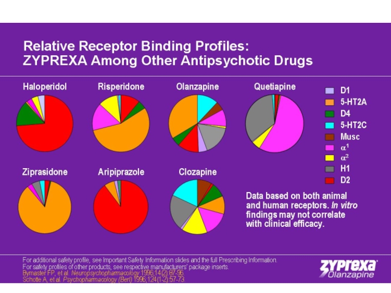
Слайд 69
Mood, emotion,
cognitive function
Motivation
Sex
Appetite
Aggression
Anxiety
Irritability
Energy
Interest
Impulsivity
Drive
Norepinephrine
Serotonin
Dopamine
Several Neurotransmitters Are Involved in Regulating Mood
Stahl SM.
Essential Psychopharmacology: Neuroscientific Basis and Practical Applications. 2nd ed. Cambridge, UK: Cambridge University Press; 2000:152.

Слайд 70Gamma-aminobutyric acid (GABA)
GABA is the main inhibitory neurotransmitter in the central
nervous system (CNS). GABAergic inhibition is seen at all levels of the CNS, including the hypothalamus, hippocampus, cerebral cortex and cerebellar cortex. As well as the large well-established GABA pathways, GABA interneurons are abundant in the brain, with 50% of the inhibitory synapses in the brain being GABA mediated.
 GABA is the main inhibitory neurotransmitter in the central nervous system (CNS). GABAergic")
Слайд 71Antianxiety Agents
GABA receptors
Valium (diazepam)
Ativan (lorazepam)
Klonopin (clonazepam)
Xanax (alprazolam)
Ativan (lorazepam)Klonopin (clonazepam)Xanax (alprazolam)")
Слайд 72Antianxiety Agents (cont)
Valium/Ativan/Klonopin/Xanax
Clumsiness
Sleepiness
Dizziness
Irritability
Unsteadiness
Confusion
Problems with memory
Valium/Ativan/Klonopin/Xanax Clumsiness Sleepiness Dizziness Irritability Unsteadiness Confusion Problems with memory")
Слайд 73Serotonin (5-HT)
The principal centers for serotonergic neurons are the rostral and
caudal raphe nuclei. From the rostral raphe nuclei axons ascend to the cerebral cortex, limbic regions and specifically to the basal ganglia. Serotonergic nuclei in the brain stem give rise to descending axons, some of which terminate in the medulla, while others descend the spinal cord.
 The principal centers for serotonergic neurons are the rostral and caudal raphe nuclei. From")
Слайд 74Antianxiety Agents (cont)
5HT Receptors
Buspar (buspirone)
MISC (MOA unknown)
Atarax (hydroxizine HCl)
Vistaril (hydroxizine
pamoate)
5HT ReceptorsBuspar (buspirone) MISC (MOA unknown)Atarax (hydroxizine HCl)Vistaril (hydroxizine pamoate)")
Слайд 75Antianxiety Agents (cont)
5HT
Buspar
Confusion, Dizziness, Disinhibition, Drowsiness
MISC
Atarax/Vistaril
Cognitive Impairments, Sedation, Blurred Vision
 5HT BusparConfusion, Dizziness, Disinhibition, Drowsiness MISC Atarax/VistarilCognitive Impairments, Sedation, Blurred Vision")
Слайд 76Norepinephrine (NE)
Many regions of the brain are supplied by the noradrenergic
systems. The principal centers for noradrenergic neurons are the locus coeruleus and the caudal raphe nuclei. The ascending nerves of the locus coeruleus project to the frontal cortex, thalamus, hypothalamus and limbic system. Noradrenaline is also transmitted from the locus coeruleus to the cerebellum. Nerves projecting from the caudal raphe nuclei ascend to the amygdala and descend to the midbrain.
 Many regions of the brain are supplied by the noradrenergic systems. The principal centers")
Слайд 77Serotonin (5-HT)
The principal centers for serotonergic neurons are the rostral and
caudal raphe nuclei. From the rostral raphe nuclei axons ascend to the cerebral cortex, limbic regions and specifically to the basal ganglia. Serotonergic nuclei in the brain stem give rise to descending axons, some of which terminate in the medulla, while others descend the spinal cord.
 The principal centers for serotonergic neurons are the rostral and caudal raphe nuclei. From")
Слайд 78Antidepressants
TCA (NE and/or 5HT reuptake presynaptic)
Elavil (amitriptyline)
Asendin (amoxapine)
Anafranil (clomipramine)
Norpramin (desipramine)
Sinequan (doxepin)
Tofranil (imipramine)
Pamelor/Aventyl (nortriptyline)
Vivactil (protriptyline)
Surmontil (trimipramine)
Elavil (amitriptyline) Asendin (amoxapine)Anafranil (clomipramine)Norpramin (desipramine)Sinequan (doxepin)Tofranil (imipramine)Pamelor/Aventyl")
Слайд 79Antidepressants (cont)
TCA
Elavil/Tofranil/Pamelor
Fatigue
Drowsiness/Insomnia
Mild Tremors
Nightmares
Restlessness
Confusion
 TCA Elavil/Tofranil/PamelorFatigueDrowsiness/InsomniaMild TremorsNightmaresRestlessnessConfusion")
Слайд 80Serotonin (5-HT)
The principal centers for serotonergic neurons are the rostral and
caudal raphe nuclei. From the rostral raphe nuclei axons ascend to the cerebral cortex, limbic regions and specifically to the basal ganglia. Serotonergic nuclei in the brain stem give rise to descending axons, some of which terminate in the medulla, while others descend the spinal cord.
 The principal centers for serotonergic neurons are the rostral and caudal raphe nuclei. From")
Слайд 81Antidepressants (cont)
SSRI (selective seratonin reuptake inhibitors)
Celexa (citalopram)
Lexapro (escitalopram)
Prozac/Sarafem (fluoxetine)
Paxil (paroxetine)
Zoloft (sertraline)
Luvox
(fluvoxamine)
Viibryd (vilazodone)
Viibryd (vilazodone)
SSRI (selective seratonin reuptake inhibitors)Celexa (citalopram)Lexapro (escitalopram)Prozac/Sarafem (fluoxetine)Paxil (paroxetine)Zoloft (sertraline)Luvox (fluvoxamine)Viibryd (vilazodone)")
Слайд 82Antidepressants (cont)
SSRI
Celexa/Prozac/Paxil/Zoloft/Lexapro/Viibryd
Agitation
Nervousness
Fatigue
Sleep Problems
Vertigo
Sexual Side Effects
 SSRI Celexa/Prozac/Paxil/Zoloft/Lexapro/ViibrydAgitationNervousnessFatigueSleep ProblemsVertigoSexual Side Effects")
Слайд 83Antidepressants (cont)
MAOI (monoamine oxidase inhibitors)
Nardil (phenelzine)
Parnate (tranylcypromine)
Marplan (isocarbozide)
MAOI (monoamine oxidase inhibitors)Nardil (phenelzine)Parnate (tranylcypromine)Marplan (isocarbozide)")
 MAOI Nardil/Parnate/MarplanDizzinessHeadacheSleep Problems")
Слайд 85Norepinephrine (NE)
Many regions of the brain are supplied by the noradrenergic
systems. The principal centers for noradrenergic neurons are the locus coeruleus and the caudal raphe nuclei. The ascending nerves of the locus coeruleus project to the frontal cortex, thalamus, hypothalamus and limbic system. Noradrenaline is also transmitted from the locus coeruleus to the cerebellum. Nerves projecting from the caudal raphe nuclei ascend to the amygdala and descend to the midbrain.
 Many regions of the brain are supplied by the noradrenergic systems. The principal centers")
Слайд 86Serotonin (5-HT)
The principal centers for serotonergic neurons are the rostral and
caudal raphe nuclei. From the rostral raphe nuclei axons ascend to the cerebral cortex, limbic regions and specifically to the basal ganglia. Serotonergic nuclei in the brain stem give rise to descending axons, some of which terminate in the medulla, while others descend the spinal cord.
 The principal centers for serotonergic neurons are the rostral and caudal raphe nuclei. From")
Слайд 87Antidepressants (cont)
MISC (MOA unclear)
Desyrel (trazodone)
Wellbutrin/Zyban (bupropion)
Effexor (venlafaxine)
Serzone (nefazodone)
Cymbalta (duloxetine)
Pristiq (desvenlafaxine)
Remeron (mirtazepine)
MISC (MOA unclear)Desyrel (trazodone)Wellbutrin/Zyban (bupropion)Effexor (venlafaxine)Serzone (nefazodone)Cymbalta (duloxetine)Pristiq (desvenlafaxine)Remeron (mirtazepine)")
Слайд 88Antidepressants (cont)
MISC
Desyrel/Wellbutrin/Effexor/Serzone/Cymbalta/
Pristiq/Remeron
Agitation
Drowsiness
Sleep Disturbance
Strange Dreams
Increased Blood Pressure
MISC Desyrel/Wellbutrin/Effexor/Serzone/Cymbalta/ Pristiq/RemeronAgitationDrowsinessSleep DisturbanceStrange DreamsIncreased Blood Pressure")

Слайд 90Gathering Information
The Initial Play Therapy Session
Observation: Medication Symptoms/Impact
Behavioral Changes
Cognitive Changes
Emotional Changes

Слайд 91Intake
Past medications: List, in chronological order, all psychotropic medications the individual
took in the past. If the list is long, print it separately and bring it to your appointment.
Age Medication Name Dose Comments
____ _____________ ________ ______________________ ____ _____________ ________ ______________________ ____ _____________ ________ ______________________
____ _____________ ________ ______________________
Age Medication Name Dose Comments
____ _____________ ________ ______________________ ____ _____________ ________ ______________________ ____ _____________ ________ ______________________
____ _____________ ________ ______________________

Слайд 92Intake
Current medications: List, in chronological order, all psychotropic medications the individual
is currently taking. Don’t forget about over-the counter medications.
Age Medication Name Dose Comments
____ _____________ ________ ______________________ ____ _____________ ________ ______________________ ____ _____________ ________ ______________________
____ _____________ ________ ______________________
Age Medication Name Dose Comments
____ _____________ ________ ______________________ ____ _____________ ________ ______________________ ____ _____________ ________ ______________________
____ _____________ ________ ______________________


Слайд 94The Initial Treatment Plan
How will you address medication side effect(s) as
part of the therapeutic process?
Can you link a skill/activity/technique to a side effect and reduce its impact on therapy?
What can you do to accomplish side effect reduction as well as therapeutic progress?
Can you link a skill/activity/technique to a side effect and reduce its impact on therapy?
What can you do to accomplish side effect reduction as well as therapeutic progress?
 as part of the therapeutic")
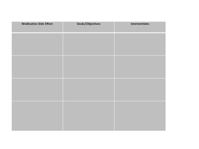
Слайд 96Addressing Medication Side Effects in the Treatment Plan
4 Presentation Types, Each
Requires Something Different
The Warm Up
The Cool Down
The Warm Up-Cool Down
The Cool Down-Cool Down
The Warm Up
The Cool Down
The Warm Up-Cool Down
The Cool Down-Cool Down

Слайд 97Left and Right Brain
LEFT BRAIN FUNCTIONS
uses logic
detail oriented
facts rule
words and language
present
and past
math and science
can comprehend
knowing
acknowledges
order/pattern perception
knows object name
reality based
forms strategies
practical
safe
RIGHT BRAIN FUNCTIONS
uses feeling
"big picture" oriented
imagination rules
symbols and images
present and future
philosophy & religion
can "get it" (i.e. meaning)
believes
appreciates
spatial perception
knows object function
fantasy based
presents possibilities
impetuous
risk taking


Слайд 99Working with Lethargy in Play Therapy (cont)
If you have an outdoor
space:
Consider the benefits of “fresh air and natural sunlight”
Walks
Hop Scotch
Swinging
Consider the benefits of “fresh air and natural sunlight”
Walks
Hop Scotch
Swinging
If you have an outdoor space:Consider the benefits of")
Слайд 100Dopamine (DA)
Dopamine is transmitted via three major pathways. The first extends
from the substantia nigra to the caudate nucleus-putamen (neostriatum) and is concerned with sensory stimuli and movement. The second pathway projects from the ventral tegmentum to the mesolimbic forebrain and is thought to be associated with cognitive, reward and emotional behavior. The third pathway, known as the tubero-infundibular system, is concerned with neuronal control of the hypothalmic-pituatory endocrine system.
 Dopamine is transmitted via three major pathways. The first extends from the substantia nigra")
Слайд 101Antipsychotics
Phenothiazine Derv. (DA receptor antagonist)
Thorazine (Chlorpromazine)
Prolixin (fluphenazine)
Serentil (mesoridazine)
Trilafon (perphenazine)
Compazine (prochlorperazine)
Stelazine (trifluoperazine)
Mellaril
(thioridazine)
Thorazine (Chlorpromazine)Prolixin (fluphenazine)Serentil (mesoridazine)Trilafon (perphenazine)Compazine (prochlorperazine)Stelazine (trifluoperazine)Mellaril (thioridazine)")
Слайд 102Antipsychotics (cont)
Phenothiazine derv.
Thorazine/Stelazine/Mellaril
Akathisia
Akinesia
Sleepiness
Cognitive Blunting
Stiffness
 Phenothiazine derv.Thorazine/Stelazine/MellarilAkathisiaAkinesiaSleepinessCognitive BluntingStiffness")
Phenylbutylpiperadine derv.Haldol (haloperidol)Orap (pimozide)")
Слайд 104Antipsychotics (cont)
Phenylbutylpiperadine derv.
Haldol/Orap
Akathisia
Akinesia
Blurred Vision
Sleepiness
Cognitive Blunting
 Phenylbutylpiperadine derv.Haldol/OrapAkathisiaAkinesiaBlurred VisionSleepinessCognitive Blunting")
Слайд 105Dopamine (DA)
Dopamine is transmitted via three major pathways. The first extends
from the substantia nigra to the caudate nucleus-putamen (neostriatum) and is concerned with sensory stimuli and movement. The second pathway projects from the ventral tegmentum to the mesolimbic forebrain and is thought to be associated with cognitive, reward and emotional behavior. The third pathway, known as the tubero-infundibular system, is concerned with neuronal control of the hypothalmic-pituatory endocrine system.
 Dopamine is transmitted via three major pathways. The first extends from the substantia nigra")
Слайд 106Serotonin (5-HT)
The principal centers for serotonergic neurons are the rostral and
caudal raphe nuclei. From the rostral raphe nuclei axons ascend to the cerebral cortex, limbic regions and specifically to the basal ganglia. Serotonergic nuclei in the brain stem give rise to descending axons, some of which terminate in the medulla, while others descend the spinal cord.
 The principal centers for serotonergic neurons are the rostral and caudal raphe nuclei. From")
Слайд 107Glutamate
In the normal brain the prominent glutamatergic pathways are: the cortico-cortical
pathways; the pathways between the thalamus and the cortex; and the extrapyramidal pathway (the projections between the cortex and striatum). Other glutamate projections exist between the cortex, substantia nigra, subthalmic nucleus and pallidum. Glutamate-containing neuronal terminals are ubiquitous in the central nervous system and their importance in mental activity and neurotransmission is considerable.

Слайд 108Acetylcholine (Ach)
There are three Acetylcholine pathways in the CNS. (a) The
Pons to thalamus and cortex, (b) Magnocellular forebrain nucleus to cortex, & (c) Septohippocampal. In the central nervous system, ACh has a variety of effects as a neuromodulator upon plasticity, arousal and reward. ACh has an important role in the enhancement of sensory perceptions when we wake up and in sustaining attention.
ACh has also been shown to promote REM sleep
ACh has also been shown to promote REM sleep
 There are three Acetylcholine pathways in the CNS. (a) The Pons to thalamus and")
Слайд 110Antipsychotics (cont)
Dibenzapine derv.
Loxitane (loxapine)
Zyprexa (olanzapine)
Seroquel (quetiapine)
Benzisoxazole derv.
Risperdal (risperidone)
Dibenzapine derv.Loxitane (loxapine)Zyprexa (olanzapine)Seroquel (quetiapine)Benzisoxazole derv.Risperdal (risperidone)")
Слайд 111Antipsychotics (cont)
Dibenzapine derv.
Loxitane/Zyprexa/Seroquel
Sedation
Cognitive Blunting
Benzisoxazole derv.
Risperdal
Drowsiness, Dizziness, Cognitive Blunting, Movement Disorders
 Dibenzapine derv.Loxitane/Zyprexa/SeroquelSedationCognitive Blunting Benzisoxazole derv.RisperdalDrowsiness, Dizziness, Cognitive Blunting, Movement Disorders")
Слайд 112Antipsychotics (cont)
Dihydroindolones
Geodone (ziprasidone)
Moban (molindone)
Quinolinone
Abilify (aripiprazole)
Benzoisothiazol derv.
Latuda (lurasidone)
MISC
Eskalith/Lithobid (lithium)
 DihydroindolonesGeodone (ziprasidone)Moban (molindone) QuinolinoneAbilify (aripiprazole) Benzoisothiazol derv.Latuda (lurasidone) MISCEskalith/Lithobid (lithium)")
Слайд 113Antipsychotics (cont)
Dihydroindolones
Geodone/Moban
Sleepiness
Confusion
Quinolinone
Abilify
Confusion
Benzoisothiazol derivatives
Latuda (lurasidone)
Drowsiness
An internal restless or jittery feeling (akathisia)
Movement
or muscle disorders
Insomnia
MISC
Lithium
Tremors
Insomnia
MISC
Lithium
Tremors
 Dihydroindolones Geodone/MobanSleepinessConfusion Quinolinone AbilifyConfusion Benzoisothiazol derivatives Latuda (lurasidone)Drowsiness An internal restless or jittery feeling (akathisia)Movement or muscle disordersInsomnia MISC LithiumTremors")
Слайд 114Working With Cognitive Cloudiness in Play Therapy
Slow Down
Consider the benefits of
“fresh air and natural sunlight”

Слайд 115Working With Cognitive Cloudiness in Play Therapy (cont)
Simple Games (still require
an attempt to focus)
Matching Games
Card Games
Matching Games
Card Games
Simple Games (still require an attempt to focus)Matching")
PuzzlesMazesGuessing GamesHangman")

Слайд 118Working With Emotional Blunting in Play Therapy (cont)
Emotions Tic Tac Toe
Emotions
Identification
Emotion Cards—identification and act out
Facial Expressions
Emotion Cards—identification and act out
Facial Expressions
Emotions Tic Tac ToeEmotions IdentificationEmotion Cards—identification and act")
Art—Guided or AbstractJokesCartoons")
Слайд 120Working with Coordination Difficulties in Play Therapy
Practice
Use Rhythm
Increase speed/intensity


Слайд 122Gross Motor Skills (cont)
Involve the following in Play Therapy:
Things that can
be manipulated, stacked, etc. but are larger.
Legos
Blocks
Dominos
Marbles
Jenga
Legos
Blocks
Dominos
Marbles
Jenga
Involve the following in Play Therapy:Things that can be manipulated, stacked, etc.")
Слайд 123Fine Motor Skills
Involve the following in Play Therapy:
Things that can be
manipulated, stacked, etc. but are smaller.
Pick up Sticks
Tiddlywinks
The game “Operation”
Ring Toss Games
Fishing Games
Pick up Sticks
Tiddlywinks
The game “Operation”
Ring Toss Games
Fishing Games

 Crafts which include:BeadsMacaroni/Shaped Pasta")

Слайд 126Dopamine (DA)
Dopamine is transmitted via three major pathways. The first extends
from the substantia nigra to the caudate nucleus-putamen (neostriatum) and is concerned with sensory stimuli and movement. The second pathway projects from the ventral tegmentum to the mesolimbic forebrain and is thought to be associated with cognitive, reward and emotional behavior. The third pathway, known as the tubero-infundibular system, is concerned with neuronal control of the hypothalmic-pituatory endocrine system.
 Dopamine is transmitted via three major pathways. The first extends from the substantia nigra")
Слайд 127CNS Stimulants
Analeptic
Provigil (modafinil)
Amphetamines
Dexedrine (dextroamphetamine)
Desoxyn (methamphetamine)
Adderall (amphetamine
mixture)
Vyvanse (lisdexamfetamine)
Vyvanse (lisdexamfetamine)
 AmphetaminesDexedrine (dextroamphetamine)Desoxyn (methamphetamine)Adderall (amphetamine mixture) Vyvanse (lisdexamfetamine)")
Слайд 128CNS Stimulants (cont)
Analeptic
Provigil
Irritability
Amphetamines
Adderall/Dexedrine/Desoxyn/Vyvanse
Agitation/Aggression
Sleep Problems
Nervousness
Restlessness
Adderall more likely to create some mood
lability and irritability than the other stimulant medications.
 Analeptic Provigil Irritability Amphetamines Adderall/Dexedrine/Desoxyn/VyvanseAgitation/AggressionSleep ProblemsNervousnessRestlessnessAdderall more likely to create some mood lability and irritability than")
Слайд 129CNS Stimulants (cont)
Non-Amphetamines
Ritalin/Concerta/Metadate/Methylin (methylphenidate)
Cylert (pemoline)
Focalin (dexmethylphenidate)
Daytrana (methylphenidate)---Patch
Non-AmphetaminesRitalin/Concerta/Metadate/Methylin (methylphenidate)Cylert (pemoline)Focalin (dexmethylphenidate) Daytrana (methylphenidate)---Patch")
Слайд 130CNS Stimulants (cont)
Non-Amphetamines
Ritalin/Concerta/Daytrana/Metadate/Methylin
Sleep Problems
Nervousness
Agitation/Aggression
Cylert
Insomnia
Depression
Irritability
Focalin
Nervousness
Sleep Problems
 Non-Amphetamines Ritalin/Concerta/Daytrana/Metadate/MethylinSleep ProblemsNervousnessAgitation/Aggression CylertInsomniaDepressionIrritability FocalinNervousnessSleep Problems")
Слайд 131Norepinephrine (NE)
Many regions of the brain are supplied by the noradrenergic
systems. The principal centers for noradrenergic neurons are the locus coeruleus and the caudal raphe nuclei. The ascending nerves of the locus coeruleus project to the frontal cortex, thalamus, hypothalamus and limbic system. Noradrenaline is also transmitted from the locus coeruleus to the cerebellum. Nerves projecting from the caudal raphe nuclei ascend to the amygdala and descend to the midbrain.
 Many regions of the brain are supplied by the noradrenergic systems. The principal centers")
Слайд 132MISC ADHD Medications
Strattera (atomoxetine) potent inhibitor of presynaptic NE transporter
 potent inhibitor of presynaptic NE transporter")
 Strattera FatigueSleep Disturbance")
Слайд 134Working with Agitation/Aggression in Play Therapy
Sandtray or Sand Play
Clay Therapy (Paul
White)
Bibliotherapy
Bibliotherapy
Bibliotherapy")
Слайд 135Working with Agitation/Aggresion in Play Therapy (cont)
Consider the benefits of “fresh
and Natural sun light”
Rhythm
Music
Natural Sounds
Rhythm
Music
Natural Sounds
Consider the benefits of “fresh and Natural sun light”RhythmMusicNatural Sounds")
Слайд 136Gamma-aminobutyric acid (GABA)
GABA is the main inhibitory neurotransmitter in the central
nervous system (CNS). GABAergic inhibition is seen at all levels of the CNS, including the hypothalamus, hippocampus, cerebral cortex and cerebellar cortex. As well as the large well-established GABA pathways, GABA interneurons are abundant in the brain, with 50% of the inhibitory synapses in the brain being GABA mediated.
 GABA is the main inhibitory neurotransmitter in the central nervous system (CNS). GABAergic")
Слайд 137Sedative/Hypnotics
(GABA)
Newer
Ambien (zolpidem)
ProSom (estazolam)
Lunesta (eszopiclone)
Sonata (zaleplon)
Older
Halcion (triazolam)
Restoril (temazepam)
 NewerAmbien (zolpidem)ProSom (estazolam)Lunesta (eszopiclone)Sonata (zaleplon) OlderHalcion (triazolam)Restoril (temazepam)")
Слайд 138Sedative/Hypnotics (cont)
GABA
Ambien/Prosom/Lunesta/Sonata/Halcion/Restoril
Fatigue
Clumsiness
 GABAAmbien/Prosom/Lunesta/Sonata/Halcion/RestorilFatigueClumsiness")
 Melatonin Rozerem (ramelteon) FatigueClumsiness")
Слайд 140Gamma-aminobutyric acid (GABA)
GABA is the main inhibitory neurotransmitter in the central
nervous system (CNS). GABAergic inhibition is seen at all levels of the CNS, including the hypothalamus, hippocampus, cerebral cortex and cerebellar cortex. As well as the large well-established GABA pathways, GABA interneurons are abundant in the brain, with 50% of the inhibitory synapses in the brain being GABA mediated.
 GABA is the main inhibitory neurotransmitter in the central nervous system (CNS). GABAergic")
Слайд 141Anticonvulsants/Psychiatric Uses
Tegretol/Carbatrol (carbamazepine)
Trileptal (oxcarbazepine)
Neurontin (gabapentin)
Topamax (topiramate)
Depakote/Depakene (valproic acid)
Lamictal (lamotrigine)
Gabitril (tiagabine)
Trileptal (oxcarbazepine)Neurontin (gabapentin)Topamax (topiramate)Depakote/Depakene (valproic acid)Lamictal (lamotrigine)Gabitril (tiagabine)")
Слайд 142Anticonvulsants/Psychiatric Uses (cont)
Tegretol/Carbatrol
Dizziness, Drowsiness, Blurred Vision
Trileptal/Neurontin/Topamax/Lamictal
Fatigue, Dizziness, Nervousness
Depakote/Depakene
Drowsiness, Lethargy
Gabitril
Fatigue, dizziness, unstable
walking, seizures
 Tegretol/CarbatrolDizziness, Drowsiness, Blurred Vision Trileptal/Neurontin/Topamax/LamictalFatigue, Dizziness, Nervousness Depakote/DepakeneDrowsiness, Lethargy GabitrilFatigue, dizziness, unstable walking, seizures")
Слайд 143Acetylcholine (Ach)
There are three Acetylcholine pathways in the CNS. (a) The
Pons to thalamus and cortex, (b) Magnocellular forebrain nucleus to cortex, & (c) Septohippocampal. In the central nervous system, ACh has a variety of effects as a neuromodulator upon plasticity, arousal and reward. ACh has an important role in the enhancement of sensory perceptions when we wake up and in sustaining attention.
ACh has also been shown to promote REM sleep
ACh has also been shown to promote REM sleep
 There are three Acetylcholine pathways in the CNS. (a) The Pons to thalamus and")
Слайд 144Antiparkinsons/Psychiatric Uses
Cogentin (bentropine)
Artane (trihexyphenidyl)
No major negative effects
Artane (trihexyphenidyl)No major negative effects")
Слайд 145MISC MISC MISC/Psychiatric Uses
Benadryl (diphenhyramine)—with older Antipsychotics
Inversine (mecamylamine)---Tourette’s
Revia (naltrexone)---Severe Behavioral Disorder
in MR, Pervasive Developmental Disorders
—with older AntipsychoticsInversine (mecamylamine)---Tourette’sRevia (naltrexone)---Severe Behavioral Disorder in MR, Pervasive Developmental Disorders")
 Benadryl Sedation, Cognitive Impairments")

Слайд 148Norepinephrine (NE)
Many regions of the brain are supplied by the noradrenergic
systems. The principal centers for noradrenergic neurons are the locus coeruleus and the caudal raphe nuclei. The ascending nerves of the locus coeruleus project to the frontal cortex, thalamus, hypothalamus and limbic system. Noradrenaline is also transmitted from the locus coeruleus to the cerebellum. Nerves projecting from the caudal raphe nuclei ascend to the amygdala and descend to the midbrain.
 Many regions of the brain are supplied by the noradrenergic systems. The principal centers")
Слайд 149MISC MISC MISC/Psychiatric Uses
Inderal (propranolol)---IED, PTSD
Catapres (clonidine)—ADHD, Conduct Disorder, Tourette’s
Tenex/Intuniv (guanfacine)---ADHD,
Tourette’s
Irritability, Tiredness, Hypotension
Irritability, Tiredness, Hypotension
---IED, PTSDCatapres (clonidine)—ADHD, Conduct Disorder, Tourette’sTenex/Intuniv (guanfacine)---ADHD, Tourette’sIrritability, Tiredness, Hypotension")
Слайд 150Antihypetensives
Inderal (propranolol)
Drowsiness, Hypotension
Catapres (clonidine)
Sedation, Drowsiness, Depression, Irritability,
Hypotension
Tenex/Intuniv (guanfacine)
Irritability, Tiredness, Hypotension
Drowsiness, HypotensionCatapres (clonidine)Sedation, Drowsiness, Depression, Irritability, HypotensionTenex/Intuniv (guanfacine)Irritability, Tiredness, Hypotension")

Слайд 152Games We Should All Have: They Accomplish Multiple Tasks
Jenga
Pick-up-Sticks
Connect 4
Tic Tac
Toe
Operation
Chutes and Ladders
Operation
Chutes and Ladders

Слайд 153Conclusion
Remember:
The goal is to go slow and be supportive. Allow the
child to push past the side effect.
When stimulated the brain/body can overcome/compensate for medication side effects.
When stimulated the brain/body can overcome/compensate for medication side effects.

Слайд 154References
Aarts, E., van Holstein, M., & Cools, R. (2011). Striatal dopamine
and the interference
between motivation and cognition. Frontiers in Psychology, 2(163), 1-11.
Adinoff, B., Devous, M. D. Sr., Cooper, D. B., Best, S. E., Chandler, P., Harris, T.,…Cullum, C. M. (2003). Resting regional cerebral blood flow and gambling task performance in cocaine-dependent subjects and healthy comparison subjects. American Journal of Psychiatry, 160(10), 1892-1892.
Anda, R. F., Felitti, V. J., Walker J., Whitfield, C. L., Bremner, J. D., Perry, B. D., Dube S.
R., & Giles, W. H. (2006). The enduring effects of abuse and related adverse
experiences in childhood: A convergence of evidence from neurobiology and
epidemiology. European Archives of Psychiatry and Clinical Neurosciences, 56(3),
174–86.
Brown, R. T., Carpenter, L. A., & Simerly, E. (2005). Mental health medications for
Children: A primer. New York: The Guilford Press.
Bush, G., Valera, E. M., & Seidman, L. J. (2005). Functional neuroimaging of attention-
deficit/hyperactivity disorder: A review and suggested future directions. Biological
Psychiatry, 57(11), 1273–1284.
. Striatal dopamine and the interference")
Слайд 155References (cont)
Centers for Disease Control and Prevention. (2012). Retrieved on August
11, 2012 from
http://www.cdc.gov/ace/images/ace_pyramid_home.jpg
Colton, D. L., & Sheridan, S. M. (1998). Conjoint behavioral consultation and social skills
training: Enhancing the play behaviors of boys with attention deficit hyperactivity
disorder. Journal of Educational and Psychological Consultation, 9(1), 3-28.
Dagher, A., & Robbins, T. W. (2009). Personality, addiction, dopamine: Insights from
Parkinson’s disease. Neuron, 61(4), 502-510.
Edwards, V. J., Anda, R. F., Dube, S. R., Dong, M., Chapman, D. F., & Felitti, V. J.
(2005). The wide-ranging health consequences of adverse childhood experiences. In K.
Kendall-Tackett & S. Giacomoni (Eds.) Victimization of children and youth: Patterns
of abuse, response strategies, Kingston, NJ: Civic Research Institute.
Centers for Disease Control and Prevention. (2012). Retrieved on August 11, 2012 from http://www.cdc.gov/ace/images/ace_pyramid_home.jpgColton, D.")
Слайд 156References (cont)
Gogtay, N., Giedd, J. N., Lusk, L., Hayashi, K. M.,
Greenstein, D., Vaituzia, A.
C.,...Thompson, P. M.. (2004). Dynamic mapping of human cortical development during childhood through early adulthood. Proceedings of the National Academy of Sciences of the United States of America (PNAS), 101, 8174-8179. Retrieved on July 5, 2011, from www.phas.org. doi:10.1073/pnas.0402680101
Greenspan, S. Dir/floortime. Retrieved from
http://www.icdl.com/dirFloortime/overview/documents/WhatisDIR.pdf
Greenspan, S. I., & Wieder, S . (1997a) ‘An integrated developmental approach to
interventions for young children with severe difficulties in relating and
communicating’, Zero to Three National Center for Infants, Toddlers,
and Families 17(5).
Greenspan, S. I., & Wieder, S . (1997b) ‘Developmental patterns and outcomes in
infants and children with disorders in relating and communication: A chart
review of 200 cases of children with autistic Spectrum Disorders’, The
Journal of Developmental and Learning Disorders, 1(1), 87–141.
Gogtay, N., Giedd, J. N., Lusk, L., Hayashi, K. M., Greenstein, D., Vaituzia, A.")
Слайд 157References (cont)
Ingersoll, R. E., Bauer, A., & Burns, L. (2004).
Children and psychotropic
medications: What role should advocacy counseling play? Journal of
Counseling and Development, 82, 337-343.
LaRue, R. H, Northup, J., Baumeister, A. A., Hawkins, M. F., Seale, L., Williams,
T., & Ridgway. (2008). An evaluation of stimulant medication on reinforcing
effect of play. Journal of Applied Behavioral Analysis, 41, 143-147.
Levy, L. M., & Degnan, A. J. (2012, January). GABA-based evaluation of
neurological conditions: MR spectroscopy. American Journal of
Neuroradiology, 1-6. Retrieved from http://dx.doi.org/10.3174/ajnr.A2902
Lundbeck Institute. CNS image bank-brain physiology-normal brain. Retrieved on
July 5, 2011, from
http://www.cnsforum.com/imagebank/section/Bp_Normal_brain/default.aspx
Martin, A., Scahill, L., & Kratochvil, C. (Eds.). (2010). Pediatric
psychopharmacology: Principles and practices (2nd ed.). New York: Oxford
University Press.
 Ingersoll, R. E., Bauer, A., & Burns, L. (2004). Children and psychotropic medications:")
Слайд 158References (cont)
Nestler, E. J., Hyman, S. E., & Malenka, R. C.
(2001). Molecular
neuropharmacology: A foundation for clinical neuroscience. New York:
McGraw-Hill.
Preston, J. D., O'Neal. J., & Talaga, M.C. (2010). Child and adolescent
psychopharmacology made simple (2nd ed.) . Oakland, CA: New Harbinger
Publications, Inc.
Robbins, T. W., & Everitt, B. J. (1995). The cognitive neurosciences. Cambridge, MA: MIT Press.
Royall, D. R., Lauterbach, E. C., Cummings, J. F., Reeves, A., Rummans, T. A.,
Kaufer, D. I.,…Coffey, C. E. (2002). Executive control function: A review of
its promise and challenges for clinical research. Journal of Neuropsychiatry and
Clinical Neurosciences, 14(4), 377-405.
Schore, A. N. (2001), The effects of early relational trauma on right brain
development, affect regulation, and infant mental health. Infant Mental Health
Journal, 22(1-2), 201-269. doi:10.1002/1097
0355(200101/04)22:1<201::AID-IMHJ8>3.0.CO;2-9
Nestler, E. J., Hyman, S. E., & Malenka, R. C. (2001). Molecular neuropharmacology: A")
Слайд 159References (cont)
Schore, A. N. (2005). Right-brain affect regulation: An essential mechanism
of
development, trauma, dissociation, and psychotherapy. In centers, D., Solomon, M., & Siegel, D. (Eds.), The healing power of emotion: Integrating
relationships, body and mind. A dialogue among scientists and clinicians (pp.
112-144). New York: WW Norton.
Schore, A. N. (2009). Relational trauma and the developing right brain. Annals of
the New York Academy of Sciences, 1159(1), 189-2003. doi:10.1111/j.1749
6632.2009.04474.
Sinacola, R. S., & Peter-Strickland, T. (2011). Basic psychopharmacology for
counselors and psychotherapists (2nd ed.). Upper Saddle River, NJ: Pearson
Higher Education, Inc.
Sweeney, D. S., & Tatum, R. J. (1995). Play therapy and psychopharmacology:
What the play therapist need to know. International Journal of Play Therapy,
4(2), 41-57.
Sweeney, D. S., & Tatum, R. J. (2001). What the play therapist needs to know
about medications. In G. L. Landreth (Ed.), Innovations in play therapy:
Issues, process, and special populations (pp. 51-64). New York: Brunner
Routledge.
Schore, A. N. (2005). Right-brain affect regulation: An essential mechanism of development, trauma, dissociation, and")
Слайд 160References (cont)
Toga, A. W., & Mazziotta, J. C. (2000). Brain mapping:
The systems: San Diego,
CA: Academic Press.
Wieder, S., & Greenspan, S. I. (2003). Climbing the symbolic ladder in the DIR
model through floor time/interactive play. Autism, 7(4), 425-435.
Willis, D. W. (March 23, 2007). Early brain development: Relational healing
from risk. Paper presented at the Substance Use and Brain Development
Conference, Eugene, OR.
Wilson, S. N (2005). The meanings of medication. American Journal of
Psychotherapy, 59(1), 19-29.
Wise, R. A. (2004). Dopamine, learning, and motivation. Nature Reviews:
Neuroscience, 5, 483-494.
van der Kolk, B. A., Roth, S., Pelcovitz, D., Sunday, S., & Spinazzola, J. (2005).
Disorders of extreme stress: The empirical foundation of complex adaptation
to trauma. Journal of Traumatic Stress, 18(5) 389-399.
Toga, A. W., & Mazziotta, J. C. (2000). Brain mapping: The systems: San Diego, CA:")
Слайд 161
Recommended videos:
Medicating Kids—Frontline (2001)
The Medicated Child—Frontline—(2008)
The Secret Life of the
Brain—PBS (2002)
Generation Meds—ABC World News—Diane Sawyer—(2011)—Over Medication of Children in Foster Care
Contact Information:
hudspee@hsu.edu
www.playtherapytraining.com
Generation Meds—ABC World News—Diane Sawyer—(2011)—Over Medication of Children in Foster Care
Contact Information:
hudspee@hsu.edu
www.playtherapytraining.com
The Medicated Child—Frontline—(2008)The Secret Life of the Brain—PBS (2002)Generation Meds—ABC World")