- Главная
- Разное
- Дизайн
- Бизнес и предпринимательство
- Аналитика
- Образование
- Развлечения
- Красота и здоровье
- Финансы
- Государство
- Путешествия
- Спорт
- Недвижимость
- Армия
- Графика
- Культурология
- Еда и кулинария
- Лингвистика
- Английский язык
- Астрономия
- Алгебра
- Биология
- География
- Детские презентации
- Информатика
- История
- Литература
- Маркетинг
- Математика
- Медицина
- Менеджмент
- Музыка
- МХК
- Немецкий язык
- ОБЖ
- Обществознание
- Окружающий мир
- Педагогика
- Русский язык
- Технология
- Физика
- Философия
- Химия
- Шаблоны, картинки для презентаций
- Экология
- Экономика
- Юриспруденция
Lymphoma. Overview презентация
Содержание
- 1. Lymphoma. Overview
- 2. Overview Concepts, classification, lymphoma genesis Epidemiology Clinical
- 3. Conceptualizing lymphoma neoplasms of lymphoid origin (lymph
- 4. Conceptualizing lymphoma Hodgkin Lymphoma – relatively uniform
- 5. B-cell development
- 6. The challenge of lymphoma classification
- 7. Principles of the WHO classification
- 8. Lymphoma classification (based on 2001 WHO) T-cell
- 9. WHO Classification 2001-2008 ?Precursor B-and T-cell
- 10. WHO/REAL Classification of Lymphoid Neoplasms B-Cell Neoplasms
- 11. Clinical classification of NHL
- 12. A practical way to think of lymphoma
- 13. Mechanisms of lymphoma genesis Genetic alterations -
- 14. Epidemiology of lymphomas 5th most frequently diagnosed
- 15. Epidemiology of lymphomas Geographic variability – B
- 16. Incidence of lymphomas in comparison with other cancers in Canada
- 17. Age distribution of new NHL cases in Canada
- 18. Non-Hodgkin lymphoma Incidence Diffuse large B-cell lymphoma Follicular lymphoma Other NHL
- 19. Age distribution of new Hodgkin lymphoma cases in Canada
- 20. Risk factors for NHL immunosuppression or immunodeficiency
- 21. Clinical manifestations Variable severity: asymptomatic to extremely
- 22. Other complications of lymphoma bone marrow failure
- 23. Diagnosis requires an adequate biopsy Diagnosis should
- 24. Staging of lymphoma – Ann Arbor system
- 25. Staging Pocedures History and physical examination Bone
- 26. Prognostic factors Histologic type Age Performance status
- 27. Prognostic models - IPI A – age
- 29. IPI: Overall Survival (OS) by Risk Strata
- 30. Diffuse Large B-Cell Lymphoma (DLCL): OS Patients
- 31. Antigen Expression in B-Cell Lineage Pre-B
- 32. CD20 Expression in B-Cell Malignancies Histology
- 33. Three types of lymphoma worth knowing about Follicular lymphoma Diffuse large B-cell lymphoma Hodgkin lymphoma
- 34. Follicular lymphoma most common type of “indolent”
- 35. defer treatment if asymptomatic (“watch-and-wait”) several chemotherapy
- 36. Treatment Chemotherapy – single agent ±
- 37. Diffuse large B-cell lymphoma most common type
- 38. National High Priority Lymphoma Study: Progression-Free Survival
- 39. GELA Phase III Trial: EFS 1.0 0.8 0.6 0.4 0.2 0 EFS P
- 40. 0 0.5 1.0 1.5 2.0
- 41. GELA Phase III Trial: Summary Significantly higher
- 42. Hodgkin lymphoma Thomas Hodgkin (1798-1866)
- 43. Hodgkin lymphoma cell of origin: germinal centre
- 44. Reed-Sternberg cell
- 45. RS cell and variants popcorn cell lacunar
- 46. Reed-Sternberg cell
- 47. A possible model of pathogenesis germinal
- 48. Hodgkin lymphoma Histological subtypes Nodular lymphocyte predominance
- 49. Epidemiology less frequent than non-Hodgkin lymphoma males
- 50. Associated (etiological?) factors EBV infection smaller family
- 51. Clinical manifestations: lymphadenopathy, mostly mediastinal contiguous spread
- 52. Treatment and Prognosis
- 53. Long term complications of treatment infertility MOPP
- 54. Case: W.P. 25 year old woman persistent
- 55. W.P. at presentation
- 56. W.P. at presentation
- 57. Case: W.P. differential diagnosis lymphoma Hodgkin
- 58. What next? Needle aspirate of LN: a
- 59. Case: W.P. lymph node biopsy
- 60. Case: W.P. lymph node biopsy
- 61. Case: W.P. lymph node biopsy
- 62. Case: W.P. staging investigations CT neck/chest/abdomen/pelvis bone
- 63. W.P. at presentation
- 64. Staging investigations bone marrow normal CT scan:
- 65. What is her diagnosis and stage? nodular sclerosis HD stage IIB with bulky mediastinal mass
- 66. Case: W.P. Treatment discussion with patient
- 67. W.P. post-chemotherapy
- 68. Case: W.P. post-ABVD response to chemo, but
- 69. Case: W.P. post-radiotherapy serial CT scans did not show progression patient remains in remission

Слайд 2Overview
Concepts, classification, lymphoma genesis
Epidemiology
Clinical presentation
Diagnosis
Staging
Three important types of lymphoma

Слайд 3Conceptualizing lymphoma
neoplasms of lymphoid origin (lymph nodes or extra nodal lymphatic
leukemia vs. lymphoma
lymphomas as clonal expansions of cells (B or T lymphocytes or NK cells) at certain developmental stages
, typically causing lymphadenopathyleukemia")
Слайд 4Conceptualizing lymphoma
Hodgkin Lymphoma – relatively uniform in histology, clinical presentation and
Non Hodgkin Lymphoma – a large and heterogeneous category with various cell origin, histology, clinical course. Comprises most of lymphomas


Слайд 7
Principles of the WHO classification
1.Morphology2.Immunophenotype3.Molecularbiology4.Genetic5.Clinicalpresentationand course
I love pathologists who can

Слайд 8Lymphoma classification
(based on 2001 WHO)
T-cell & NK-cell neoplasms
Precursor T-cell neoplasms (3)
Mature
T-cell proliferation of uncertain malignant potential (1)
Hodgkin lymphoma
Classical Hodgkin lymphomas (4)
Nodular lymphocyte predominant Hodgkin lymphoma (1)
B-cell neoplasms
Precursor B-cell neoplasms (2 types)
Mature B-cell neoplasms (19)
B-cell proliferations of uncertain malignant potential (2)
T-cell & NK-cell neoplasmsPrecursor T-cell neoplasms (3)Mature T-cell and NK-cell")
Слайд 9WHO Classification 2001-2008
?Precursor B-and T-cell neoplasms
?Mature B cell neoplasms?
Mature T-cell and
Hodgkin lymphoma?
Immunodeficiency associated lymphoproliferativedisorders
?Histiocyticand dendritic cell neoplasms

Слайд 10WHO/REAL Classification of Lymphoid Neoplasms
B-Cell Neoplasms
Precursor B-cell neoplasm
Precursor B-lymphoblastic leukemia/lymphoma
(precursor
Mature (peripheral) B-neoplasms
B-cell chronic lymphocytic leukemia / small lymphocytic lymphoma
B-cell prolymphocytic leukemia
Lymphoplasmacytic lymphoma‡
Splenic marginal zone B-cell lymphoma
(+ villous lymphocytes)*
Hairy cell leukemia
Plasma cell myeloma/plasmacytoma
Extranodal marginal zone B-cell lymphoma of MALT type
Nodal marginal zone B-cell lymphoma
(+ monocytoid B cells)*
Follicular lymphoma
Mantle cell lymphoma
Diffuse large B-cell lymphoma
Mediastinal large B-cell lymphoma
Primary effusion lymphoma†
Burkitt’s lymphoma/Burkitt cell leukemia§
T and NK-Cell Neoplasms
Precursor T-cell neoplasm
Precursor T-lymphoblastic leukemia/lymphoma
(precursor T-acute lymphoblastic leukemia
‡ Formerly known as lymphoplasmacytoid lymphoma or immunocytoma
II Entities formally grouped under the heading large granular lymphocyte
leukemia of T- and NK-cell types
* Provisional entities in the REAL classification
Mature (peripheral) T neoplasms
T-cell chronic lymphocytic leukemia / small
lymphocytic lymphoma
T-cell prolymphocytic leukemia
T-cell granular lymphocytic leukemiaII
Aggressive NK leukemia
Adult T-cell lymphoma/leukemia (HTLV-1+)
Extranodal NK/T-cell lymphoma, nasal type#
Enteropathy-like T-cell lymphoma**
Hepatosplenic γδ T-cell lymphoma*
Subcutaneous panniculitis-like T-cell lymphoma*
Mycosis fungoides/Sézary syndrome
Anaplastic large cell lymphoma, T/null cell,
primary cutaneous type
Peripheral T-cell lymphoma, not otherwise characterized
Angioimmunoblastic T-cell lymphoma
Anaplastic large cell lymphoma, T/null cell,
primary systemic type
Hodgkin’s Lymphoma (Hodgkin’s Disease)
Nodular lymphocyte predominance Hodgkin’s lymphoma
Classic Hodgkin’s lymphoma
Nodular sclerosis Hodgkin’s lymphoma (grades 1 and 2)
Lymphocyte-rich classic Hodgkin’s lymphoma
Mixed cellularity Hodgkin’s lymphoma
Lymphocyte depletion Hodgkin’s lymphoma
† Not described in REAL classification
§ Includes the so-called Burkitt-like lymphomas
** Formerly known as intestinal T-cell lymphoma
# Formerly know as angiocentric lymphoma
 Mature (peripheral)")


Слайд 13Mechanisms of lymphoma genesis
Genetic alterations - lack of apoptosis (bcl-2), proliferation
Infection – viral (EBV, HCV, HTLV-1), bacterial – H. Pylori
Environmental factors – chemicals, diet
Immunosuppression – AIDS, post transplant (solid organs, BMT)
Chronic antigen stimulation - autoimmunity
Family history – 3.3 times increase risk
, proliferation (c-myc)Infection – viral (EBV,")
Слайд 14Epidemiology of lymphomas
5th most frequently diagnosed cancer, ±4% of all cancers
males > females
whites > other races
incidence
NHL increasing over time
Hodgkin lymphoma stable

Слайд 15Epidemiology of lymphomas
Geographic variability – B cell lymphoma common in Western


Слайд 20Risk factors for NHL
immunosuppression or immunodeficiency
connective tissue disease
family history of lymphoma
infectious
chemicals
dietary
ionizing radiation

Слайд 21Clinical manifestations
Variable
severity: asymptomatic to extremely ill
time course: evolution over weeks, months,
Systemic manifestations
Weakness, fever, night sweats, weight loss, anorexia, pruritus
Local manifestations
lymphadenopathy, splenomegaly - most common
any tissue potentially can be infiltrated

Слайд 22Other complications of lymphoma
bone marrow failure (infiltration)
CNS infiltration
immune hemolysis or thrombocytopenia
compression
pleural/pericardial effusions, ascites
CNS infiltrationimmune hemolysis or thrombocytopeniacompression of structures (eg spinal")
Слайд 23Diagnosis requires an adequate biopsy
Diagnosis should be biopsy-proven before treatment is
Need enough tissue to assess cells and architecture, immunopenotyping, cytogenetics and molecular studies
- open vs core needle biopsy vs FNA

Слайд 24Staging of lymphoma – Ann Arbor system
A: absence of B symptoms
B:

Слайд 25Staging Pocedures
History and physical examination
Bone marrow aspiration and biopsy
Imaging – anatomical:

Слайд 26Prognostic factors
Histologic type
Age
Performance status
Ann Arbor stage
Size of tumor mass
Extranodal involvement
LDH, β2-microglobulin
Molecular
Response to treatment

Слайд 27Prognostic models - IPI
A – age > 60 ► 1 pt.
P
L – LDH ↑ ► 1 PT.
E – extranodal sites > 1 ► 1 pt.
S – stage ≥ 3 ► 1 pt.

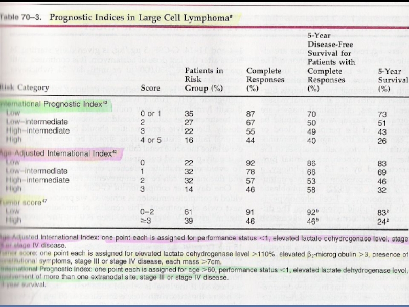
Слайд 29IPI: Overall Survival (OS)
by Risk Strata
The Non-Hodgkin's Lymphoma Pathologic Classification
100
75
50
25
0
0
2
4
6
8
10
H
HI
LI
L
Patients (%)
Year
 by Risk Strata The Non-Hodgkin's Lymphoma Pathologic Classification Project. Cancer. 1982;49:2112.10075502500246810HHILILPatients (%)Year")
Слайд 30Diffuse Large B-Cell
Lymphoma (DLCL): OS
Patients (%)
Year
Adapted from Armitage. J Clin Oncol.
100
60
40
20
0
0
2
5
6
7
8
3
4
1
80
IPI 0-1
IPI 2-3
IPI 4-5
P<0.001
: OSPatients (%)YearAdapted from Armitage. J Clin Oncol. 1998;16:2780.100604020002567834180IPI 0-1IPI 2-3IPI 4-5P")
Слайд 31Antigen Expression in B-Cell Lineage
Pre-B
Early B
Mature B
Plasmacytoid B
±CD5
CD19
CD20
CD22
CD52
Plasma
Intermediate B
?
?
?
Stem
Burkitt’s, FL, DLCL, HCL
ALL CLL, PLL
WM
MM

Слайд 32CD20 Expression
in B-Cell Malignancies
Histology
0 100 200 300 400 500
Burkitt’s lymphoma
CLL
CLL/PLL
Follicular small
Hairy cell
Large cell
LP/Waldenström’s
Mantle cell
Marginal zone
Small cleaved
Adapted with permission from G.D. Maloney.
Mean channel fluorescence

Слайд 33Three types of lymphoma worth knowing about
Follicular lymphoma
Diffuse large B-cell lymphoma
Hodgkin

Слайд 34Follicular lymphoma
most common type of “indolent” lymphoma in the Western world
usually
often asymptomatic
not curable (some exceptions)
associated with BCL-2 gene rearrangement [t(14;18)]
cell of origin: germinal center B-cell

Слайд 35defer treatment if asymptomatic (“watch-and-wait”)
several chemotherapy options if symptomatic
median survival: years
although
transformation to aggressive lymphoma can occur
several chemotherapy options if symptomaticmedian survival: yearsalthough considered “indolent”, morbidity and")
Слайд 36Treatment
Chemotherapy – single agent ± corticosteroids, combination – CVP, CHOP
Monoclonal Ab – anti-CD20, anti-CD22, anti-CD30, anti-CD25, anti-CD52 etc.
Combination of chemotherapy and monoclonal antibodies
Radiotherapy - involved field, extended, adjuvant
Radioimmunotherapy

Слайд 37Diffuse large B-cell lymphoma
most common type of “aggressive” lymphoma
usually symptomatic
extranodal involvement
cell of origin: germinal center B-cell
treatment should be offered
curable in ~ 40%

Слайд 38National High Priority Lymphoma
Study: Progression-Free Survival
Adapted from Fisher. N Engl J
Patients (%)
Year
100
80
60
40
20
0
0
1
2
3
4
5
6
CHOP
m-BACOD
ProMACE-CytaBOM
MACOP-B
Year1008060402000123456CHOPm-BACODProMACE-CytaBOMMACOP-B")

Слайд 40
0
0.5
1.0
1.5
2.0
2.5
3.0
0
0.2
0.4
0.6
0.8
1.0
GELA Phase III Trial: OS
R-CHOP
CHOP
P=0.007
R-CHOP
CHOP
Survival
Years
No. at Risk
Coiffier et al. N Engl

Слайд 41GELA Phase III Trial: Summary
Significantly higher CR/CRu rate with Rituxan® +
Significantly longer EFS and OS rates with Rituxan® + CHOP
Rituxan® does not increase apparent toxicity of CHOP
Coiffier et al. N Engl J Med. 2002;346:235.

")
Слайд 43Hodgkin lymphoma
cell of origin: germinal centre B-cell
Reed-Sternberg cells (or RS
most cells in affected lymph node are polyclonal reactive lymphoid cells, not neoplastic cells
 in the affected")

Слайд 45RS cell and variants
popcorn cell
lacunar cell
classic RS cell
(mixed cellularity)
(nodular sclerosis)
(lymphocyte
predominance)
(nodular sclerosis)(lymphocytepredominance)")

Слайд 47A possible model of pathogenesis
germinal
center
B cell
transforming
event(s)
loss of apoptosis
RS cell
inflammatory
response
EBV?
cytokines
loss of apoptosisRS cellinflammatoryresponseEBV?cytokines")
Слайд 48Hodgkin lymphoma
Histological subtypes
Nodular lymphocyte predominance Hodgkin lymphoma
Classical Hodgkin lymphoma
nodular sclerosis (most
mixed cellularity
lymphocyte-rich
lymphocyte depleted
mixed cellularitylymphocyte-richlymphocyte depleted")
Слайд 49Epidemiology
less frequent than non-Hodgkin lymphoma
males 3.5/100000; females 2.5/100000
peak incidence in 3rd
Stage at Diagnosis, Proportion
Stage I - 24.4%
Stage II - 30.8% Stage III - 15.4% Stage IV - 12.8% Stage not known - 16.7%

Слайд 50Associated (etiological?) factors
EBV infection
smaller family size
higher socio-economic status
Caucasian > non-Caucasian
possible genetic
other: HIV? occupation? herbicides?
 factorsEBV infectionsmaller family sizehigher socio-economic statusCaucasian > non-Caucasianpossible genetic predispositionother: HIV? occupation? herbicides?")
Слайд 51Clinical manifestations:
lymphadenopathy, mostly mediastinal
contiguous spread
extra nodal sites relatively uncommon except in
“B” symptoms
very rare causes obstruction, like superior vena cava syndrome


Слайд 53Long term complications of treatment
infertility
MOPP > ABVD; males > females
sperm banking
premature menopause
secondary malignancy
skin, AML, lung, MDS, NHL, thyroid, breast...
cardiac disease

Слайд 54Case: W.P.
25 year old woman
persistent dry cough
fever, night sweats, weight loss
left cervical lymphadenopathy (2 cm)
left supraclavicular node (2 cm)
no splenomegaly


Слайд 57
Case: W.P. differential diagnosis
lymphoma
Hodgkin
non-Hodgkin
lung cancer
other neoplasms: thyroid, germ cell
non-neoplastic causes less
sarcoid, TB, ...

Слайд 58What next?
Needle aspirate of LN: a few necrotic cells
Needle biopsy of


Слайд 62Case: W.P. staging investigations
CT neck/chest/abdomen/pelvis
bone marrow
PET scan
Blood work: normal CBC, ESR,


Слайд 64Staging investigations
bone marrow normal
CT scan: Lt. supraclavicular adenopathy; large mediastinal mass;
PET avid


Слайд 66Case: W.P. Treatment
discussion with patient
treatment with ABVD x 6 cycles
constitutional
bulky mediastinal mass is a special situation that merits additional radiation after chemotherapy


Слайд 68Case: W.P. post-ABVD
response to chemo, but residual mediastinal/hilar mass
repeat PET scan
proceed with radiotherapy as originally planned

Слайд 69Case: W.P. post-radiotherapy
serial CT scans did not show progression
patient remains in