- Главная
- Разное
- Дизайн
- Бизнес и предпринимательство
- Аналитика
- Образование
- Развлечения
- Красота и здоровье
- Финансы
- Государство
- Путешествия
- Спорт
- Недвижимость
- Армия
- Графика
- Культурология
- Еда и кулинария
- Лингвистика
- Английский язык
- Астрономия
- Алгебра
- Биология
- География
- Детские презентации
- Информатика
- История
- Литература
- Маркетинг
- Математика
- Медицина
- Менеджмент
- Музыка
- МХК
- Немецкий язык
- ОБЖ
- Обществознание
- Окружающий мир
- Педагогика
- Русский язык
- Технология
- Физика
- Философия
- Химия
- Шаблоны, картинки для презентаций
- Экология
- Экономика
- Юриспруденция
Leishmaniasis. Department of Infectious Diseases Leishmaniasis презентация
Содержание
- 1. Leishmaniasis. Department of Infectious Diseases Leishmaniasis
- 2. Leishmaniasis Leishmaniasis is a zoonosis.
- 3. Leishmaniasis Leishmania donovani (complex) (VL) Leishmania
- 4. Three important Species
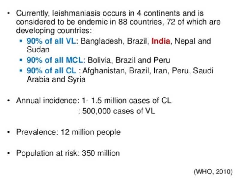
- 5. Geographical distribution of leishmaniasis
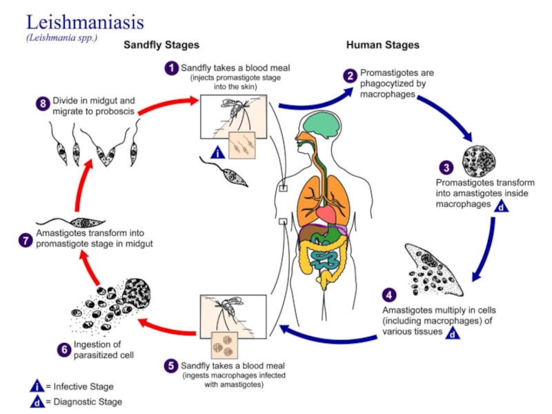
- 8. Life Cycle of leishmaniasis Promastigote
- 9. Promastigote stage flagella Promastigote stage inside the
- 10. Promastigotes in rosettes in a culture of an orient sore on N.N.N. medium (Giemsa stain).
- 11. Ovoid small intracellular parasites in a bone
- 12. Life cycle
- 14. Bite of sand fly Bite of
- 15. Transmission of Leishmaniasis _ by sand
- 16. Cutaneous Leishmaniasis Cutaneous forms of the disease
- 17. A cutaneous leishmaniasis lesion on the arm.
- 18. Cutaneous Leishmaniasis
- 19. The Baghdad
- 20. Leishmania tropica Causes ulceration of the skin
- 21. Mucocutaneous Leishmaniasis Mucocutaneous leishmaniasis (Espundia) Leishmania braziliensis & L . maxicana
- 22. mucocutaneous forms of leishmaniasis , lesions can
- 23. Visceral Leishmaniasis Visceral disease (Kala-azar)
- 24. Visceral disease (Kala-azar) Most severe form of
- 25. Post Kala Azar Dermal Leishmanoid Normally develops
- 26. Hepatosplenomegaly Post Kala Azar Dermal Leishmanoid
- 27. Dogs can act as reservoirs of Leishmania parasites. They also exhibit symptoms of infection.
- 28. Diagnosis Diagnosing Leishmaniasis can be difficult Sometimes
- 29. Diagnosis 1. Clinical Diagnosis: signs & symptoms 2. Laboratory Diagnosis : Patient history (travel, vectors)
- 30. Cutaneous leishmaniasis : Tissue sample (scraping, aspirate
- 31. Animal inoculation Inoculate serum of infected person in lab. animals.
- 32. Cutaneous and mucocutaneous treatment Antimony components
- 33. Visceral leishmaniasis treatment Pentostam or
- 34. Visceral leishmaniasis treatment (con.) Miltefosine (Impavido) (2.5
- 36. Leishmaniasis control Vector control insecticides insecticide

Слайд 2
Leishmaniasis
Leishmaniasis is a zoonosis.
Transmitted among mammalian hosts by female sand

Слайд 3Leishmaniasis
Leishmania donovani (complex) (VL)
Leishmania tropica (CL)
Leishmania major (CL)
Leishmania aethiopica (CL)
Leishmania
Leishmania brazilliensis (complex) (MCL)
Species Pathogenic in Humans
 (VL)Leishmania tropica (CL)Leishmania major (CL)Leishmania aethiopica (CL)Leishmania mexicana (Complex) (CL)Leishmania brazilliensis")
Слайд 4Three important Species
Leishmania donovani (VL )
VISCERAL LEISHMANIASIS :
Leishmania tropica (CL)
OLD WORLD CUTANEOUS LEISHMANIASIS : involving epithelial cells the skin at the site of a sand fly bite.
Leishmania brazilliensis (MCL)
NEW WORLD MUCO CUTANEOUS LEISHMANIASIS : involving mucous membranes of the mouth and nose after spread from a nearby cutaneous lesion.
 VISCERAL LEISHMANIASIS : involving endothelial tissue liver, spleen,")


Слайд 9Promastigote stage
flagella
Promastigote stage inside the Sandfly
Sand fly : Vectors Intermediate

.")
Слайд 11Ovoid small intracellular parasites in a bone marrow aspirate. The typical
Leishmania sp.
amastigote stage


Слайд 14
Bite of sand fly
Bite of sand fly
Leishmania Morphology
Digenetic Life Cycle
Promastiogte stage
_inside
Amastigote stage
_Mammalian stage
_Non-motile
_Intracellular

Слайд 15Transmission of Leishmaniasis
_ by sand flies.
_ artificial transmission of leishmania
Rarely, Leishmaniasis is spread from a pregnant woman to her baby (Materno-fetal transplacental transmission).
Blood transfusion or contaminated needles also can spread Leishmaniasis.

Слайд 16Cutaneous Leishmaniasis
Cutaneous forms of the disease normally produce skin ulcers on

Слайд 17A cutaneous leishmaniasis lesion on the arm.
The skin sores will
Some people have swollen lymph glands near the sores. For example, the glands under the arm can swell if the sores are on the arm or hand.


Слайд 20Leishmania tropica
Causes ulceration of the skin called Cutaneous Leshmaniasis
Dry or urban
Dry sore that may persist for several months before healing, then person is immune
Some people “vaccinate” their children against Leshmaniasis.
Rarely can cause infections of the viscera

Слайд 21Mucocutaneous Leishmaniasis
Mucocutaneous leishmaniasis (Espundia)
Leishmania braziliensis & L . maxicana
Leishmania braziliensis & L . maxicana")
Слайд 22mucocutaneous forms of leishmaniasis , lesions can lead to partial or
Mucocutaneous Leishmaniasis
Nasal stuffiness, runny nose , bleeding of nose, rectum &vagina.
Ulcer & erosion of mouth, nose, rectum, lips, gums, vaginal

")
Слайд 24Visceral disease (Kala-azar)
Most severe form of disease, the disease typically starts
Since leishmaniasis is primarily a disease of the reticulo-endothelial system,
replacement of infected cells produces hyperplasia and consequent enlargement of
the visceral organs associated with the system (e.g., spleen and liver) .
Hepatosplenomegaly
Most severe form of disease, the disease typically starts with irregular bouts of")
Слайд 25Post Kala Azar Dermal Leishmanoid
Normally develops
Some people recover spontaneously
Some people who were treated later develop Post-Kala- azar dermal leishmanoid


Слайд 27Dogs can act as reservoirs of Leishmania parasites.
They also exhibit

Слайд 28Diagnosis
Diagnosing Leishmaniasis can be difficult Sometimes the Lab tests are negative

Слайд 29Diagnosis
1. Clinical Diagnosis: signs & symptoms
2. Laboratory Diagnosis :
Patient history (travel,
")
Слайд 30Cutaneous leishmaniasis :
Tissue sample (scraping, aspirate or punch biopsy) for smear
Visceral leishmaniasis :
Bone marrow biopsy or splenic aspirate for smear and culture.(N.N.N) V.L.(anemia , leukopenia , glubuline/albumine is high (Hypergammaglobulinia)
Serology ( ELISA ) ( IFAT ).
PCR
Skin test
Inoculate serum of infected person in lab. animals.
Laboratory Diagnosis of leishmaniasis :
 for smear and culture Visceral leishmaniasis")

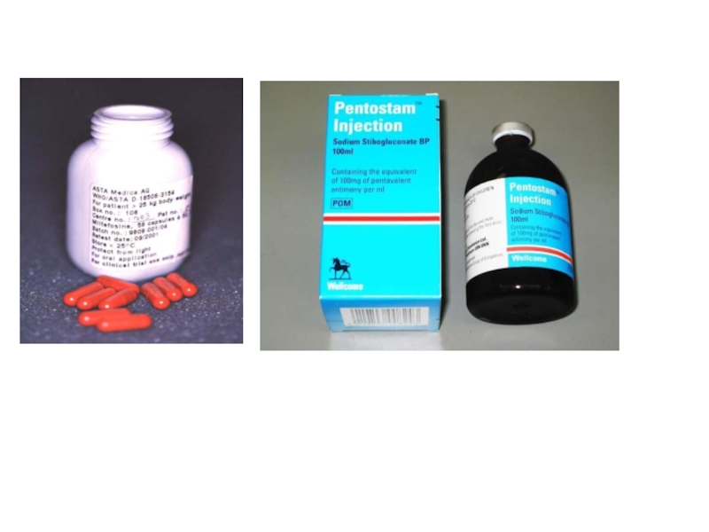
Слайд 32Cutaneous and mucocutaneous treatment
Antimony components : Meglumine antimoniate (Glucantime) and
20 mg/kg/d IV or IM for 20d
Pentamidine, Paromomycin are alternative drugs for CL
Amphotricine B for antimony resistant MCL
Fluconazole may decrease healing time
 and Sodium stibogluconate (Pentostam) are")
Слайд 33Visceral leishmaniasis treatment
Pentostam or Glucantime 20 mg /kg/d
Amphotricin B: 0.5-1 mg/kg IV daily 15-20d
Liposomal Amphotricin B (Ambisome): 3 mg/kg/d IV on days 1-5, day 14 and day 21
Low toxicity and high stability, better delivery
Alternative: Pentamidine (4mg/kg three times weekly, between 5-25 weeks ), Parmomycine

Слайд 34Visceral leishmaniasis treatment (con.)
Miltefosine (Impavido) (2.5 mg/kg /d p.o. for 28
It was developed for cancer therapy at first
The only oral drug
safer and more tolerable drug (less toxicity for bone marrow and haematopoietic progenitor cells)
teratogenic
Miltefosine (Impavido) (2.5 mg/kg /d p.o. for 28 d)It was developed for")
Слайд 36Leishmaniasis control
Vector control
insecticides
insecticide impregnated bed nets (IIB)
Case finding treatment
Aniaml
Treatment or killing of seropositive dogs
Rodent killing
Decrease of susceptibility: Childhood age, malnutrition and Immunosuppression are susceptibility factors for VL.
eliminating of childhood malnutrition
try to produce an efficient vaccine
Case finding treatment Aniaml reservoir controlTreatment or killing")