- Главная
- Разное
- Дизайн
- Бизнес и предпринимательство
- Аналитика
- Образование
- Развлечения
- Красота и здоровье
- Финансы
- Государство
- Путешествия
- Спорт
- Недвижимость
- Армия
- Графика
- Культурология
- Еда и кулинария
- Лингвистика
- Английский язык
- Астрономия
- Алгебра
- Биология
- География
- Детские презентации
- Информатика
- История
- Литература
- Маркетинг
- Математика
- Медицина
- Менеджмент
- Музыка
- МХК
- Немецкий язык
- ОБЖ
- Обществознание
- Окружающий мир
- Педагогика
- Русский язык
- Технология
- Физика
- Философия
- Химия
- Шаблоны, картинки для презентаций
- Экология
- Экономика
- Юриспруденция
Influenza презентация
Содержание
- 1. Influenza
- 2. INFLUENZA is an acute infectious disease
- 3. Influenza takes the leading position in
- 4. History The first documented pandemic of influenza
- 5. History In 1890 M.I. Afanasiev and in
- 6. Etiology The family Orthomyxoviridae comprises: influenza
- 7. Etiology The virions are spherical 80-120 nm
- 8. Etiology Influenza viruses bind to cells by
- 9. One of the most prominent features of
- 10. Nomenclature. The World Health Organization system of
- 11. A / Scotland / 42/89 (H3N2)
- 12. Nomenclature There are 15 different H antigens
- 13. Nomenclature However, in 1997 an outbreak, with
- 14. Physical characteristics The influenza viruses withstands slow
- 15. Physical characteristics Exposure to heat for 30
- 16. Epidemiology Epidemics, which must have been caused
- 17. Epidemiology In 1933 Christopher Andrewes and others
- 18. Epidemiology The mayor pandemics are associated with
- 19. Epidemiology Until 1977, when H1N1 reappeared, it
- 20. Epidemiology Influenza B viruses do not undergo
- 21. Epidemiology A sick person is the only
- 22. Epidemiology The influenza infection is spread with
- 23. Epidemiology The viable influenza virus can live
- 24. Epidemiology The speed of influenza spreading depends
- 25. Pathogenesis Pathogencity of influenza viruses is multifactirial
- 26. Pathogenesis The virus multiplication cycle lasts 7-10
- 27. Pathogenesis The cell, in which virus reproduces,
- 28. Pathogenesis The cells affected by a virus
- 29. Pathogenesis When the process spreads in the
- 30. Pathogenesis Under these conditions the influenza virus
- 31. Pathogenesis Besides, it is a fact that
- 32. Pathogenesis The earliest response is the synthesis
- 33. Clinical features. In classic influenza A: The
- 34. Clinical features The incubation period at influenza
- 35. Clinical features The first symptoms are chilliness,
- 36. Clinical features The main physical finding is
- 37. Clinical features Adynamia, malaise can be considerable
- 38. Clinical features of influenza
- 39. Clinical features Many patients have both upper
- 40. Clinical features The affection of the upper
- 41. Clinical features During fauces examination hyperemia of
- 42. Clinical features Diverse changes in the cardiovascular
- 43. Clinical features There are various affections of
- 44. Clinical features. In classic influenza B: Symptoms
- 45. Clinical features. In classic influenza C:
- 46. Complications of influenza: Primary influenza pneumonia may
- 47. The changes in the hemogram are manifested
- 48. Laboratory diagnosis The virusological methods of
- 49. Laboratory diagnosis The serological diagnostics of influenza
- 50. Laboratory diagnosis The immunefluorescent method is recommended
- 51. immune fluorescent method The essence of the
- 52. Treatment During the epidemics 10-25% of
- 53. Treatment Even in case of a moderate
- 54. Treatment Oral amantadine hydrochloride was introduced in
- 55. Treatment The usage of rimantadinum is quite
- 56. "Rimantadinum" appoints under the circuit: 1-st
- 57. More recently neuraminidase inhibitor Oseltamivir. Oseltamivir uses
- 58. There is fast elimination of intoxication and
- 59. Such preparations as 5-20% albumin solution, native
- 60. Immunity After an attack of influenza the

Слайд 2INFLUENZA
is an acute infectious disease which occurs in epidemics and
is caused by a virus, it is characterized by an abrupt onset and such manifesta-tions as general intoxication and affection of the respi-ratory tract mucosa.

Слайд 3 Influenza takes the leading position in the human pathology.
The
main thing is that besides relatively mild cases of the disease, there are severe cases resulting in disability and sometimes death when children or old people contract the disease.
Influenza and other acute respiratory diseases constitute about 75% of all infectious diseases.
According to the USA statistics influenza takes the tenth position in fatal outcomes.

Слайд 4History
The first documented pandemic of influenza (retrospectively - influenza type A
virus) occurred in 1889.
It is supposed to have begun in China and then spread to all the countries of the world in the following 1,5-2 years.
It is supposed to have begun in China and then spread to all the countries of the world in the following 1,5-2 years.
 occurred in 1889.")
Слайд 5History
In 1890 M.I. Afanasiev and in 1892 the German doctor R.
Pfeiffer isolated small bacilli from the sputum of sick people, most specialists considered them to be influenza pathogens for 20 -25 years.

Слайд 6Etiology
The family Orthomyxoviridae comprises: influenza A, B, C.
Influenza A
viruses can infect a variety of different host species, an ability that is of great importance in determining their ability to cause pandemic infection in humans. Influenza virus type A was the first to be isolated in 1933, by intranasal inoculation of the ferret. In 1940 influenza virus type B was isolated.

Слайд 7Etiology
The virions are spherical 80-120 nm in diameter. They have a
nucleocapsid comprising RNA, RNA-polymerase. The nucleocapsid is surrounded by an M1 protein shell, immediately exterior to which is a lipid envelope derived from the host cell. The M2 protein projects through the envelope to form ion channels, which allow pH changes in the endosome (pay attention: only influenza virus type A has M2 protein). Two types of spike project from the envelope, the haemagglutinin (H) and the neuraminidase (N).

Слайд 8Etiology
Influenza viruses bind to cells by the haemagglutinin interacting with membrane
recep-tors.
Neuraminidase activity is important in the final stages of release of new virus particles from infected cells.
Neuraminidase activity is important in the final stages of release of new virus particles from infected cells.

Слайд 9One of the most prominent features of the influenza viruses is
their ability to change antigenically either gradually (antigenic drift) or suddenly (antigenic shift).
Only influenza A virus has the potential to shift, whereas A, B and C may drift antigenically, although only very minor changes have been demonstrated in influenza virus type C.
Only influenza A virus has the potential to shift, whereas A, B and C may drift antigenically, although only very minor changes have been demonstrated in influenza virus type C.

Слайд 10Nomenclature. The World Health Organization system of nomenclature includes:
the host
of origin;
geographical origin,
strain number and year of isolation;
then follows in parentheses the antigenic description of the haemagglutinin and the neuraminidase and other.
geographical origin,
strain number and year of isolation;
then follows in parentheses the antigenic description of the haemagglutinin and the neuraminidase and other.

")
Слайд 12Nomenclature
There are 15 different H antigens and 9 N antigens. But
only H1-H3 and N1-N2 have been found in epidemic / pandemic viruses from humans, the others being recovered from animals and birds.

Слайд 13Nomenclature
However, in 1997 an outbreak, with high case mortality, occurred in
humans in Hong Kong;
the virus was an avian strain, H5N1, which appeared to transmit directly from chickens to humans.
No human to human spread was demonstrated.
the virus was an avian strain, H5N1, which appeared to transmit directly from chickens to humans.
No human to human spread was demonstrated.

Слайд 14Physical characteristics
The influenza viruses withstands slow drying at room temperature on
articles such as blankets and glass; it has been demonstrated in dust after an interval as long as 2 weeks.
Virus can survive in cold sea water for long period at -70°C, and remains viable indefinitely when freeze-dried.
Virus can survive in cold sea water for long period at -70°C, and remains viable indefinitely when freeze-dried.

Слайд 15Physical characteristics
Exposure to heat for 30 min at 56°C is sufficient
to inactivate most strains.
The viruses are inactivated by a variety of substances, such as 20 % ether in the cold, phenol, formaldehyde, soaps and many others.
The viruses are inactivated by a variety of substances, such as 20 % ether in the cold, phenol, formaldehyde, soaps and many others.

Слайд 16Epidemiology
Epidemics, which must have been caused by influenza viruses, have been
described for over 2000 years. Typically, there is a sudden appearance of cases of respiratory disease; these occur for several weeks and then suddenly cease. The epidemics occur frequently at irregular intervals. The great pandemic of 1918-1919 was particularly severe, killing between 20 million and 40 million people as it spread around the world.

Слайд 17Epidemiology
In 1933 Christopher Andrewes and others isolated influenza A virus. Continued
isolation studies and analysis of isolates have given an understanding of how the epidemic behaviors relates to changes in the virus. Also, it is possible to deduce which viral antigens circulated before virus isolation was possible by virtue of the phenomenon of original antigenic sin.

Слайд 18Epidemiology
The mayor pandemics are associated with antigenic shifts – when the
viral H or N, or both, are changed. These is too extensive to be the result of mutation, and analysis of the viral indicates that shift results from the acquisition of a complete new RNA segment.
A “new” virus can result from the process of reassortment. All the H and N antigenic subtypes are found in aquatic birds (both seabirds and ducks). The genetic reassortment may take place in pigs that have receptors for both human and avian strains, and may act as a mixing vessel from which certain subtypes may transmit to humans.
A “new” virus can result from the process of reassortment. All the H and N antigenic subtypes are found in aquatic birds (both seabirds and ducks). The genetic reassortment may take place in pigs that have receptors for both human and avian strains, and may act as a mixing vessel from which certain subtypes may transmit to humans.

Слайд 19Epidemiology
Until 1977, when H1N1 reappeared, it was the rule that when
a “new” virus appeared, the “old” one disappeared, but since that time two subtypes have been circulating concurrently, namely H3N2 and H1N1. The latter antigens had not been found since the 1950s and since they were antigenically very similar to viruses from the 1957 pandemic may have reappeared from a frozen source. There is no evidence of latent or persistent infection of humans.

Слайд 20Epidemiology
Influenza B viruses do not undergo antigenic shift as there is
no animal reservoir and, although epidemics do occur at 3-6 year intervals, they never reach pandemic proportions, and their extent is usually limited to small communities such as boarding schools or residences for the elderly. The antigenic changes result from mutation, as do those seen in influenza A after the appearance of “new” virus strains; the changes are the cause of antigenic drift.

Слайд 21Epidemiology
A sick person is the only source of the disease. The
virus quickly multiplies in the epithelial tissue of the respiratory tract mucous membrane of a sick person and in 24-48 hours there is an aerosol cloud with a great concentration of influenza virus around a patient in sneezing and coughing. As the immunity of a specific type forms very quickly, the virus disappears from the organism of a sick person on the fifth day of the disease.

Слайд 22Epidemiology
The influenza infection is spread with the help of small particle
aerosol dispersion. The mechanism of virus spreading is based on the condition that the virus is in the air for a long time, it has an ability to keep its infectious force under unfavorable conditions of the environment and the ability of virus particles to move with air at long distances and penetrate different parts of respiratory tracts infecting a person.

Слайд 23Epidemiology
The viable influenza virus can live and be infectious in the
air for 2 -3 hours. It can live for 1 -2 days on the furniture and other surfaces. The ultraviolet rays, humidity decrease and temperature increase and other factors shorten the virus life time. The virus lives within the limits of 1 -3 meters.

Слайд 24Epidemiology
The speed of influenza spreading depends on the speed of people
moving on the territory. The considerable increase of tranffic, the movement of great numbers of people within separate countries, between countries and continents ensures a constant possibility of the virus spreading at considerable distances and the ability to infect people in any part of the globe.

Слайд 25Pathogenesis
Pathogencity of influenza viruses is multifactirial and may involve viral, host
and environment factors. After penetrating the respiratory tracts, the virus sticks to the epithelial cells which have receptors. When the virus fixed on the cell surface receptors some complex enzymatic processes begin to occur, they ensure its penetration a cell in which it reproduces. This complex multistage process results in the cell destruction, and new virions born in the cells occupy new areas of the mucous membranes.

Слайд 26Pathogenesis
The virus multiplication cycle lasts 7-10 hours. Every virion which penetrated
a cell gives birth to 1000 virions. That's why the influenza incubation period is so short.

Слайд 27Pathogenesis
The cell, in which virus reproduces, produces and secretes interferon. This
interferon gets into the neighboring cells and after that they are not defenseless against the virus invasion. Interferon prevents virus protein from synthesis. The further development of virus infection depends on the struggle of these two forces -virus genome and cell interferon: either it stops at the very beginning and the disease lasts a short time and a patient gets well or the infection spreads in the lungs and fatal pneumonia develops.

Слайд 28Pathogenesis
The cells affected by a virus are rejected and the products
of their decomposition are absorbed, causing a general feverish disease. At the same time in the submucous membrane there develop inflammatory processes with distinctive circulatory disorders, that are clinically manifested by hemorrhage syndrome.

Слайд 29Pathogenesis
When the process spreads in the lung tissue, there are signs
of general edema with scattered or fused foci of hemorrhage in severe cases with the development of influenza pneumonia.

Слайд 30Pathogenesis
Under these conditions the influenza virus easily penetrates the blood and
virusemia develops. However, virusemia in influenza does not last long, as the virus quickly dies under the influence of nonspecific immunity factors - interferon, complement, properdin, β-lysines, etc.
The affection of the internals organs in influenza is associated with virusemia. However, in the pathogenesis of affections the leading role does not belong to the cytopathogenic phenomena, it belongs to the organism response to toxic products or other substances, which appear during the influenza virus reproduction process.
The affection of the internals organs in influenza is associated with virusemia. However, in the pathogenesis of affections the leading role does not belong to the cytopathogenic phenomena, it belongs to the organism response to toxic products or other substances, which appear during the influenza virus reproduction process.

Слайд 31Pathogenesis
Besides, it is a fact that even in the mild cases
of the disease there are signs of considerable depression of the organism hemopoietic and immune systems. The number leukocytes in blood decreases and their functions are suppressed. Macrophages become less active. Due to it bacteria become more active and the concomitant diseases take an acute form.

Слайд 32Pathogenesis
The earliest response is the synthesis and release of interferons from
the infected cells: these can diffuse to and protect both adjacent and more distant cells before the virus arrives. Specific antibody will help to limit the extracellular spread of the virus, while T-cell responses are directed against the viral glycoproteins on the surface of infected cells, leading to their destruction by cytotoxic T-cells and also by antibody-dependent cell cytotoxicity.

Слайд 33Clinical features.
In classic influenza A:
The incubation period is short, 2 days,
but it may vary from 1 to 4 days;
The illness is characterized by a sudden onset of systemic symptoms such as chills, fever, headache, myalgia and anorexia;
Respiratory symptoms are also common but take second place to the systemic effects, espessially early in the illness.
The illness is characterized by a sudden onset of systemic symptoms such as chills, fever, headache, myalgia and anorexia;
Respiratory symptoms are also common but take second place to the systemic effects, espessially early in the illness.

Слайд 34Clinical features
The incubation period at influenza in short: from several hours
to 2 days. Its duration depends on the dose and toxic characteristics of the virus. The incubation period is short if the dose is big and the virulence is considerable. The disease develops so fast that a practically healthy person becomes seriously ill in several minutes or hours.

Слайд 35Clinical features
The first symptoms are chilliness, high temperature, headaches, dizziness, a
syncope condition, malaise, pains in different parts of the body i.e. the symptoms of general intoxication.
The headache is located in the forehead, temples and over the brows, it can be of different intensity.
There is an early distinctive symptom - pain in the eye pupils especially intense in the eye movement, hyperemia of the conjunctiva and sometimes scleras.
The headache is located in the forehead, temples and over the brows, it can be of different intensity.
There is an early distinctive symptom - pain in the eye pupils especially intense in the eye movement, hyperemia of the conjunctiva and sometimes scleras.

Слайд 36Clinical features
The main physical finding is pyrexia, which rises rapidly to
a peak of 38-41°C within 12 h of onset. Fever usually lasts 3 days, but it can present for 1-5 days. The 'two-humped' character of the temperature is associated with the condition when the chronic infection takes an acute form or with development of secondary flora. Such symptoms as unconsciousness, delirium, convulsions and meningeal manifestations are characteristic of intense toxicosis.

Слайд 37Clinical features
Adynamia, malaise can be considerable and are manifested since the
first day of the disease. The skin on the face is hyperemic during the first 2-3 days, in severe cases they become pale with cyanotic shade. It is often a bad prognostic sign. Sweating is a characteristic feature. Intoxication is a characteristic feature of influenza. There is hemorrhage syndrome, in 10 -20% of cases, its symptoms are nasal bleeding, sometimes reciprocal, hemorrhage in the fauces, metrorrhagia, short hemoptysis and gum bleeding.


Слайд 39Clinical features
Many patients have both upper and lower respiratory tract infection,
ofen with a troublesome, dry cough. Cough develops during the first days of the disease, dry, excruciating, hacking which is accompanied by the feeling of tickling, scratching behind the breastbone. Almost all patients have a catarrhal syndrome, which has such symptoms as rhinitis, pharyngitis, tracheitis. There are often such combined affections of the mucous membrane as rhinopharyngitis, laryngotracheitis, tracheobronchitis, etc. They usually develop in the first days of the disease.

Слайд 40Clinical features
The affection of the upper respiratory tracts is accompanied with
hyperemia and swelling of the mucous membrane, sometimes with slight hemorrhages. There is nasal obstruction, rough breathing, and discharge of different nature and consistence: mucous, mucopurulent and sanguinolent. During rhinoscopy swelling and hyperemia of the mucous membrane can be seen. At the same time accessory nasal sinus can be affected with different nature of affection - from catarrhal to purulent.

Слайд 41Clinical features
During fauces examination hyperemia of the tonsils, uvula palatina and
posterior wall of the throat can be found. Sometimes there are granules with vascular injection and hemorrhages on the soft palate.

Слайд 42Clinical features
Diverse changes in the cardiovascular system have been described. The
pulse very often corresponds to the fever, there is sometimes tachycardia, especially at the beginning of the disease, in some cases there is bradycardia. The heart sounds are dull, heart borders are widened, slight systolic murmur and sometimes extrasystoles appear. All these manifestations disappear when the general condition of the patient becomes better.

Слайд 43Clinical features
There are various affections of the nervous system during the
influenza infection. The functional disorders of the autonomic nervous system are distinctively manifested (such as sweating, changes of the pulse rate, dizziness, etc). However, all these changes quickly disappear. At the same time serious affections of the central and peripheral nervous systems are observed, they are manifested as meningitis, meningoencephalitis, radiculitis, neuritis, etc.

Слайд 44Clinical features.
In classic influenza B:
Symptoms closely resemble those associated with influenza
A infection, consisting of a 3-day febrile illness with predominantly systemic symptoms. Overall, the infection is somewhat milder; some studies have shown more involvement of the gastro-intestinal tract, with the coining of the term “gastric flu”.

Слайд 45Clinical features.
In classic influenza C:
Clinically, influenza C causes an afebrile
upper respiratory tract infection usually confined to young children: outbreaks are not recognized.

Слайд 46Complications of influenza:
Primary influenza pneumonia may occur, especially in young adults
during an outbreak, and can be fatal after a very short illness of sometimes less then 1 day. A similar rapid illness can occur in the elderly.
More commonly a bacterial pneumonia caused by Staphylococcus aureus or Streptococcus pneumoniae occurs late in the course of the illness, often after a period of improvement, resulting in a classical biphasic fever pattern.
More commonly a bacterial pneumonia caused by Staphylococcus aureus or Streptococcus pneumoniae occurs late in the course of the illness, often after a period of improvement, resulting in a classical biphasic fever pattern.

Слайд 47The changes in the hemogram
are manifested as leukopenia or normocytosis.
If
there are no complications and concomitant diseases, there is absence or decrease of eosinophils, neutropenia and relative lymphocytosis in the hemogram in influenza (the percentage of lymphocytes increases whereas their absolute number is the same). The bacterial complications is accompanied with leukocytosis and neutrophilia.

Слайд 48Laboratory diagnosis
The virusological methods of diagnostics are used to isolate
and identify the influenza virus. As a rule these methods are used to find out the nature of the outbreaks but not the sporadic cases of the disease because they are very laborious and less sensitive as compared with the serologic methods. The infection of the chicken embryos is a universal method of the primary isolation and cultivation of influenza virus.

Слайд 49Laboratory diagnosis
The serological diagnostics of influenza ensures an accurate determination of
etiology by revealing the quantitative growth of specific antibodies in the disease dynamics in blood. The serological diagnosis is especially important in case of the atypical or symptomless course of the influenza infection. Among the methods of influenza serological diagnostic the reaction of hemagglutination inhibition and the reaction of complement banding is the most common.

Слайд 50Laboratory diagnosis
The immunefluorescent method is recommended as one of the reliable
means of quick deciphering of the etiology of acute respiratory diseases. The sorting of patients with acute respiratory diseases is done on the bases of the immunefluorescent method data, it is especially important for the prevention of the cross infection. Being widely used this method is an important and reliable means of control of the etiological structure of the acute respiratory diseases in different periods according to the epidemic situation.

Слайд 51immune fluorescent method
The essence of the immune fluorescent method is in
specific reactions of antigen-antibody which reveal the presence of viral antigens in the cells by attached antibodies, the antibodies are chemically connected with the fluorescent mark, which shines in the ultraviolet rays.

Слайд 52Treatment
During the epidemics 10-25% of the adult population fall ill
with influenza, 1-2% of them need hospitalization. Most patients are treated in polyclinics.
During the out-patient reception or home visiting it is always necessary to find out if one or another patient needs to be hospitalized or can be treated at home.
The severity of the patients’ condition determined by the intoxication degree, complications and the presence or acute conditions of the preceding diseases must be taken into account. A patient with such symptoms as high temperature, consciousness disorders, convulsion syndrome, repeated vomiting, meningeal symptoms, hemorrhagic syndrome, respiratory and cardiovascular insufficiency should be hospitalized.

Слайд 53Treatment
Even in case of a moderate severe influenza form together with
an unfavorable premorbid condition in the form of ischemic heart disease, chronic nonspecific lung diseases, nervous system diseases and others the patients should be treated in hospital.
It is necessary to keep the bed even in case of influenza without any complications.
Taking into consideration the fact that the temperature reaction in influenza has a certain compensation-adaptation meaning - the suppression of virus replication, stimulation of endogenic interferon formation and mobilization of other defensive mechanisms of the organism, it is necessary to reduce it to normal values only in the patients who have problems with high temperature.

Слайд 54Treatment
Oral amantadine hydrochloride was introduced in the early 1980s, followed later
by a derivative, rimantadine. These drug work by blocking the ion channels in the envelope, thus preventing the pH changes that precede the membrane fusion step essential for nucleocapsid release. Unfortunately, these compounds only have activity against influenza virus type A but not B, C or other respiratory viruses.

Слайд 55Treatment
The usage of rimantadinum is quite effective especially during the first
days of the disease. The antiviral action of remantadinum is the most effective at the early stages of the infection development. The preparation usage from the first day of the disease results in the decrease of the expressiveness and duration of fever and other intoxication symptoms.

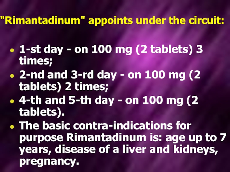
Слайд 56"Rimantadinum" appoints under the circuit:
1-st day - on 100 mg (2
tablets) 3 times;
2-nd and 3-rd day - on 100 mg (2 tablets) 2 times;
4-th and 5-th day - on 100 mg (2 tablets).
The basic contra-indications for purpose Rimantadinum is: age up to 7 years, disease of a liver and kidneys, pregnancy.
2-nd and 3-rd day - on 100 mg (2 tablets) 2 times;
4-th and 5-th day - on 100 mg (2 tablets).
The basic contra-indications for purpose Rimantadinum is: age up to 7 years, disease of a liver and kidneys, pregnancy.
Слайд 57More recently neuraminidase inhibitor Oseltamivir. Oseltamivir uses influenza A, B and
H5N1 infections. It can reduce the duration of symptoms by 1-3 days if given within 36 h of onset of illness.
"Oseltamivir" appoints under the circuit:
On 75 mg (1 capsule) 2 times a day during 5-7 days.
Purpose "Oseltamivir" is contra-indicated at chronic renal for insufficiency, pregnancy, feeding by a breast.

Слайд 58There is fast elimination of intoxication and noticeable improvement of the
patients’ general condition observed when lymphocytic interferon is inserted in the nasal path ways during the first days of the disease both by simple dripping and with a help of individual inhaler.

Слайд 59Such preparations as 5-20% albumin solution, native or dried plasma 150-200
ml, haemodesum 200 ml, rheopolyglucinum up to 500 ml, physiological salt solution are used for detoxification. The liquid must be administered with a strict control to prevent acute edema of the brain and lungs. The administration of lasix, anti-histamine preparations, rutinum, vitamin C, correlation of the acid-base and water-electrolyte balance are advisable. Antibiotics and sulfanilamides are the preparations for treatment of complications or aggravations of chronic infections which are caused bacterial flora.

Слайд 60Immunity
After an attack of influenza the ensuing immunity to the particular
subtype of infecting virus is of long duration. It is related to the amount of local antibody (Ig A) in the mucous secretions of the respiratory tract together with the specific IgG serum antibody concentration. Immunity to infection, especially with type A., is subtype-specific, giving little or no protection against subtypes possessing immunologically distinct H or N proteins.