- Главная
- Разное
- Дизайн
- Бизнес и предпринимательство
- Аналитика
- Образование
- Развлечения
- Красота и здоровье
- Финансы
- Государство
- Путешествия
- Спорт
- Недвижимость
- Армия
- Графика
- Культурология
- Еда и кулинария
- Лингвистика
- Английский язык
- Астрономия
- Алгебра
- Биология
- География
- Детские презентации
- Информатика
- История
- Литература
- Маркетинг
- Математика
- Медицина
- Менеджмент
- Музыка
- МХК
- Немецкий язык
- ОБЖ
- Обществознание
- Окружающий мир
- Педагогика
- Русский язык
- Технология
- Физика
- Философия
- Химия
- Шаблоны, картинки для презентаций
- Экология
- Экономика
- Юриспруденция
Immunophysiology of reproductive system презентация
Содержание
- 1. Immunophysiology of reproductive system
- 2. Over 50 years ago, there was the
- 3. Integrational view of the immune system during
- 4. Role of the placenta as a modulator
- 7. Trophoblasts are specialized cells of the placenta
- 8. During normal pregnancy, the human decidua contains
- 10. Comparison and contrast between a cellular response
- 11. The interaction between the trophoblast HLA molecules
- 12. Cytokines produced by uNK cells at
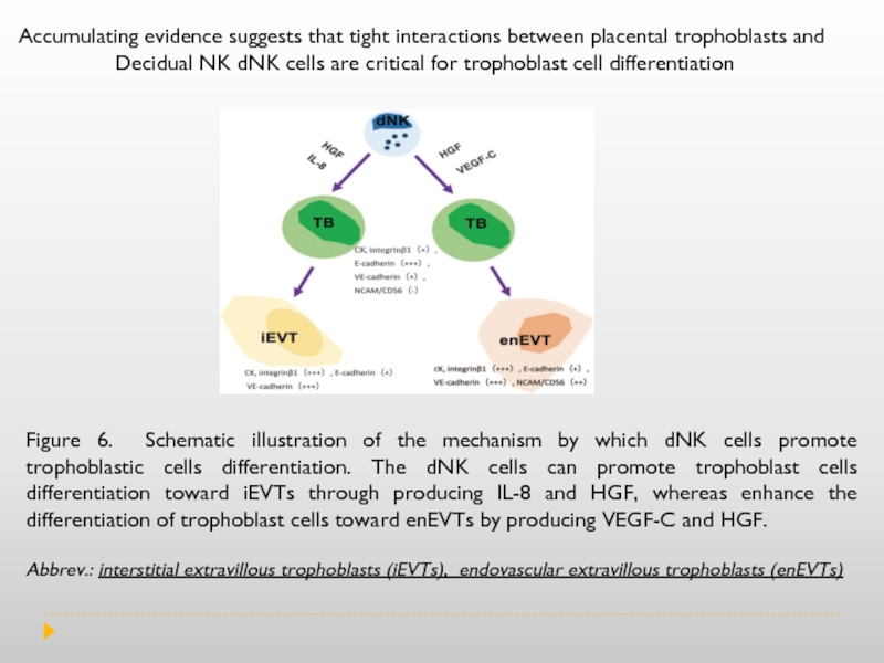
- 13. Figure 6. Schematic illustration of the mechanism
- 14. During gestation, uNK (uterine NK cells) are
- 15. Pre-eclampsia (PE) is a disorder of pregnancy characterized by the
Слайд 2Over 50 years ago, there was the assumption that the placenta
The immunology of pregnancy is the result of the combination of signals and responses originated from the maternal immune system and the fetal–placental immune system. The signals originated in the placenta will modulate the way the maternal immune system will behave in the presence of potential dangerous signals. The immune system of the mother should not be thought of as suppressed, but rather modulated and streamlined to focus on pathogen recognition, communication, trafficking and repair. This suggests that the mother’s immune system is still able to mount an attack, but only when absolutely necessary. Such modulated mechanisms allow the mother to maintain a well-balanced immune system.
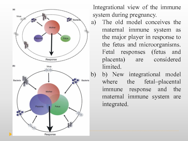
Слайд 3Integrational view of the immune system during pregnancy.
The old model
b) New integrational model where the fetal–placental immune response and the maternal immune system are integrated.
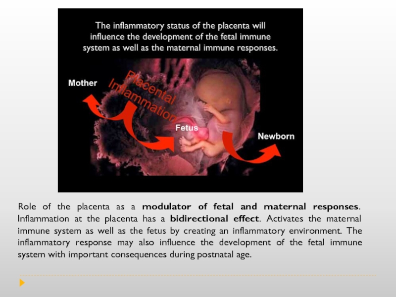
Слайд 4Role of the placenta as a modulator of fetal and maternal
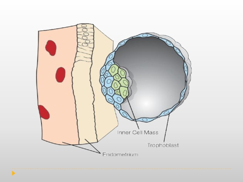
Слайд 7Trophoblasts are specialized cells of the placenta that play an important
In addition, cytotrophoblast can differentiate into another type of trophoblast called the extravillous trophoblast and penetrate into the decidualised uterus. This process is essential not only for physically attaching the placenta to the mother, but also for altering the vasculature in the uterus to allow it to provide an adequate blood supply to the growing fetus as pregnancy progresses. Some of these trophoblast even replace the endothelial cells in the uterine spiral arteries as they remodel these vessels into wide bore conduits that are independent of maternal vasoconstriction. This ensures the fetus receives a steady supply of blood, and the placenta is not subjected to fluctuations in oxygen that could cause it damage.
Decidualization is a process that results in significant changes to cells of the endometrium in preparation for, and during, pregnancy.
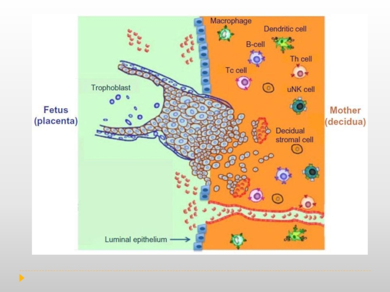
Слайд 8During normal pregnancy, the human decidua contains a high number of
These data further support the idea that the fetal–maternal immune interaction is more complex than the comparison to transplant allograft.
Consequently, the presence of immune cells at the implantation site is not associated with a response to the ‘foreign’ fetus but to facilitate and protect the pregnancy. Therefore, the immune system at the implantation site is not suppressed, on the contrary it is active, functional and is carefully controlled.
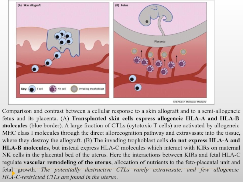
Слайд 10Comparison and contrast between a cellular response to a skin allograft
Слайд 11The interaction between the trophoblast HLA molecules and the KIR receptors
Unlike CTLs, however, and unlike conventional NK cells, uNK (uterine NK cells) cells are not good killers and do not destroy the bearer of allogeneic MHC molecules (the trophoblast) but respond by producing soluble factors that promote placentation. NK receptors are expressed by uNK cells and may also regulate interactions with trophoblasts as well with other uterine leukocytes and stromal cells. uNK cells are a unique subset that populates the uterine mucosa and which, contrary to their blood counterparts whose evocative name they share, keep their killer instinct under control and instead help the growing placenta and fetus. uNK cells, by producing growth factors, chemokines, and cytokines , contribute to regulating trophoblast invasion and vascular remodeling in the uterus, a vital process for the placenta to sustain fetal growth.
Слайд 12
Cytokines produced by uNK cells at the human fetal-maternal interface include
In normal pregnancies, recognition of fetal HLA-C by receptor KIR-BB of uNK triggers the release of TGF-? s by uNK cells. TGF-? - transforming growth factor beta, whose participation in immunoregulation and angiogenesis has been well-established.
Слайд 13Figure 6. Schematic illustration of the mechanism by which dNK cells
Abbrev.: interstitial extravillous trophoblasts (iEVTs), endovascular extravillous trophoblasts (enEVTs)
Accumulating evidence suggests that tight interactions between placental trophoblasts and
Decidual NK dNK cells are critical for trophoblast cell differentiation
Слайд 14During gestation, uNK (uterine NK cells) are in intimate contact with
Mechanisms by which immune cells (focus: uNK cells) regulate key early events in establishment of pregnancy: implantation, angiogenesis, and vascular remodeling.
Слайд 15Pre-eclampsia (PE) is a disorder of pregnancy characterized by the onset of high blood pressure and
DCs and macrophages, present in the human endometrium, play a role in decidualization and implantation. After uNK, macrophages are the second most abundant population in the maternofetal interphase in both implantation and early pregnancy development. Macrophages congregate around spiral arteries, while the placenta develops and supports vascular remodeling by releasing proangiogenic factors, such as VEGF and MMPs and by removing apoptotic cells. The polarization of decidual macrophages toward M2, found in normal pregnancies, indicates that their immunosuppressive activities are critical for maintaining immunological homeostasis during pregnancy. Uterine DCs play a key role in embryo implantation, when they show an immature phenotype. Immature DC’s have been associated with the initiation and maintenance of peripheral tolerance and their presence in large numbers in the uterine decidua has been associated with the establishment of healthy pregnancies in women.
Tregs are essential for pregnancy maintenance, and low levels have been found in pregnancy complications. Thus not only women with unexplained recurrent spontaneous abortions, but also patients with preeclampsia display low levels of Tregs in both maternal blood and placenta. The prevalence of Th17 cells and the Th17/Treg ratio increases in peripheral blood in preeclampsia as compared with normal pregnancy.
