ALMATY 2015
- Главная
- Разное
- Дизайн
- Бизнес и предпринимательство
- Аналитика
- Образование
- Развлечения
- Красота и здоровье
- Финансы
- Государство
- Путешествия
- Спорт
- Недвижимость
- Армия
- Графика
- Культурология
- Еда и кулинария
- Лингвистика
- Английский язык
- Астрономия
- Алгебра
- Биология
- География
- Детские презентации
- Информатика
- История
- Литература
- Маркетинг
- Математика
- Медицина
- Менеджмент
- Музыка
- МХК
- Немецкий язык
- ОБЖ
- Обществознание
- Окружающий мир
- Педагогика
- Русский язык
- Технология
- Физика
- Философия
- Химия
- Шаблоны, картинки для презентаций
- Экология
- Экономика
- Юриспруденция
Hemangioma презентация
Содержание
- 1. Hemangioma
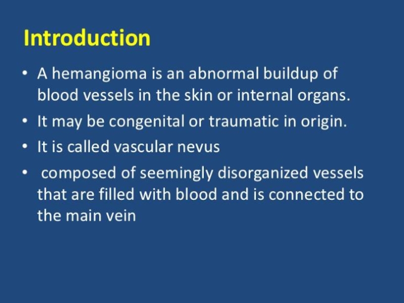
- 2. PLAN Introduction The main part Reference
- 5. Simple hemangioma is usually red or
- 6. Cavernous hemangioma is usually located under
- 7. Combined hemangioma usually a combination
- 8. Extensive capillary-cavernous hemangioma of the left half
- 9. Capillary hemangioma of the child Hemangioma of the humerus
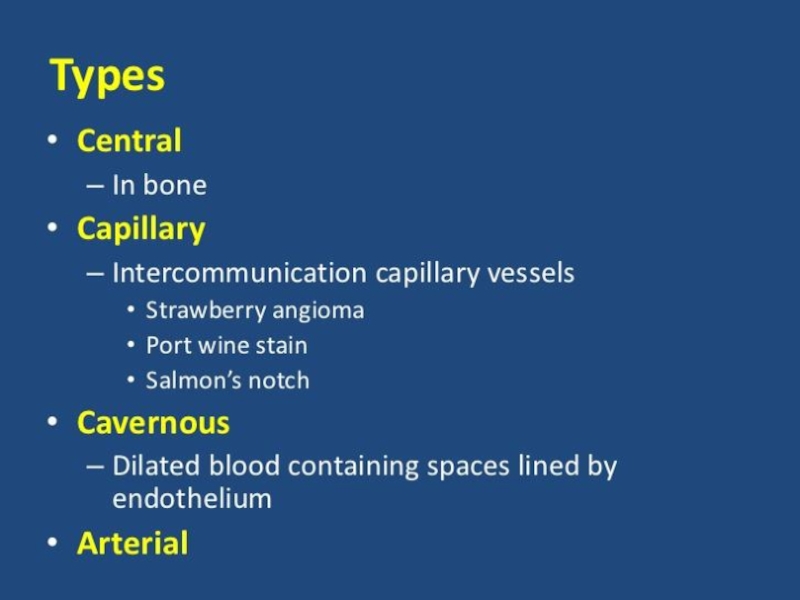
- 10. Hemangioma arterial (h. Arteriale) - hemangioma, arterial blood vessels from growing type.
- 11. HEMANGIOMA CAN BE LIVER KIDNEY VERTEBRAE LIPS
- 13. Microscopic structure of capillary hemangioma. The wall
- 14. Microscopic finding of the giant hemangioma.
- 15. Cavernous Hemangioma of the Maxillary andEthmoid Sinus
- 16. Gross appearance of the sclerosed hemangioma of
- 19. REFERENCE http://www.ayzdorov.ru/lechenie_gemangioma_chto.php http://razvitie-krohi.ru/zdorove-rebenka/vse-o-gemangiomah-u-novorozhdennyih-detey.html https://ru.wikipedia.org/wiki/%D0%93%D0%B5%D0%BC%D0%B0%D0%BD%D0%B3%D0%B8%D0%BE%D0%BC%D0%B0 https://www.google.kz/search?q=Microscopic+structure+of+capillary+hemangioma&es_sm=93&source=lnms&tbm=isch&sa=X&ei=xrA2VaOcF-ehyAPo-oLIBA&ved=0CAcQ_AUoAQ&biw=1366&bih=643#tbm=isch&q=Microscopic+structure+hemangioma http://synapse.koreamed.org/DOIx.php?id=10.3350/kjhep.2010.16.4.410&vmode=PUBREADER#!po=91.6667
Слайд 1HEMANGIOMA
MADE BY KALAMBEKOV MEREY
CHECKED SAPARGALIEVA A.D.
KAZAKH NATIONAL MEDICAL UNIVERSITY
NAMED AFTER
S.D.ASFENDIYAROV


Слайд 5Simple hemangioma
is usually red or blue-purple color, is located on
the surface, clearly delineated boundaries, affects the skin and a few millimeters of subcutaneous fat layer, usually grows in hand. Rare hemangioma uneven, slightly protruding above the skin (usually smooth). One symptom of hemangiomas is that if you push on it, it is for a short period of time fades, then again takes its color.

Слайд 6Cavernous hemangioma
is usually located under the skin, is a limited
nodular formation, soft-elastic consistency. Consists of various cavities - caverns filled with blood. Look cavernous hemangioma as tumor formation, on top of the usual skin color, sometimes bluish. With the growth of the tumor skin becomes blue-purple color. When pressed on the hemangioma she falls and thus a bit pale (due to the outflow of blood). When you cry and cough hemangioma increases.

Слайд 7Combined hemangioma
usually a combination of surface and subcutaneous hemangiomas
(simple and cavernous). Detected by the prevalence of one or the other of the tumor vasculature. Appearance and consistency, again, depends on its constituent tissues.
. Detected by")
Слайд 8Extensive capillary-cavernous hemangioma of the left half of the head with
a pronounced exophytic growth
Cavernous hemangioma


 - hemangioma, arterial blood vessels from growing type.")

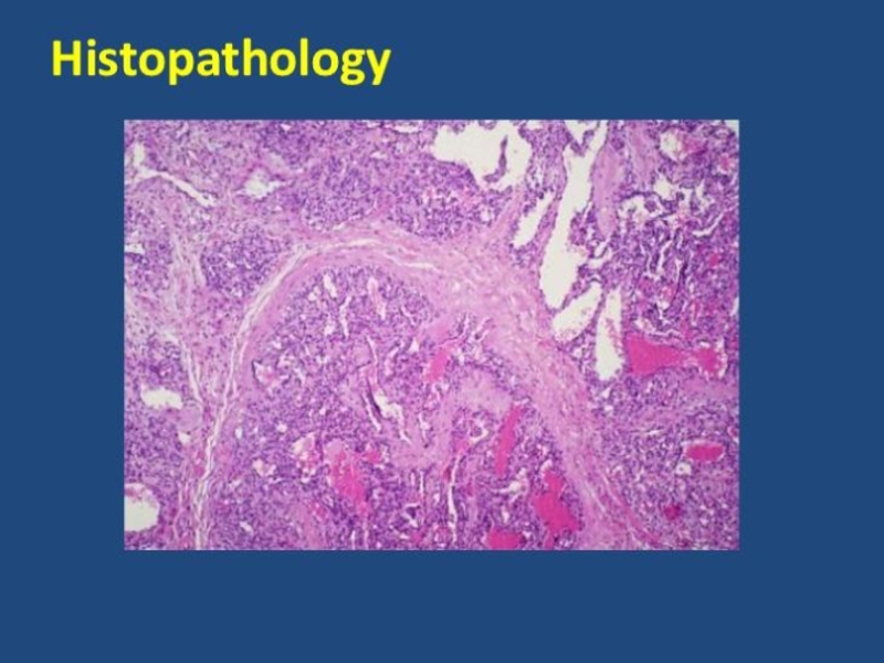
Слайд 13Microscopic structure of capillary hemangioma. The wall of the capillaries represented
two-three-layer endothelium (tissue atypia), the cavity often filled with blood.
,")
Слайд 14
Microscopic finding of the giant hemangioma. (A) Low-power view disclosing proliferation
of the cavernous vascular structures. (B) Each vascular space is lined by flat endothelial cells and filled with fresh red blood cells. (A: H-E, ×12.5; B: H-E, ×100).
 Low-power view disclosing proliferation of the cavernous vascular")

Слайд 16Gross appearance of the sclerosed hemangioma of liver. A gray-white sunken
nodule is noted in the subcapsular area (A). The cut surface discloses a well-demarcated homogenous gray-white solid nodule, measuring 2.1 cm at its greatest dimension, with tiny red spots (B).

Слайд 19REFERENCE
http://www.ayzdorov.ru/lechenie_gemangioma_chto.php
http://razvitie-krohi.ru/zdorove-rebenka/vse-o-gemangiomah-u-novorozhdennyih-detey.html
https://ru.wikipedia.org/wiki/%D0%93%D0%B5%D0%BC%D0%B0%D0%BD%D0%B3%D0%B8%D0%BE%D0%BC%D0%B0
https://www.google.kz/search?q=Microscopic+structure+of+capillary+hemangioma&es_sm=93&source=lnms&tbm=isch&sa=X&ei=xrA2VaOcF-ehyAPo-oLIBA&ved=0CAcQ_AUoAQ&biw=1366&bih=643#tbm=isch&q=Microscopic+structure+hemangioma
http://synapse.koreamed.org/DOIx.php?id=10.3350/kjhep.2010.16.4.410&vmode=PUBREADER#!po=91.6667