- Главная
- Разное
- Дизайн
- Бизнес и предпринимательство
- Аналитика
- Образование
- Развлечения
- Красота и здоровье
- Финансы
- Государство
- Путешествия
- Спорт
- Недвижимость
- Армия
- Графика
- Культурология
- Еда и кулинария
- Лингвистика
- Английский язык
- Астрономия
- Алгебра
- Биология
- География
- Детские презентации
- Информатика
- История
- Литература
- Маркетинг
- Математика
- Медицина
- Менеджмент
- Музыка
- МХК
- Немецкий язык
- ОБЖ
- Обществознание
- Окружающий мир
- Педагогика
- Русский язык
- Технология
- Физика
- Философия
- Химия
- Шаблоны, картинки для презентаций
- Экология
- Экономика
- Юриспруденция
GU tumors. Renal cell carcinoma презентация
Содержание
- 1. GU tumors. Renal cell carcinoma
- 2. Renal cell carcinoma ETIOLOGY: CIGARETTE SMOKING OBESITY
- 3. Renal cell carcinoma Clinical presentation:
- 4. Renal cell carcinoma
- 6. Biology of RCC Von Hippel-Lindau (VHL) syndrome
- 7. Motzer. Five variables as risk factors for
- 8. Renal cell carcinoma Radiographic evaluation: CT
- 9. Renal cell carcinoma - treatment Localized RCC
- 10. Renal cell carcinoma - treatment Chemotherapy -
- 11. Renal cell carcinoma - treatment VEGF Targeted
- 12. immunotherapy Opdivo (Nivolumab) - anti PD1
- 14. Bladder cancer Pathology - transitional cell carcinoma
- 15. Bladder cancer Clinical presentations: gross
- 17. Bladder cancer - treatment Ta, Tis, T1
- 18. Bladder cancer - treatment Muscularis propria-invasive disease
- 20. Bladder cancer - treatment Adjuvant chemotherapy?
- 22. Risk factors GENETIC FACTORS two-fold
- 23. Risk factors AGE :rarely occurs before the age of 45 RACE, ETHNICITY
- 24. BRCA1/2 mutations The
- 25. Dr.Neiman Victoria
- 26. PRETREATMENT STAGING Serum PSA
- 27. 27.09.2017 Dr.Neiman Victoria TNM staging
- 28. Dr.Neiman Victoria PREDICTING ORGAN CONFINED DISEASE Biopsy Gleason grade
- 29. Pretreatment Risk Assessment in Localized Disease
- 30. The most effective therapy for
- 31. Increased PSA After Radical Prostatectomy
- 33. OTHER THERAPIES Cryotherapy Laparoscopic and robotic prostatectomy
- 34. Pure germ cell tumor – one site
- 35. Cancer of Testis
- 36. Cancer of Testis - Staging T1- without
- 37. Cancer of Testis - Staging M1a –
- 38. Cancer of Testis - Staging St I
- 39. Cancer of Testis – Prognostic Group
- 40. Seminoma St I
- 41. Seminoma St II- Low- tumor burden (St IIA-B =
- 42. Seminoma St II - III – (High
- 43. Seminoma St II-III High- tumor burden
- 44. Seminoma metast – inferiority of carbo vs
- 45. Non-Seminoma Good and interm progn: testis/retroperitoneal
- 46. Non-Seminoma St I RPLND bilateral

Слайд 2Renal cell carcinoma
ETIOLOGY:
CIGARETTE SMOKING
OBESITY
ANALGESIC ABUSE (phenacetin)
INDUSTRIAL SOLVENT, TRICHLOROETHYLENE
EXPOSURE TO CADMIUM
ACQUIRED CYSTIC
INDUSTRIAL SOLVENT, TRICHLOROETHYLENEEXPOSURE TO CADMIUMACQUIRED CYSTIC DISEASE")
Слайд 3Renal cell carcinoma
Clinical presentation:
- Pain
- Hematuria
- Flank
metastatic disease – 30% (75% - lung mets)
locally advanced - 25%
localized disease - 45%


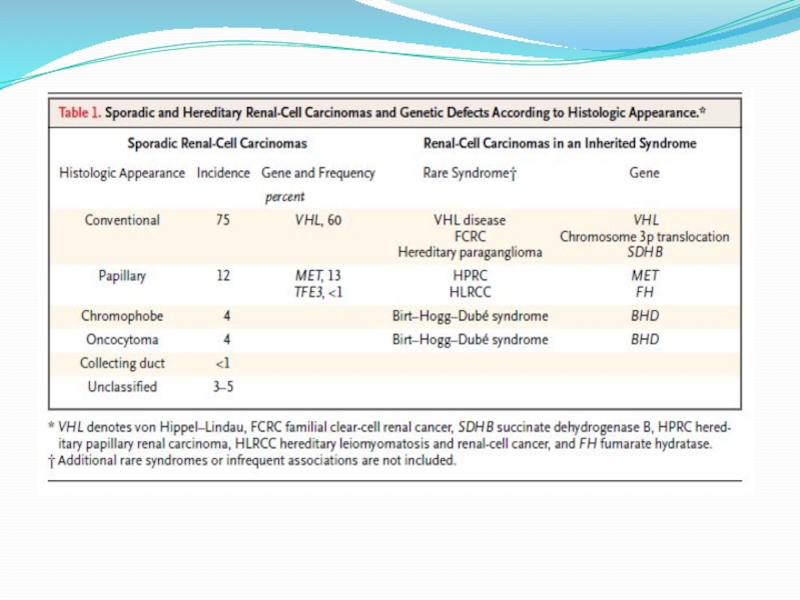
Слайд 6Biology of RCC
Von Hippel-Lindau (VHL) syndrome is characterized by germline mutation
Noninherited clear-cell RCC characterized by VHL gene tumor suppressor gene inactivation, leads to
Constitutive expression of oxygen-regulated transcription factor (HIFa)
Induction of hypoxia-inducible genes, including vascular endothelial growth factor (VEGF)
VEGF overexpression promotes tumor angiogenesis
 syndrome is characterized by germline mutation of chromosome 3p, development")
Слайд 7Motzer. Five variables as risk factors for short survival
Low KPS (
Low hemoglobin
High corrected serum calcium (>10mg/dL)
Time of metastatic desease from diagnosis (less than a year)
Low hemoglobinHigh corrected")
Слайд 8Renal cell carcinoma
Radiographic evaluation:
CT is the modality of choice for imaging
MRI
US
Renal arteriography

Слайд 9Renal cell carcinoma - treatment
Localized RCC
- surgical treatment
Metastatic RCC
- resection of metastasis (lung)

Слайд 10Renal cell carcinoma - treatment
Chemotherapy -
Chemotherapy currently has

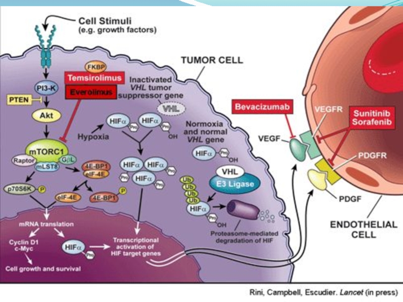
Слайд 11Renal cell carcinoma - treatment
VEGF Targeted therapy
VEGF receptor:
surafenib
Pazopanib
Axitinib
VEGF ligand:
Bevacizumab

 - anti PD1")
Слайд 14Bladder cancer
Pathology - transitional cell carcinoma (TCC) – 90%
squamous Cell carcinoma
Risk factors – gene abnormalities (protooncogene Ras p21 protein)
chemical exposure
chronic irritation (SqCC)
 – 90%")
Слайд 15Bladder cancer
Clinical presentations:
gross painless hematuria
Workup:
cytology
upper truct study (CT)
Clinical stage of the primary tumor - TURBT

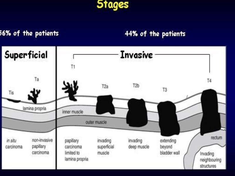
Слайд 17Bladder cancer - treatment
Ta, Tis, T1 – 70%
TURBT
Intravesical drug
BCG
MITOMYCIN C
DOXORUBICIN
GEMCITABINE
THIOTEPA

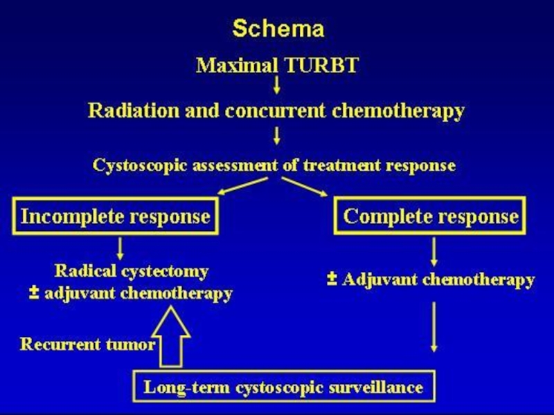
Слайд 18Bladder cancer - treatment
Muscularis propria-invasive disease
Radical cystectomy
Complications of Cystectomy (ileal Conduit):
Metabolic
Increase Cl
Decrease K,CA, MG
Bladder Preservation treatment
:Metabolic acidosisIncrease ClDecrease K,CA, MGBladder")
Слайд 20Bladder cancer - treatment
Adjuvant chemotherapy?
4 cycles of Cisplatin plus
Metastatic Bladder Cancer
MVAC MS - 15.2 m
gemcitabine/cisplatin –MS - 14.0 m (more less toxicity)

Слайд 21
Prostate cancer
Prostate cancer is the most common cancer in American

Слайд 22
Risk factors
GENETIC FACTORS
two-fold elevated in men with an affected first
trend toward increasing risk with a greater number of affected family members; men with two or three affected first-degree relatives had a 5- and 11-fold increased risk of prostate cancer
In a study of 45,000 Scandinavian twin pairs, concordance for cancer in identical twins was higher for prostate cancer than either breast or colorectal cancer
,")

Слайд 24
BRCA1/2 mutations
The presence of BRCA1/2 mutations may increase the risk


Слайд 26
PRETREATMENT STAGING
Serum PSA
Biopsy of the tumor
Digital rectal examination
to detect the presence of extraprostatic extension or seminal vesicle invasion
Computed tomography (CT) of the abdomen and pelvis and radionuclide bone scan are used selectively
endorectal coil MRI may be useful in selected patients


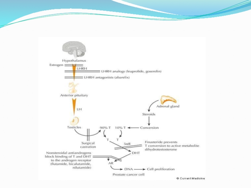
Слайд 30
The most effective therapy for clinically localized prostate cancer
Surgery
radiation therapy
androgen deprivation therapy (ADT)
observation (also termed watchful waiting).
 androgen deprivation therapy")
Слайд 31
Increased PSA After Radical Prostatectomy
Risks Factor for Clinical Relapse
1. Doubling time
The shorter the time, the higher the risk
2. Time to biochemical failure
The shorter the time, the higher the risk
3. Gleason score
higher scores reflect more aggressive tumors


Слайд 34Pure germ cell tumor – one site of hystology
Mixed germ cell
SEMINOMA
NON-SEMINOMA: - embrional carcinoma
- teratoma
- choriocarcinoma
- yolk sac tumor
Cancer of Testis

Слайд 35Cancer of Testis
Good progn 55% 90%
5y PFS 90% 80%
5y OS 92% 85%
Interm progn 30% 10%
5y PFS 75% 67%
5y OS 80% 72%
Poor progn 15%
5y PFS 40%
5y OS 50%

Слайд 36Cancer of Testis - Staging
T1- without involv of tunica vaginalis
T2 –vascular/lumphovascul
T3- spermatic cord inv.
T4- scrotum
c N – number of LN not important, size!:
C N1 <2cm
C N2 2-5 cm
C N3 >5 cm
PN- number and size important!:
P N1- 1-5 LN-s , <2cm
PN2- single 2-5 cm, or 2-5 : <5cm
PN3->5cm

Слайд 37Cancer of Testis - Staging
M1a – non-regional nodes oo pulmonary mts
M1b
S0- normal markers
S1 LDH < 1.5 X UNL; HCG < 5000; AFP<1000
S2 LDH 1.5-10XUNL; HCG 5 000-50 000; AFP1000-10 000
S3 LDH > 10 X UNL; HCG >50 000; AFP>10 000
Normal LDH 60 – 225 90 – 337 S2
T1/2 AFP 5-7 days
T1/2 HCG 1-2 days

Слайд 38Cancer of Testis - Staging
St I – N0
St IA – pT1
St IB – p T2-4 N0 M0 S0
St IS – any T N0 M0 S1-3
St II – N1-3
St IIA – any T N1 M0 S0 -1
St IIB – any T N2 M0 S0 -1
St IIC – any T N3 M0 S0 -1
St III – M1 or S2-3
St IIIA – any T any N M1a S0 -1
St IIIB - //-// N1-3 M0 S2
//-// any N M1a S2
St IIIC //-// N1-3 M0 S3
//-// any N M1a S3
//-// any N M1b S3

Слайд 39Cancer of Testis – Prognostic Group
Any primary, Normal alfa-FP, any HCG,
Good prognosis
No non-pulmonary visceral metastasis – whole exclude M1b
Intermediate prognosis
Yes non-pulmonary visceral metastasis - M1b

Слайд 40Seminoma St I
RT para-aortic (*Fossa) (*Jones)
or
Carbo-single dose (*Oliver)
or
 (*Jones) or Carbo-single dose (*Oliver) or sirveillance (*Ward)")
Слайд 41Seminoma St II- Low- tumor burden (St IIA-B =
Dog-leg 25-30 Gy + boost 5 -7.5 Gy

Слайд 42Seminoma St II - III – (High tumor burden= N3, supradiaphragm LN,
*de Wit JCO 2001 812 pts
2y DFS 2y DFS
BEP X 3 90.4% 3 days 88.8%
BEP X 4 89.4% 5 days 89.7%
(1% differ) (0.9% diff)
5 day: Bleo 30mg d1, 8, 15 Conclusion:
Etoposide 500mg/m2 (100mg/m2 d1-5) BEPX3 sufficient for good
Platinum 100mg/m2 (20mg/m2 d1-5) prognosis;
3-day –administration not
3 day: Bleo 30mg d1, 8, 15 decrease effect.
Etoposide 500mg/m2 (165mg/m2 d1-3)
Platinum 100 mg/m2 (50mg/m2 d1-2)
 Good")
Слайд 43Seminoma St II-III High- tumor burden
Chemo +/- surgery RPLND
* good prognosis
*interm -risk (nonpulmonary visceral metastasis) - BEPX4 (VIPX4)
Residual retroperitoneal disease:
<3cm- observed
>=3cm=>PET=> positive =>surgery
Residual lung, mediast tumor- resection
*interm -risk (nonpulmonary")
Слайд 44Seminoma metast – inferiority of carbo vs cis
Bokemeyer Br J Cancer
361 pts
cisplat-based vs carbo-single
5y PFS 92% 72%
5y OS 94% 89% - 5% infer

Слайд 45Non-Seminoma
Good and interm progn:
testis/retroperitoneal primary
And
No nonpulmonary visceral metastasis
And :
S1 for
S2 for interm
Poor progn:
Mediast primary or
Yes non-pulmonary visceral metastasis or
S3

Слайд 46
Non-Seminoma St I
RPLND bilateral +/- chemo
or
Chemo BEP x 2– not
or
Surveillance (for low risk St IA - T1 S0)
Non-Seminoma St II – Low tumor burden
* <3 cm ipsilat. solitary LN- RPLND
*>=3cm , increas markers, bilater- initial chem => RPLND,
-For >6 +LN-s, >2cm, extracaps extens => BEP or EP x 2
Non-Seminoma St II - III – (High tumor burden= N3, supradiaphragm LN, visceral mts) Good progn. Group--- BEP X3
*de Wit BEP x 4 vs PE x 4 – inferiority 8% in DFS
*Horwich BEP x 4 vs CEB x 4 – inferiority 7% of carbo in 3y OS
Non-Seminoma St II - III – (High tumor burden= N3, supradiaphragm LN, visceral mts) Poor progn. Group--- BEP X 4
CT => PET => +/- RPLND; if viable malignancy in specimen => PEX2 post-op.