- Главная
- Разное
- Дизайн
- Бизнес и предпринимательство
- Аналитика
- Образование
- Развлечения
- Красота и здоровье
- Финансы
- Государство
- Путешествия
- Спорт
- Недвижимость
- Армия
- Графика
- Культурология
- Еда и кулинария
- Лингвистика
- Английский язык
- Астрономия
- Алгебра
- Биология
- География
- Детские презентации
- Информатика
- История
- Литература
- Маркетинг
- Математика
- Медицина
- Менеджмент
- Музыка
- МХК
- Немецкий язык
- ОБЖ
- Обществознание
- Окружающий мир
- Педагогика
- Русский язык
- Технология
- Физика
- Философия
- Химия
- Шаблоны, картинки для презентаций
- Экология
- Экономика
- Юриспруденция
Gonorrhoea. Nongonorrhoeal urethritis презентация
Содержание
- 1. Gonorrhoea. Nongonorrhoeal urethritis
- 2. Theoretical part Etiology Gonorrhoea is caused by
- 3. Theoretical part The study of ultra-fine sections
- 4. Transmission of the infection The basic
- 5. Pathogenesis Neither congenital, no acquired immunity to
- 6. Clinical features of gonorrhoea Present classification
- 7. Clinical features of gonorrhoea Clinical features of
- 8. Clinical features of gonorrhoea Gonorrhoea in small
- 9. Complications of gonorrhoeal urethritis in males. Acute
- 10. Complications of gonorrhoeal urethritis in males. Abscess
- 11. Complications of gonorrhoeal urethritis in males. Littre's
- 12. Male genital organs and localization of frequent
- 13. Male genital organs and localization of frequent
- 14. Male genital organs and localization of frequent
- 15. Diagnosis of gonorrhoea The diagnosis of
- 16. Diagnosis of gonorrhoea In acute fresh gonorrhoea
- 17. Diagnosis of gonorrhoea For the identification of
- 18. Treatment of gonorrhoea Gonorrhoea is managed
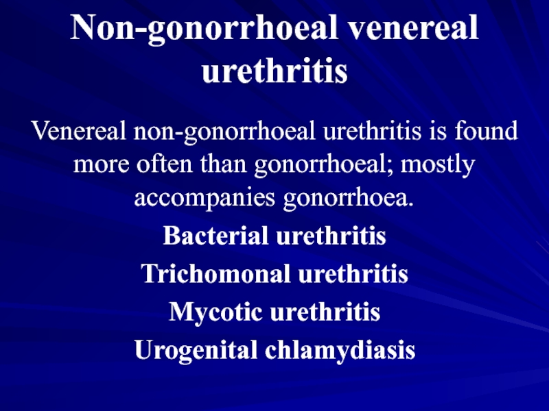
- 19. Non-gonorrhoeal venereal urethritis Venereal non-gonorrhoeal urethritis is
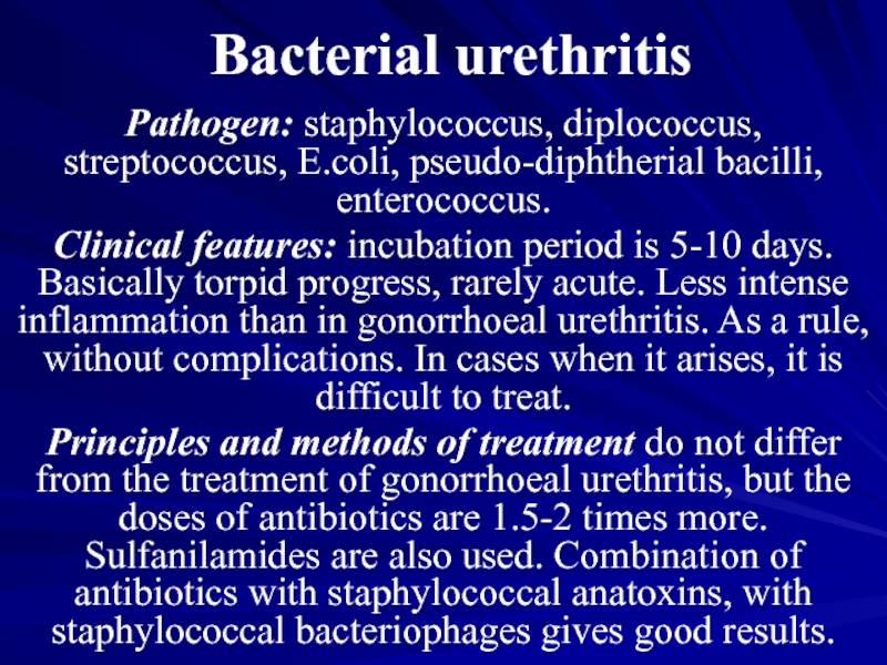
- 20. Bacterial urethritis Pathogen: staphylococcus, diplococcus, streptococcus, E.coli,
- 21. Trichomonal urethritis Pathogen: Trichomonas vaginalis. Pathogenesis:
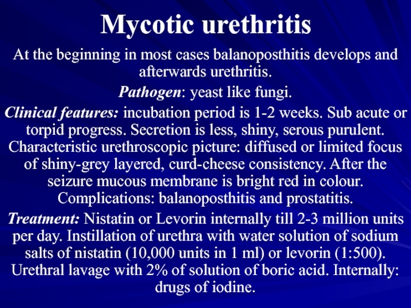
- 22. Mycotic urethritis At the beginning in most
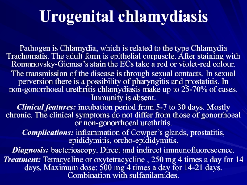
- 23. Urogenital chlamydiasis Pathogen is Chlamydia, which is
Слайд 2Theoretical part
Etiology
Gonorrhoea is caused by the Gram-negative diplococci. Gonococci are lentil-shaped
cocci about 1.5 um long and 0.75 um wide, arranged in pairs with their concave surfaces facing each other. They stain readily by all aniline dyes. The gonococci change their morphological and tinctorial properties under the effect of unfavourable factors to the point of becoming L-shaped. These L-shaped cocci may appear not only in laboratory cultures, but directly in the human body when chemotherapeutic agents are used or when the disease takes a chronic course.
Слайд 3Theoretical part
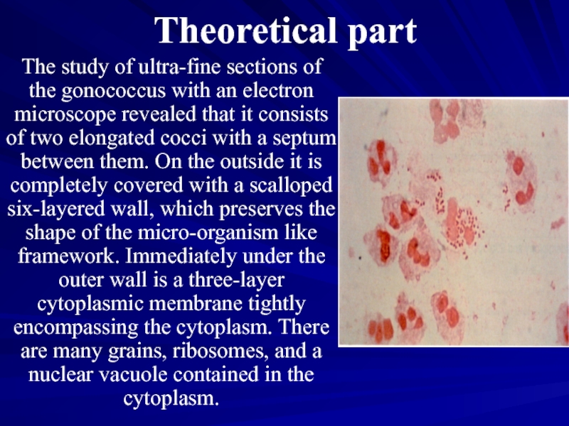
The study of ultra-fine sections of the gonococcus with an
electron microscope revealed that it consists of two elongated cocci with a septum between them. On the outside it is completely covered with a scalloped six-layered wall, which preserves the shape of the micro-organism like framework. Immediately under the outer wall is a three-layer cytoplasmic membrane tightly encompassing the cytoplasm. There are many grains, ribosomes, and a nuclear vacuole contained in the cytoplasm.
Слайд 4Transmission of the infection
The basic way of transmission for males is
normal sexual intercourse and sexual perversion. In the later case there may be infection of rectum or nasopharynx. Women and girls can be infected by sexual contacts as well as by domestic ways, through infected objects of domestic use. There is a possibility of infection of eyes in adults, as well as in children.
Слайд 5Pathogenesis
Neither congenital, no acquired immunity to gonococcus develops in humans. The
formed antibodies do not have defensive activity. Phagocytosis is complete, if gonococci were weakened by the use of drugs. The distribution of infection in the organism takes place through lymphatic and blood vessels. The pathogen cannot live in the blood as it has bacteriocidal properties.
Слайд 6Clinical features of gonorrhoea
Present classification of gonorrhoea:
Fresh:
acute,
subacute,
torpid.
Chronic.
Слайд 7Clinical features of gonorrhoea
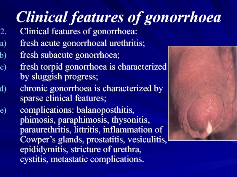
Clinical features of gonorrhoea:
fresh acute gonorrhoeal urethritis;
fresh subacute
gonorrhoea;
fresh torpid gonorrhoea is characterized by sluggish progress;
chronic gonorrhoea is characterized by sparse clinical features;
complications: balanoposthitis, phimosis, paraphimosis, thysonitis, paraurethritis, littritis, inflammation of Cowper’s glands, prostatitis, vesiculitis, epididymitis, stricture of urethra, cystitis, metastatic complications.
Слайд 8Clinical features of gonorrhoea
Gonorrhoea in small girls. As a result of
anatomical and physiological peculiarities of the genitals of small girls the inflammation of vulva, vagina, urethra, rectum may occur. In elder girls gonorrhoea is same as in women. Acute vulvovaginitis progresses with intense clinical signs.
Gonorrhoeal pharyngitis. In sexual perversion there may be a development of gonorrhoeal pharyngitis and tonsillitis. Clinically resembles catarrhal and banal inflammation, almost without any subjective feelings. Can lead to gonococcal sepsis.
Gonorrhoeal pharyngitis. In sexual perversion there may be a development of gonorrhoeal pharyngitis and tonsillitis. Clinically resembles catarrhal and banal inflammation, almost without any subjective feelings. Can lead to gonococcal sepsis.
Слайд 9Complications of gonorrhoeal urethritis in males.
Acute gonorrhoeal urethritis, especially in males
with a long and narrow prepuce, may be complicated by inflammation of its inner fold and the glans penis (balanoposthitis) and inflammatory phimosis which follow the same course as similar processes of non-gonococcal origin
Слайд 10Complications of gonorrhoeal urethritis in males.
Abscess of the preputial gland is
a rare local complication which is manifested by a moderately tender red swelling of the frenulum of the glans penis. Sometimes the gonococci penetrate into the paraurethral ducts where they are less accessible to the effect of drugs and may therefore become the cause of inefficient treatment. Inflammation of the paraurethral ducts is detected by thorough examination of the penis because they may open around the external urethral orifice, on the glans penis or in the corona glandis or at any other site. The affected paraurethral duct is palpated as a firm cord. When it is compressed a drop of pus is discharged from it. In some cases the inflamed paraurethral duct has a punctate, mildly infiltrated and hyperaemic opening on the urethral lips.
Слайд 11Complications of gonorrhoeal urethritis in males.
Littre's alveolar, tubular mucous glands and
Morgagni's lacunae found in the urethra are practically always affected by gonococci (littritis and morgagnitis). Littritis is marked by the appearance of peculiar comma-shaped purulent threads in the first portion of urine, which are impressions of the ducts of the urethral glands. In obstruction of the excretory duct by the inflammatory infiltrate, small pseudoabscesses are formed. They are felt as tender thickenings, slightly smaller than a pin-head when examined on a bougie or on the tube of the urethroscope. In some cases this pseudoabscess grows to a considerable size. In timely and proper treatment, the inflammatory infiltrate usually resolves, but in some cases purulent melting with the formation of a periurethral abscess occurs. When this abscess is opened or ruptures spontaneously gonococci are not always identified in the escaping pus. It is possible that pyogenic bacteria attendant to gonococci also contribute to the origin of this complication.
Слайд 12Male genital organs and localization of frequent complications of gonorrhoea.
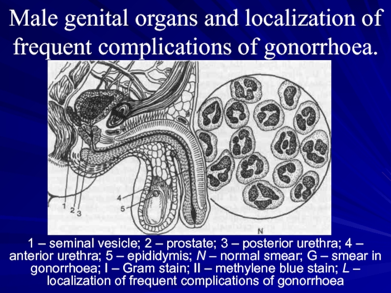
1 –
seminal vesicle; 2 – prostate; 3 – posterior urethra; 4 – anterior urethra; 5 – epididymis; N – normal smear; G – smear in gonorrhoea; I – Gram stain; II – methylene blue stain; L – localization of frequent complications of gonorrhoea
Слайд 13Male genital organs and localization of frequent complications of gonorrhoea.
Epididymitis, inflammation
of the epididymis, was formerly encountered in gonorrhoea much more frequently than now. Gonococci evidently penetrate into the epididymis from the posterior urethra through the deferent duct, though it is quite possible that the infectious agent is brought here with the blood or lymph. Inflammation of the duct itself develops not in all cases of epididymitis, and it is therefore assumed that antiperistaltic contractions of the deferent duct in affection of the prostatic urethra, and especially the seminal colliculus, contribute to its pathogenesis. Epididymitis is sometimes attended with effusion into the testicular coats.
Слайд 14Male genital organs and localization of frequent complications of gonorrhoea.
Chronic prostatitis,
on the contrary, is very common in the patients with protracted fresh or chronic gonorrhoea. It may be consequent upon acute prostatitis or occur directly in the form of chronic inflammation. The pathogenesis of chronic prostatitis in gonorrhoea is complicated. Gonococci are detected comparatively rarely in the secretions of the affected prostate even in untreated patients. The inflammatory process in the prostate is not usually liquidated after complete destruction of the gonococci in the patient's body by means of antibacterial agents. It is assumed that in such postgonorrhoeal diseases the inflammation is sustained by secondary infection, neurodystrophic changes in the tissues, and phenomena of autoaggression.
Слайд 15Diagnosis of gonorrhoea
The diagnosis of gonorrhoea may be established only when
the causative agent has been identified in the smears or cultures. Serological tests as well as the skin-allergic test with the gonococcal vaccine are merely of auxiliary importance, but can serve neither as proof gonococcal infection in the given patient nor as a criterion of cure.
Слайд 16Diagnosis of gonorrhoea
In acute fresh gonorrhoea the causative agents are usually
detected easily by microscopy of smears stained in parallel by the Gram-method and methylene blue. In torpid and chronic gonorrhoea, however, the results of bacterioscopy are less reliable. In case with the corresponding medical history and clinical picture, a negative result of one microscopic examination does not allow the diagnosis of gonorrhoea to be ruled out. The reliability of bacterioscopy is somewhat increased by repeated examination, including that after provocation, i.e. after artificial exacerbation of the inflammatory process. Combined provocation is also necessary in ascertaining the cure. Growth of cultures on artificial nutrient media in combination with microscopy practically double the number of gonorrhoeal patients detected, particularly those with the chronic form, those who had been treated earlier, those with involvement of the rectum, and others.
Слайд 17Diagnosis of gonorrhoea
For the identification of gonococci by microscopy and in
cultures, the pathological material is collected from the urethra, prostate and seminal vesicles of males, from the urethra, cervical canal, rectum and, if indicated, from the glands of the vestibule of the vagina of women, and from the vagina, urethra and rectum of girls.
Quite often other pathogenic micro-organisms are found together with gonococci in smears of the secretions, which may be transmitted during sexual intercourse.
The mixed infection makes it difficult to detect the gonococci and is reflected in the clinical picture of gonorrhoea: the duration of the incubation period increases and complications are more frequent. The gonococci phagocytosed by the urogenital trichomonads do not perish within the protozoon but are, to a certain extent, protected from the effect of the antigonorrhoeal agents. This explains some failure experienced in the treatment of gonorrhoea. The penicillin-resistant trichomonads, haemophilic vaginal bacilli, Candida fungi and Chlamydia are capable of sustaining inflammation of the urogenital organs after the death of the gonococci.
Слайд 18Treatment of gonorrhoea
Gonorrhoea is managed by means of antigonococcal agents, methods
for stimulating specific and non-specific immunity, as well as by different methods of local therapy the character of which is determined by the localization and type of focal changes in the tissues and involved organs. In acute fresh uncomplicated gonorrhoea antibiotic therapy only is applied. A complex of measures is needed in protracted, complicated and chronic forms.
The type and doses of antigonorrhoeal agents are established by periodically revised instructions in the treatment and prophylaxis of gonorrhoea endorsed by Ministry of Public Health. All physicians must follow these instructions elaborated on the basis of the latest scientific medical data and the experience of clinical institutions.
The type and doses of antigonorrhoeal agents are established by periodically revised instructions in the treatment and prophylaxis of gonorrhoea endorsed by Ministry of Public Health. All physicians must follow these instructions elaborated on the basis of the latest scientific medical data and the experience of clinical institutions.
Слайд 19Non-gonorrhoeal venereal urethritis
Venereal non-gonorrhoeal urethritis is found more often than gonorrhoeal;
mostly accompanies gonorrhoea.
Bacterial urethritis
Trichomonal urethritis
Mycotic urethritis
Urogenital chlamydiasis
Bacterial urethritis
Trichomonal urethritis
Mycotic urethritis
Urogenital chlamydiasis
Слайд 20Bacterial urethritis
Pathogen: staphylococcus, diplococcus, streptococcus, E.coli, pseudo-diphtherial bacilli, enterococcus.
Clinical features: incubation
period is 5-10 days. Basically torpid progress, rarely acute. Less intense inflammation than in gonorrhoeal urethritis. As a rule, without complications. In cases when it arises, it is difficult to treat.
Principles and methods of treatment do not differ from the treatment of gonorrhoeal urethritis, but the doses of antibiotics are 1.5-2 times more. Sulfanilamides are also used. Combination of antibiotics with staphylococcal anatoxins, with staphylococcal bacteriophages gives good results.
Principles and methods of treatment do not differ from the treatment of gonorrhoeal urethritis, but the doses of antibiotics are 1.5-2 times more. Sulfanilamides are also used. Combination of antibiotics with staphylococcal anatoxins, with staphylococcal bacteriophages gives good results.
Слайд 21Trichomonal urethritis
Pathogen: Trichomonas vaginalis. Pathogenesis: reactive condition of the organism; accompanying
diseases contribute to the development of the disease. It stops the development of acidic medium and resistance of mucous membrane of the urethra. Immunity is absent.
Clinical features: Incubation period is 5-15 days. Chronic progress. Symptoms are not much expressed. Less mucous secretion, sometimes translucent, foam. Itch. Complications – epididymitis, rarely prostatitis. There is a possibility of mixed trichomonas infection. In this case at first trichomoniasis is treated, afterwards antibiotics are prescribed.
Treatment: ethiopathogenetic and symptomatic: 25% of solution of bile of cattle in 0.25% of solution of Novocain in the form of instillation 8-10 ml during 4-5 days; instillation with 5% of solution of gastric juice, 5% of suspension of acetarsol. Internal: flagil 250 mg 2 times a day for 10 days; fazigin: 4 tablets at a time. Simultaneous treatment of sexual partner.
Слайд 22Mycotic urethritis
At the beginning in most cases balanoposthitis develops and afterwards
urethritis.
Pathogen: yeast like fungi.
Clinical features: incubation period is 1-2 weeks. Sub acute or torpid progress. Secretion is less, shiny, serous purulent. Characteristic urethroscopic picture: diffused or limited focus of shiny-grey layered, curd-cheese consistency. After the seizure mucous membrane is bright red in colour. Complications: balanoposthitis and prostatitis.
Treatment: Nistatin or Levorin internally till 2-3 million units per day. Instillation of urethra with water solution of sodium salts of nistatin (10,000 units in 1 ml) or levorin (1:500). Urethral lavage with 2% of solution of boric acid. Internally: drugs of iodine.
Слайд 23Urogenital chlamydiasis
Pathogen is Chlamydia, which is related to the type Chlamydia
Trachomatis. The adult form is epithelial corpuscle. After staining with Romanovsky-Giemsa’s stain the ECs take a red or violet-red colour.
The transmission of the disease is through sexual contacts. In sexual perversion there is a possibility of pharyngitis and prostatitis. In non-gonorrhoeal urethritis chlamydiasis make up to 25-70% of cases. Immunity is absent.
Clinical features: incubation period from 5-7 to 30 days. Mostly chronic. The clinical symptoms do not differ from those of gonorrhoeal or non-gonorrhoeal urethritis.
Complications: inflammation of Cowper’s glands, prostatitis, epididymitis, orcho-epididymitis.
Diagnosis: bacterioscopy. Direct and indirect immunofluorescence.
Treatment: Tetracycline or oxytetracycline , 250 mg 4 times a day for 14 days. Maximum dose: 500 mg 4 times a day for 14-21 days. Combination with sulfanilamides.
