- Главная
- Разное
- Дизайн
- Бизнес и предпринимательство
- Аналитика
- Образование
- Развлечения
- Красота и здоровье
- Финансы
- Государство
- Путешествия
- Спорт
- Недвижимость
- Армия
- Графика
- Культурология
- Еда и кулинария
- Лингвистика
- Английский язык
- Астрономия
- Алгебра
- Биология
- География
- Детские презентации
- Информатика
- История
- Литература
- Маркетинг
- Математика
- Медицина
- Менеджмент
- Музыка
- МХК
- Немецкий язык
- ОБЖ
- Обществознание
- Окружающий мир
- Педагогика
- Русский язык
- Технология
- Физика
- Философия
- Химия
- Шаблоны, картинки для презентаций
- Экология
- Экономика
- Юриспруденция
GI Hemorrhage презентация
Содержание

Слайд 2LOWER GI BLEEDING
Definition: LGIB is defined as bleeding from a source
distal to the ligament of Treitz
Incidence rate: 20.5 patients/ 100000/year
Incidence rate: 20.5 patients/ 100000/year

Слайд 3LGI hemorrhage
Sites
Colon – 95-97%
Small bowel – 3-5%
Only 15% of massive GI
bleeding
Finding the site
Intermittent bleeding common
Up to 42% have multiple sites
Finding the site
Intermittent bleeding common
Up to 42% have multiple sites

Слайд 4LGI hemorrhage
Etiology
Diverticulosis – 40-55%
Right sided lesions > left
90% stop spontaneously
10% rebleed
in 1st year and 25% at 4 years
Angiodysplasia – 3-20%
Most common cause of SB bleeding in >50 y/o
>50% are in right colon
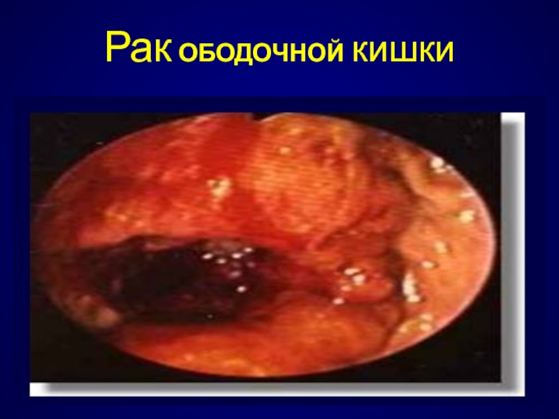
Neoplasia
Typically bleed slowly
Inflammatory conditions
15% of UC patients, 1% of chron’s patients
Radiation, infectious, AIDS rarely
Vascular
Hemorrhoids
>50% have hemorrhoids, but only 2% of bleeding attributed to them
Others
Angiodysplasia – 3-20%
Most common cause of SB bleeding in >50 y/o
>50% are in right colon
Neoplasia
Typically bleed slowly
Inflammatory conditions
15% of UC patients, 1% of chron’s patients
Radiation, infectious, AIDS rarely
Vascular
Hemorrhoids
>50% have hemorrhoids, but only 2% of bleeding attributed to them
Others

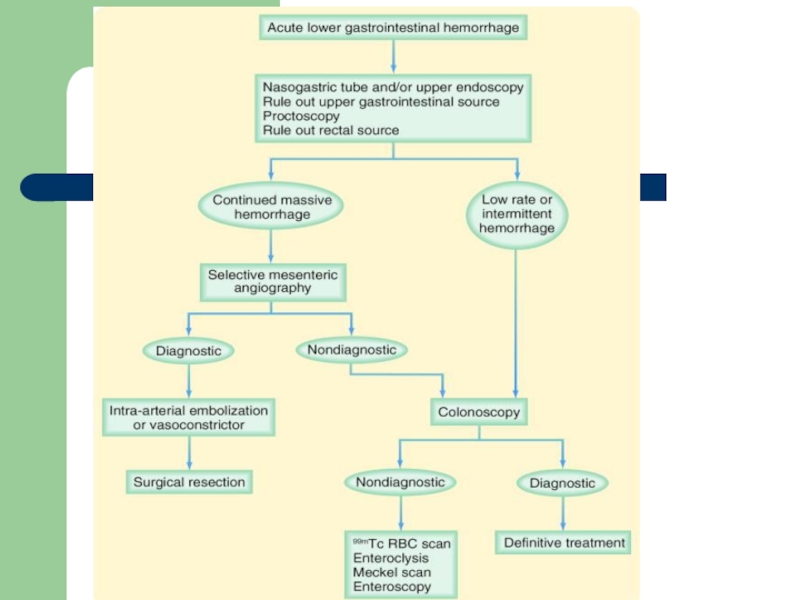
Слайд 5LGI hemorrhage diagnostics
Large caliber NGT on admission
Colonoscopy
Within 12 hours in stable
patients without large amounts of bleeding
Selective viseral angiography
Need >0.5 ml/min bleeding
40-75% sensitive if bleeding at time of exam
Tagged RBC scan
Can detect bleeding at 0.1 ml/min
85% sensitive if bleeding at time of exam
Not accurate in defining left vs right colon
Selective viseral angiography
Need >0.5 ml/min bleeding
40-75% sensitive if bleeding at time of exam
Tagged RBC scan
Can detect bleeding at 0.1 ml/min
85% sensitive if bleeding at time of exam
Not accurate in defining left vs right colon

Слайд 7CONCLUSION
LGIB requires pre op localization to detect the bleeding source
, including rectoscopy, colonoscopy,angography and nuclear scan.
Interventional treatment by colonoscopy and selective angiographic catheterization and embolization shows good results and low bleeding rates.
If an interventional therapy is not possible, a directed limited colonic or small bowel resection should be considered.
Interventional treatment by colonoscopy and selective angiographic catheterization and embolization shows good results and low bleeding rates.
If an interventional therapy is not possible, a directed limited colonic or small bowel resection should be considered.

Слайд 8CONCLUSION
Positive pre op localization of bleeding results in limited colonic or
small bowel resection when interventional therapy failed to stop bleeding.
Negative pre op localization of bleeding site results in subtotal/total colectomy in massive low GI BLEEDING.
Negative pre op localization of bleeding site results in subtotal/total colectomy in massive low GI BLEEDING.