- Главная
- Разное
- Дизайн
- Бизнес и предпринимательство
- Аналитика
- Образование
- Развлечения
- Красота и здоровье
- Финансы
- Государство
- Путешествия
- Спорт
- Недвижимость
- Армия
- Графика
- Культурология
- Еда и кулинария
- Лингвистика
- Английский язык
- Астрономия
- Алгебра
- Биология
- География
- Детские презентации
- Информатика
- История
- Литература
- Маркетинг
- Математика
- Медицина
- Менеджмент
- Музыка
- МХК
- Немецкий язык
- ОБЖ
- Обществознание
- Окружающий мир
- Педагогика
- Русский язык
- Технология
- Физика
- Философия
- Химия
- Шаблоны, картинки для презентаций
- Экология
- Экономика
- Юриспруденция
Common Laboratory Tests презентация
Содержание
- 1. Common Laboratory Tests
- 2. Let’s look at some nuances of 3
- 3. CBC Complete blood count With or without
- 4. What is measured? Red blood cell data
- 5. Total Red Blood Cell Count Count of
- 6. Hemoglobin The hemoglobin concentration is a measure
- 7. Hematocrit Hematocrit is a measure of the
- 8. Centrifuged blood (normal)
- 9. Centrifuged blood (adult male or female) What
- 10. Calculating the Hematocrit More commonly the
- 11. Mean Corpuscular Volume The MCV is a
- 12. Cell Size (fl) Number Of
- 13. Use of MCV Result The MCV is
- 14. Cell Size (fl) Number Of
- 15. Red Blood Cell Distribution Width RDW is
- 16. White Blood Cell Count A count
- 17. WBC Differential When a differential is ordered,
- 18. Manual Differentials “Manual” WBC differentials are performed
- 19. Automated Differentials The clinical laboratory may perform
- 20. Platelet Count (PLT) A count of the
- 21. CBC as reported by LUMC Lab in
- 22. MCH and MCHC Note: Both MCH and
- 23. Interpretation? Essentially normal CBC WBC, Hgb, Hct,
- 24. Absolute numbers (#) of various cell types
- 25. Interpret this CBC CBC WBC 19.5
- 26. Common Clinical Uses of CBC CBC demonstrates
- 27. One final CBC pearl Clinicians have a
- 28. BMP (Basic Metabolic Panel)
- 29. BMP The BMP is a chemistry panel
- 30. BMP Peripheral venous blood can be collected
- 31. Sodium Sodium is the major cation in
- 32. Potassium Potassium is the major intracellular cation
- 33. Chloride Chloride is the major extracellular anion
- 34. Carbon Dioxide Content The carbon dioxide content
- 35. Blood Urea Nitrogen The BUN measures the
- 36. Creatinine The creatinine test measures the amount
- 37. Glucose Plasma glucose levels should be evaluated
- 38. Diagnosing Diabetes The criteria for the diagnosis
- 39. Total Calcium The total serum calcium is
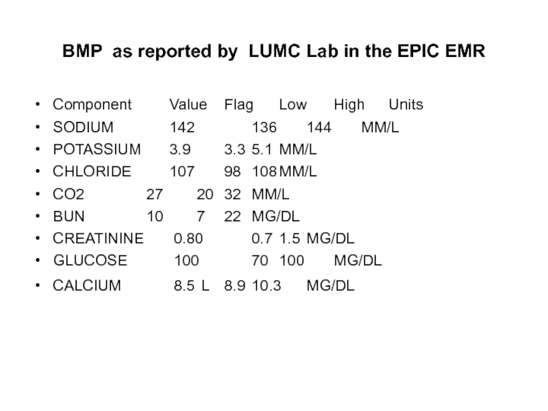
- 40. BMP as reported by LUMC Lab in
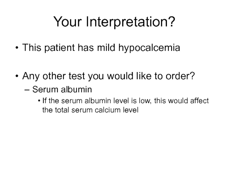
- 41. Your Interpretation? This patient has mild hypocalcemia
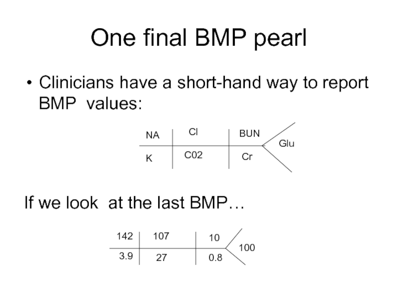
- 42. One final BMP pearl Clinicians have a
- 43. CMP (Complete Metabolic Panel)
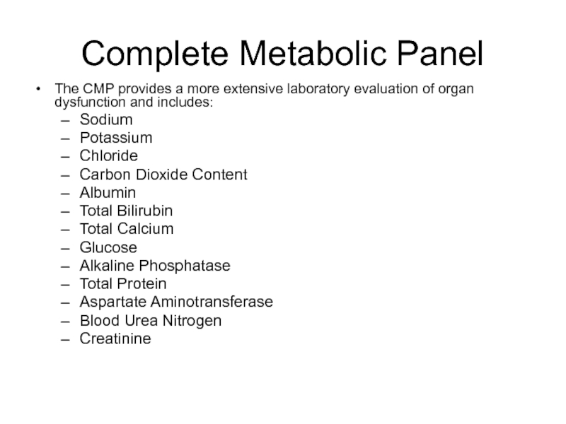
- 44. Complete Metabolic Panel The CMP provides a
- 45. Total Protein Albumin and globulin constitute most
- 46. Albumin Albumin comprises ~ 60% of the
- 47. Alkaline Phosphatase (Alk Phos or ALP)
- 48. Bilirubin, Total The total serum bilirubin level
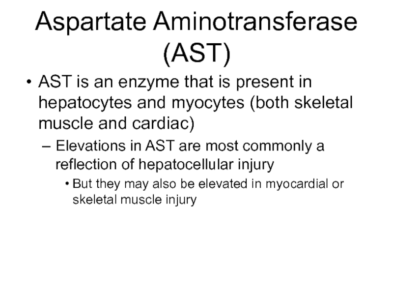
- 49. Aspartate Aminotransferase (AST) AST is an enzyme
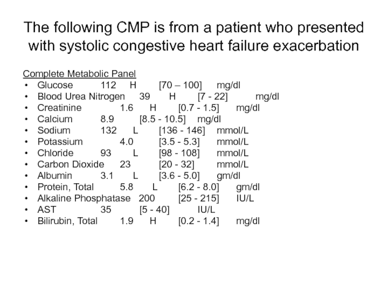
- 50. The following CMP is from a patient
- 51. Interpretion? (do not fret, you will begin
- 52. Final Comments… Excessive laboratory tests can cause
- 53. Final Comments… No laboratory test should ever be ordered unless it is medically necessary
Слайд 2Let’s look at some nuances of 3 of most commonly ordered
CBC (Complete Blood Count)
with or without differential
BMP (Basic Metabolic Panel)
CMP (Comprehensive Metabolic Panel)
Слайд 3CBC
Complete blood count
With or without differential
Peripheral venous blood is collected in
Unacceptable specimen:
Clotted or greater than 48 hours old
Methodology of testing:
Whole blood analyzer
How often is the test available for hospitalized patients?
7 days/week (24/7)
Слайд 4What is measured?
Red blood cell data
Total red blood cell count (RBC)
Hemoglobin
Hematocrit (Hct)
Mean corpuscular volume (MCV)
Red blood cell distribution width (RDW)
White blood cell data
Total white blood cell (leukocyte) count (WBC)
A white blood cell count differential may also be ordered
Platelet Count (PLT)
Слайд 5Total Red Blood Cell Count
Count of the number of circulating red
Слайд 6Hemoglobin
The hemoglobin concentration is a measure of the amount of Hgb
Hgb constitutes over 90% of the red blood cells
Decrease in Hgb concentration =
anemia
Increase in Hgb concentration =
polycythemia
Слайд 7Hematocrit
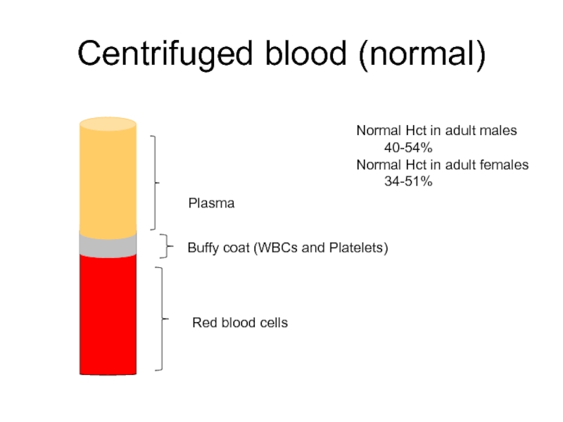
Hematocrit is a measure of the percentage of the total blood
The hematocrit can be determined directly by centrifugation (“spun hematocrit”)
The height of the red blood cell column is measured and compared to the column of the whole blood
Слайд 8Centrifuged blood (normal)
Red blood cells
Buffy coat (WBCs and Platelets)
Plasma
Normal Hct
40-54%
Normal Hct in adult females
34-51%
Слайд 9Centrifuged blood (adult male or female)
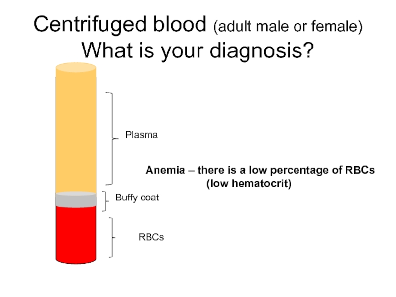
What is your diagnosis?
Anemia – there
(low hematocrit)
RBCs
Buffy coat
Plasma
Слайд 10Calculating the Hematocrit
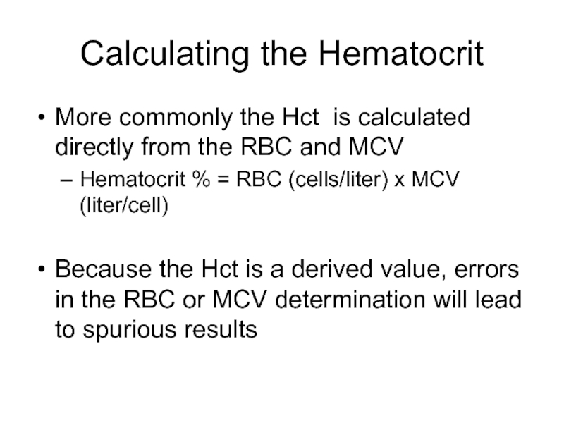
More commonly the Hct is calculated directly from
Hematocrit % = RBC (cells/liter) x MCV (liter/cell)
Because the Hct is a derived value, errors in the RBC or MCV determination will lead to spurious results
Слайд 11Mean Corpuscular Volume
The MCV is a measure of the average volume,
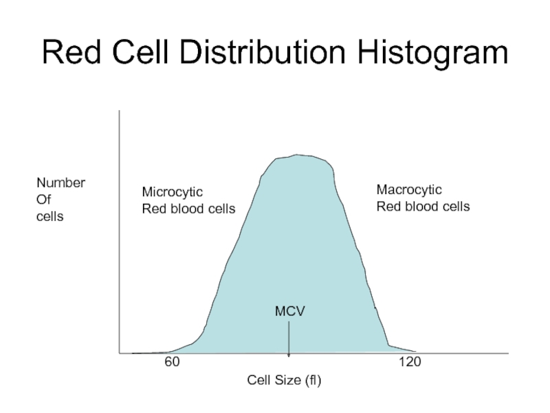
It is determined by the distribution of the red blood cell histogram
The mean of the red blood cell distribution histogram is the MCV
Слайд 13Use of MCV Result
The MCV is important in classifying anemias
Normal MCV
Decreased MCV = microcytic anemia
Increased MCV = macrocytic anemia
Слайд 14
Cell Size (fl)
Number
Of
cells
60
120
MCV
Red Cell Distribution Histogram
Microcytic
Red blood cells
Macrocytic
Red
Слайд 15Red Blood Cell Distribution Width
RDW is an indication of the variation
It is derived from the red blood cell histogram and represents the coefficient of variation of the curve
In general, an elevated RDW (indicating more variation in the size of RBCs) has been associated with anemias with various deficiencies, such as iron, B12, or folate
Thalassemia is a microcytic anemia that characteristically has a normal RDW
Слайд 16White Blood Cell Count
A count of the total WBC, or leukocyte,
A decrease in the number of WBCs =
Leukopenia
An increase in the number of WBCs =
Leukocytosis
Слайд 17WBC Differential
When a differential is ordered, the percentage of each type
Name the types of leukocytes
Neutrophils (includes bands)
Lymphocytes
Monocytes
Eosinophils
Basophils
WBC differentials are either performed manually or by an automated instrument
Слайд 18Manual Differentials
“Manual” WBC differentials are performed by trained medical technologists who
In addition to the differential count, evaluation of the smear provides the opportunity to morphologically evaluate all components of the peripheral blood, including red blood cells, white blood cells and platelets
The manual differential allows for the detection of disorders that might otherwise be lost in a totally automated system
This applies to < 20% of specimens
The instrument is programmed with criteria to flag an operator when a manual differential should be performed
Слайд 19Automated Differentials
The clinical laboratory may perform an “automated differential”
Via instruments
Usually based on the determination of different leukocyte cellular characteristics that permit separation into subtypes by using flow-cytometric techniques
Слайд 20Platelet Count (PLT)
A count of the number of platelets (thrombocytes) per
A decreased number of platelets =
Thrombocytopenia
An increased number of platelets =
Thrombocytosis
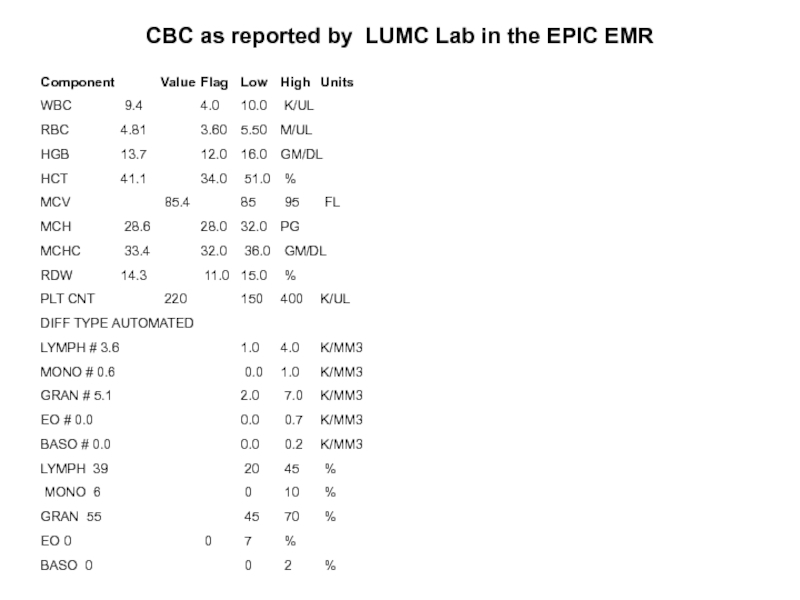
Слайд 21CBC as reported by LUMC Lab in the EPIC EMR
Component Value
WBC 9.4 4.0 10.0 K/UL
RBC 4.81 3.60 5.50 M/UL
HGB 13.7 12.0 16.0 GM/DL
HCT 41.1 34.0 51.0 %
MCV 85.4 85 95 FL
MCH 28.6 28.0 32.0 PG
MCHC 33.4 32.0 36.0 GM/DL
RDW 14.3 11.0 15.0 %
PLT CNT 220 150 400 K/UL
DIFF TYPE AUTOMATED
LYMPH # 3.6 1.0 4.0 K/MM3
MONO # 0.6 0.0 1.0 K/MM3
GRAN # 5.1 2.0 7.0 K/MM3
EO # 0.0 0.0 0.7 K/MM3
BASO # 0.0 0.0 0.2 K/MM3
LYMPH 39 20 45 %
MONO 6 0 10 %
GRAN 55 45 70 %
EO 0 0 7 %
BASO 0 0 2 %
Слайд 22MCH and MCHC
Note:
Both MCH and MCHC are of little clinical diagnostic
MCH is the hemoglobin concentration per cell
MCHC is the average hemoglobin concentration per total red blood cell volume
Слайд 23Interpretation?
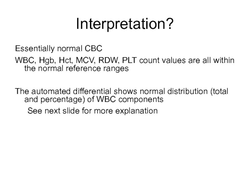
Essentially normal CBC
WBC, Hgb, Hct, MCV, RDW, PLT count values are
The automated differential shows normal distribution (total and percentage) of WBC components
See next slide for more explanation
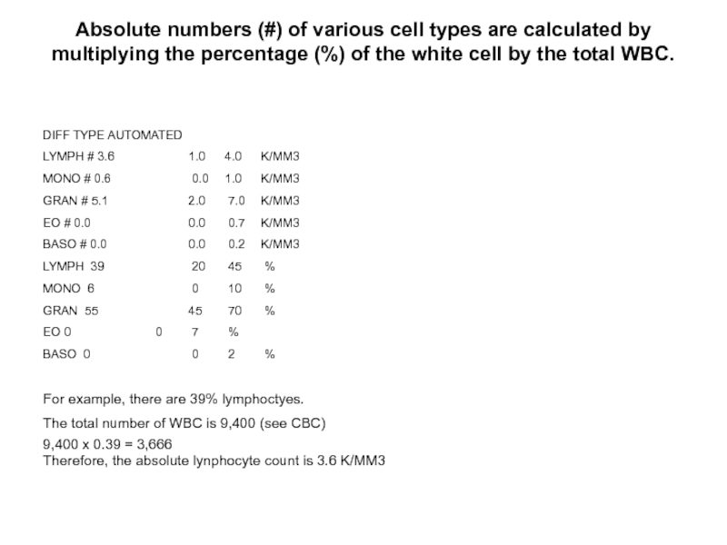
Слайд 24Absolute numbers (#) of various cell types are calculated by multiplying
DIFF TYPE AUTOMATED
LYMPH # 3.6 1.0 4.0 K/MM3
MONO # 0.6 0.0 1.0 K/MM3
GRAN # 5.1 2.0 7.0 K/MM3
EO # 0.0 0.0 0.7 K/MM3
BASO # 0.0 0.0 0.2 K/MM3
LYMPH 39 20 45 %
MONO 6 0 10 %
GRAN 55 45 70 %
EO 0 0 7 %
BASO 0 0 2 %
For example, there are 39% lymphoctyes.
The total number of WBC is 9,400 (see CBC)
9,400 x 0.39 = 3,666
Therefore, the absolute lynphocyte count is 3.6 K/MM3
Слайд 25Interpret this CBC
CBC
WBC 19.5 [4.0-10.0] k/ul
RBC 3.49
Hgb 10.4 [12.0-16.0] gm/dl
Hct 31.2 [34.0-51.0] %
MCV 82 [85-95] fl
MCH 28.3 [28.0-32.0] pg
MCHC 33.3 [32.0-36.0] gm/dl
RDW 16.6 [11.0-15.0] %
Plt Count 98 [150-400] k/ul
Слайд 26Common Clinical Uses of CBC
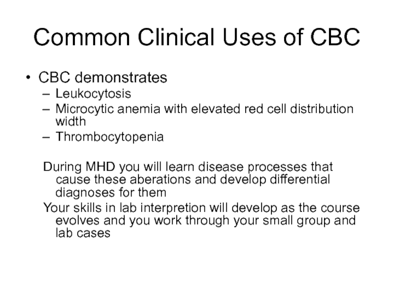
CBC demonstrates
Leukocytosis
Microcytic anemia with elevated red cell
Thrombocytopenia
During MHD you will learn disease processes that cause these aberations and develop differential diagnoses for them
Your skills in lab interpretion will develop as the course evolves and you work through your small group and lab cases
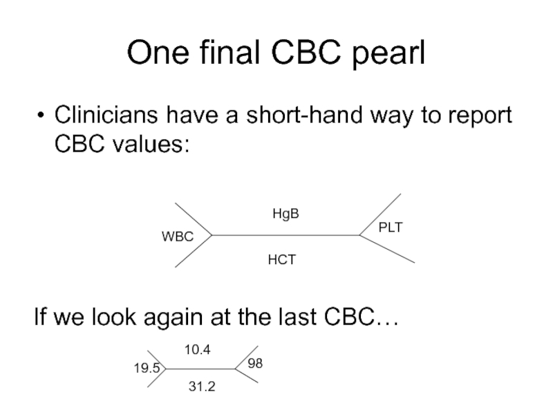
Слайд 27One final CBC pearl
Clinicians have a short-hand way to report CBC
If we look again at the last CBC…
WBC
HgB
HCT
PLT
19.5
10.4
31.2
98
Слайд 29BMP
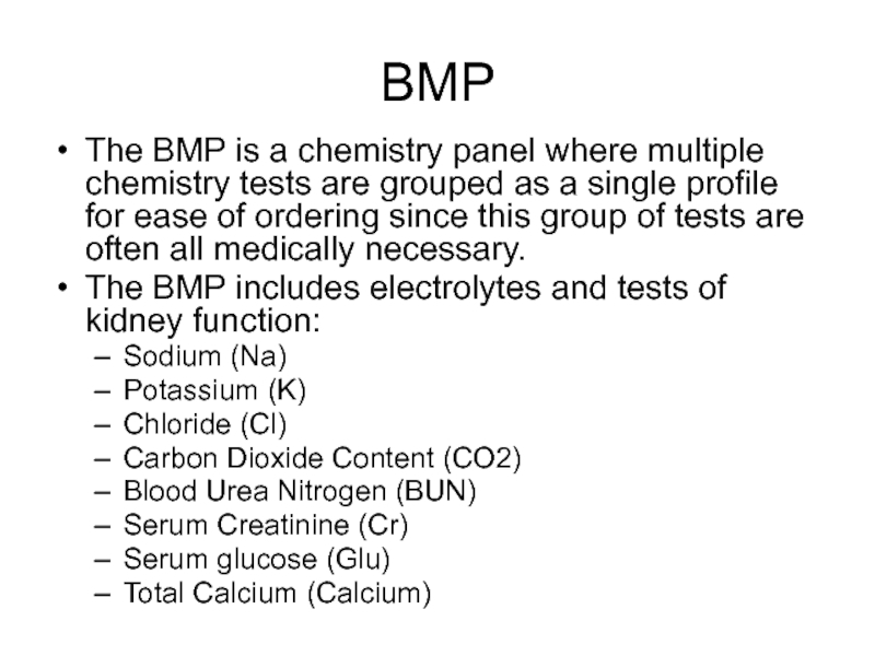
The BMP is a chemistry panel where multiple chemistry tests are
The BMP includes electrolytes and tests of kidney function:
Sodium (Na)
Potassium (K)
Chloride (Cl)
Carbon Dioxide Content (CO2)
Blood Urea Nitrogen (BUN)
Serum Creatinine (Cr)
Serum glucose (Glu)
Total Calcium (Calcium)
Слайд 30BMP
Peripheral venous blood can be collected in several types of tube
Light
Plasma separating tube (PST) with the anticoagulant lithium heparin
Gold SST
Serum separating tube (SST) contains a gel at the bottom to separate blood cellular components from serum on centrifugation
Red
No Additives – blood clots and serum is separated by centrifugation
How often is the lab test available for hospitalized patients?
7 days/week (24/7)
Слайд 31Sodium
Sodium is the major cation in the extracellular space where serum
Sodium salts are major determinants of extracellular osmolality.
Increased serum sodium level =
Hypernatremia
Decreased serum sodium level =
Hyponatremia
Слайд 32Potassium
Potassium is the major intracellular cation with levels of ~ 4
Elevated serum potassium level =
Hyperkalemia
Decreased serum potassium level =
Hypokalemia
*note – if a specimen is hemolyzed (such as by traumatic venipuncture or drawing blood with a needle that is too small) potassium levels may be “falsely” elevated. Why?
There are high concentrations of K in red blood cells. If RBCs are lysed during phlebotomy, K is released into the serum resulting in elevated measured levels.
Слайд 33Chloride
Chloride is the major extracellular anion with serum concentration of ~
Hyperchloremia and hypochloremia are rarely isolated phenomena.
Usually they are part of shifts in sodium or bicarbonate to maintain electrical neutrality.
Слайд 34Carbon Dioxide Content
The carbon dioxide content (CO2) measures the H2CO3, dissolved
Because the amounts of H2CO3 and dissolved CO2 in the serum are so small, the CO2 content is an indirect measure of the HCO3 anion
Therefore, clinicians most often refer to the CO2 measurement in the BMP as the “bicarbonate level” or “bicarb level”
Слайд 35Blood Urea Nitrogen
The BUN measures the amount of urea nitrogen in
Urea is formed in the liver as the end product of protein metabolism and is transported to the kidneys for excretion.
Nearly all renal diseases can cause an inadequate excretion of urea, which causes the blood concentration to rise above normal.
The BUN is interpreted in conjunction with the creatinine test – these tests are referred to as “renal function studies”.
Слайд 36Creatinine
The creatinine test measures the amount of creatinine in the blood.
Creatinine
Creatinine, as with blood urea nitrogen, is excreted entirely by the kidneys and blood levels are therefore proportional to renal excretory function.
Слайд 37Glucose
Plasma glucose levels should be evaluated in relation to a patient’s
i.e., postprandial vs fasting
Elevated glucose levels may also be indicative of diabetes mellitus
Glucose is the most commonly measured test in the laboratory
Слайд 38Diagnosing Diabetes
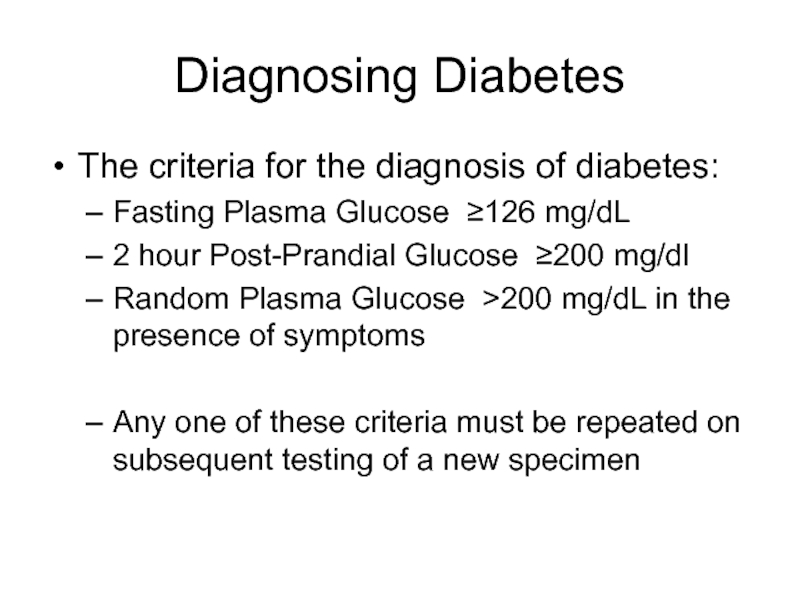
The criteria for the diagnosis of diabetes:
Fasting Plasma Glucose ≥126
2 hour Post-Prandial Glucose ≥200 mg/dl
Random Plasma Glucose >200 mg/dL in the presence of symptoms
Any one of these criteria must be repeated on subsequent testing of a new specimen
Слайд 39Total Calcium
The total serum calcium is a measure of both
Free (ionized)
Protein bound (usually to albumin) calcium
Therefore, the total serum calcium level is affected by changes in serum albumin
As a rule of thumb, the total serum calcium level decreases by approximately 0.8mg for every 1gram decrease in the serum albumin level.
Слайд 40BMP as reported by LUMC Lab in the EPIC EMR
Component Value
SODIUM 142 136 144 MM/L
POTASSIUM 3.9 3.3 5.1 MM/L
CHLORIDE 107 98 108 MM/L
CO2 27 20 32 MM/L
BUN 10 7 22 MG/DL
CREATININE 0.80 0.7 1.5 MG/DL
GLUCOSE 100 70 100 MG/DL
CALCIUM 8.5 L 8.9 10.3 MG/DL
Слайд 41Your Interpretation?
This patient has mild hypocalcemia
Any other test you would like
Serum albumin
If the serum albumin level is low, this would affect the total serum calcium level
Слайд 42One final BMP pearl
Clinicians have a short-hand way to report BMP
If we look at the last BMP…
NA
K
Cl
C02
BUN
Cr
Glu
142
3.9
107
27
10
0.8
100
Слайд 44Complete Metabolic Panel
The CMP provides a more extensive laboratory evaluation of
Sodium
Potassium
Chloride
Carbon Dioxide Content
Albumin
Total Bilirubin
Total Calcium
Glucose
Alkaline Phosphatase
Total Protein
Aspartate Aminotransferase
Blood Urea Nitrogen
Creatinine
Слайд 45Total Protein
Albumin and globulin constitute most of the protein within the
Слайд 46Albumin
Albumin comprises ~ 60% of the total protein within the extracellular
Albumin’s major effect within the blood is to maintain colloid osmotic pressure
Transports many important blood constituents
drugs, hormones, enzymes
Albumin is synthesized in the liver and therefore is a measure of hepatic function
Слайд 47Alkaline Phosphatase
(Alk Phos or ALP)
Alkaline phosphatase is an enzyme present
Isoenzymes are forms of an enzyme that catalyze the same reaction, but are slightly different in structure
The two major circulating alkaline phosphatase isoenzymes are bone and liver.
Therefore elevation in serum alkaline phosphatase is most commonly a reflection of liver or bone disorders.
Levels of alk phos are increased in both extrahepatic and intrahepatic obstructive biliary disease
Слайд 48Bilirubin, Total
The total serum bilirubin level is the sum of the
Normally the unconjugated bilirubin makes up 70-85% of the total bilirubin
Remember that bilirubin metabolism begins with the breakdown of red blood cells in the reticuloendothelial system and bilirubin metabolism continues in the liver
Elevation in total bilirubin may therefore be a reflection of any aberrations in bilirubin metabolism or increased levels of bilirubin production (such as hemolysis)
Слайд 49Aspartate Aminotransferase
(AST)
AST is an enzyme that is present in hepatocytes and
Elevations in AST are most commonly a reflection of hepatocellular injury
But they may also be elevated in myocardial or skeletal muscle injury
Слайд 50The following CMP is from a patient who presented with systolic
Complete Metabolic Panel
Glucose 112 H [70 – 100] mg/dl
Blood Urea Nitrogen 39 H [7 - 22] mg/dl
Creatinine 1.6 H [0.7 - 1.5] mg/dl
Calcium 8.9 [8.5 - 10.5] mg/dl
Sodium 132 L [136 - 146] mmol/L
Potassium 4.0 [3.5 - 5.3] mmol/L
Chloride 93 L [98 - 108] mmol/L
Carbon Dioxide 23 [20 - 32] mmol/L
Albumin 3.1 L [3.6 - 5.0] gm/dl
Protein, Total 5.8 L [6.2 - 8.0] gm/dl
Alkaline Phosphatase 200 [25 - 215] IU/L
AST 35 [5 - 40] IU/L
Bilirubin, Total 1.9 H [0.2 - 1.4] mg/dl
Слайд 51Interpretion? (do not fret, you will begin learning this skill as MHD
BUN and creatinine are elevated with a BUN:Creat ratio greater than 20:1 consistent with pre-renal azotemia, the result of inadequate renal perfusion and resulting reduced urea clearance.
Hepatic congestion leads to hypoxia and altered function of the liver cells. Bilirubin, especially the indirect fraction, and enzymes, like alkaline phosphatase, may be elevated. Total protein may decline at the expense of the decreased albumin produced in the liver.
The electrolyte changes, especially hyponatremia, reflect a dilutional effect with water retention and decreased glomerular filtration rate (poor perfusion)
Hyperglycemia is present but it is not known whether this was a fasting or random sample
Слайд 52Final Comments…
Excessive laboratory tests can cause iatrogenic anemia!
Although the goal of
Every test ordered, including lab tests, on a patient should be assessed for its benefits, risks and true need.



![Interpret this CBCCBCWBC 19.5 [4.0-10.0] k/ul RBC 3.49 [3.60-5.50] m/ul Hgb 10.4](/img/tmb/5/490234/e4211c7596028722a981d123276e4be7-800x.jpg)
