with basis of law
Atherosclerosis (AS) Hypertension disease (HD) Symptomatic arterial hypertension (SAH)
Lecture on pathological anatomy
for the 3-rd year students
- Главная
- Разное
- Дизайн
- Бизнес и предпринимательство
- Аналитика
- Образование
- Развлечения
- Красота и здоровье
- Финансы
- Государство
- Путешествия
- Спорт
- Недвижимость
- Армия
- Графика
- Культурология
- Еда и кулинария
- Лингвистика
- Английский язык
- Астрономия
- Алгебра
- Биология
- География
- Детские презентации
- Информатика
- История
- Литература
- Маркетинг
- Математика
- Медицина
- Менеджмент
- Музыка
- МХК
- Немецкий язык
- ОБЖ
- Обществознание
- Окружающий мир
- Педагогика
- Русский язык
- Технология
- Физика
- Философия
- Химия
- Шаблоны, картинки для презентаций
- Экология
- Экономика
- Юриспруденция
Atherosclerosis. Hypertension disease. Symptomatic arterial hypertension презентация
Содержание
- 1. Atherosclerosis. Hypertension disease. Symptomatic arterial hypertension
- 2. Atherosclerosis is a chronic disease,
- 3. Risk factors of atherosclerosis:
- 4. Stages of pathogenesis
- 5. Morphogenesis
- 7. Evolution of arterial wall changes in the
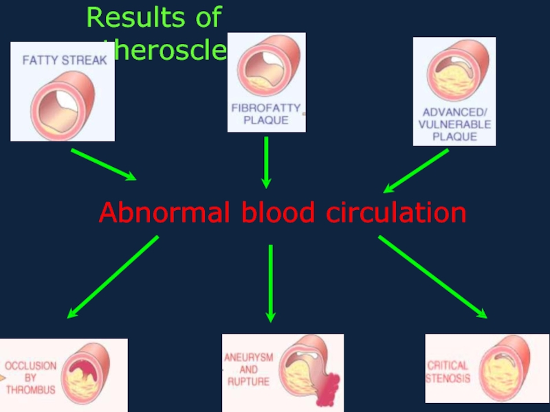
- 9. Results of atherosclerosis
- 10. Microscopically, the aortic atheromatous plaque is thicker
- 11. Clinic-anatomic forms of atherosclerosis
- 13. True aneurysms False aneurysms Classification of aneurysms
- 14. Hypertension
- 17. In
- 18. Clinical-morphological stages
- 19. Clinical-morphological stages
- 20. This is a different kind of arterio-
- 21. Clinical-morphological forms of HD
- 22. Clinical-morphological forms of HD
Слайд 1
ZAPORIZHZHIAN STATE MEDICAL UNIVERSITY
The department of pathological anatomy and forensic medicine
Слайд 2
Atherosclerosis is a chronic
disease, that is stereotyped response to injury featuring
the accumulation of cholesterol-rich fat in the intimae of the large and medium-sized arteries of the body.
These masses form plaques
These masses form plaques
Слайд 3
Risk factors of atherosclerosis:
High levels of LDL cholesterol and HDL.
Micro-trauma of
intimae by: cytokines, toxins (exo- and endo-genous), immune complexes...
High blood pressure.
Pathology of lipid exchange at diabetes
mellitus, thyroxin lack.
Law level of muscle activity (lack of
exercise).
Biochemical lesions that promote thrombosis.
Some heredity stations:
Familial combined hyper-lipidemia
Tangier disease.
High blood pressure.
Pathology of lipid exchange at diabetes
mellitus, thyroxin lack.
Law level of muscle activity (lack of
exercise).
Biochemical lesions that promote thrombosis.
Some heredity stations:
Familial combined hyper-lipidemia
Tangier disease.
Слайд 4
Stages of pathogenesis
The development of focal areas of chronic endothelial injury,
with resulting increased endothelial permeability or other evidence of endothelial dysfunction.
Increased insudation of lipoprotein, mainly LDL with its high content of cholesterol, and also VLDL.
Cellular interactions in foci of injury involving endothelial cells, monocytes/macrophages, T- lymphocytes, and smooth muscle cells of intimal or medial origin.
Proliferation of smooth muscle cells in the intima with the formation of connective tissue
Слайд 5Morphogenesis
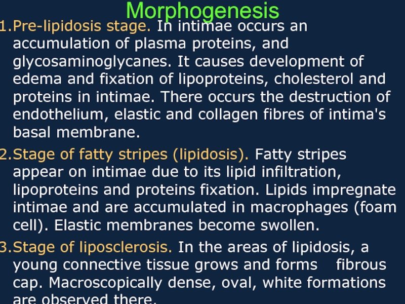
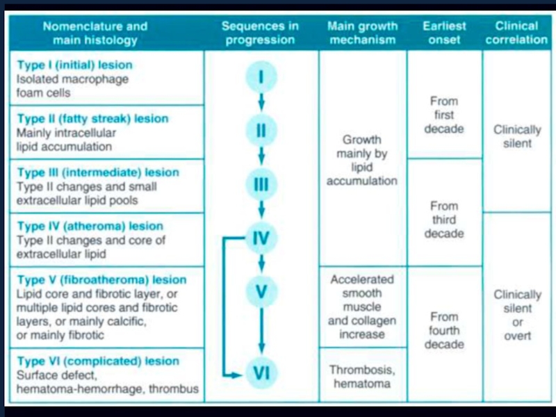
Pre-lipidosis stage. In intimae occurs an accumulation of plasma proteins, and
glycosaminoglycanes. It causes development of edema and fixation of lipoproteins, cholesterol and proteins in intimae. There occurs the destruction of endothelium, elastic and collagen fibres of intima's basal membrane.
Stage of fatty stripes (lipidosis). Fatty stripes appear on intimae due to its lipid infiltration, lipoproteins and proteins fixation. Lipids impregnate intimae and are accumulated in macrophages (foam cell). Elastic membranes become swollen.
Stage of liposclerosis. In the areas of lipidosis, a young connective tissue grows and forms fibrous cap. Macroscopically dense, oval, white formations are observed there.
Слайд 6
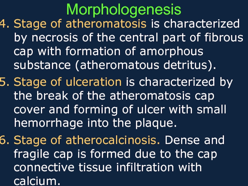
Stage of atheromatosis is characterized by necrosis of the central part
of fibrous cap with formation of amorphous substance (atheromatous detritus).
Stage of ulceration is characterized by the break of the atheromatosis cap cover and forming of ulcer with small hemorrhage into the plaque.
Stage of atherocalcinosis. Dense and fragile cap is formed due to the cap connective tissue infiltration with calcium.
Stage of ulceration is characterized by the break of the atheromatosis cap cover and forming of ulcer with small hemorrhage into the plaque.
Stage of atherocalcinosis. Dense and fragile cap is formed due to the cap connective tissue infiltration with calcium.
Morphologenesis
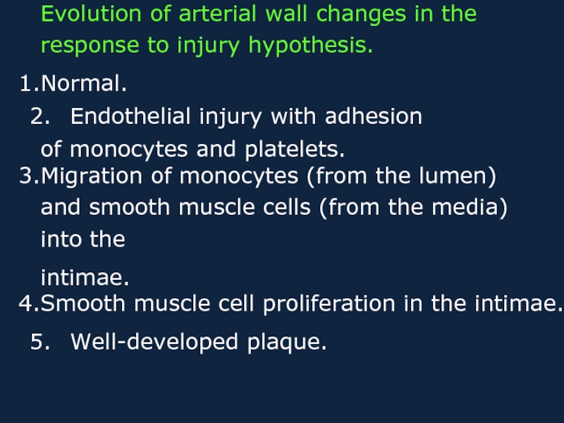
Слайд 7Evolution of arterial wall changes in the response to injury hypothesis.
Normal.
Endothelial
injury with adhesion
of monocytes and platelets.
Migration of monocytes (from the lumen) and smooth muscle cells (from the media) into the
intimae.
Smooth muscle cell proliferation in the intimae.
Well-developed plaque.
of monocytes and platelets.
Migration of monocytes (from the lumen) and smooth muscle cells (from the media) into the
intimae.
Smooth muscle cell proliferation in the intimae.
Well-developed plaque.
Слайд 10Microscopically, the aortic atheromatous plaque is thicker than the remaining media.
The plaque contains amorphous pink material with slit-like "cholesterol clefts" of lipid material. There is overlying recent hemorrhage. Thrombus may form on top of such a plaque
Слайд 11
Clinic-anatomic forms of atherosclerosis
Atherosclerosis of aorta - the most frequent form.
Usually it is not complicated by the thrombosis, thromboembolism and embolism of lower extremities. Development of aortal aneurysm is possible.
Atherosclerosis of coronary arteries of heart
- ischemic heart disease (IHD).
Atherosclerosis of renal arteries leads to atrophy of parenchyma, or infarction. Outcome is atherosclerotic nefro-cirrhosis.
Atherosclerosis of arteries of an intestine is complicated by the thrombosis, leading to gangrene or infarction of bowel.
Слайд 12
Atherosclerosis of arteries of cerebrum.
The results are ischemic infarctions of brain
or brain haemorrhage. Atherosclerosis of carotides leads to acute local ischemia and cerebral softening (infarction)
Atherosclerosis of arteries of extremities, very often this process is located in femoral arteries. The thrombosis with gangrene of lower extremity is possible.
Atherosclerosis of arteries of extremities, very often this process is located in femoral arteries. The thrombosis with gangrene of lower extremity is possible.
Clinic-anatomic forms of atherosclerosis
Слайд 13True aneurysms
False aneurysms
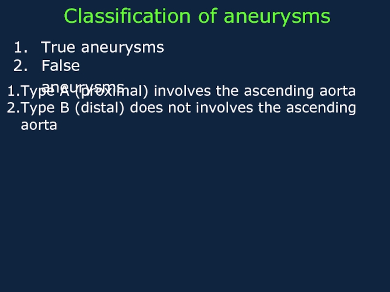
Classification of aneurysms
Type A (proximal) involves the ascending aorta
Type
B (distal) does not involves the ascending aorta
Слайд 14Hypertension
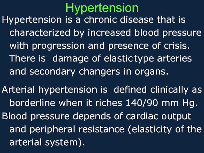
Hypertension is a chronic disease that is characterized by increased blood
pressure with progression and presence of crisis. There is damage of elastic type arteries and secondary changers in organs.
Arterial hypertension is defined clinically as
borderline when it riches 140/90 mm Hg.
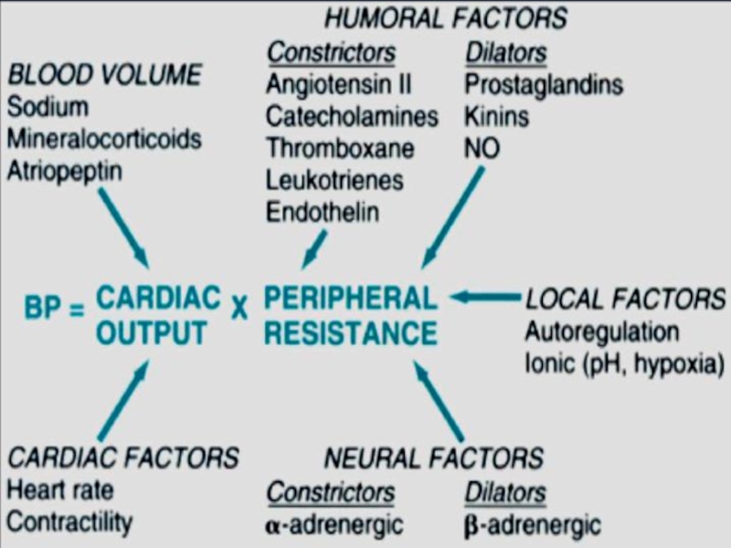
Blood pressure depends of cardiac output and peripheral resistance (elasticity of the arterial system).
Arterial hypertension is defined clinically as
borderline when it riches 140/90 mm Hg.
Blood pressure depends of cardiac output and peripheral resistance (elasticity of the arterial system).
Слайд 16
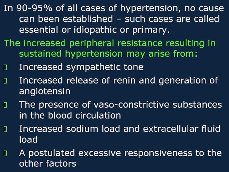
In 90-95% of all cases of hypertension, no cause can been
established – such cases are called essential or idiopathic or primary.
The increased peripheral resistance resulting in sustained hypertension may arise from:
Increased sympathetic tone
Increased release of renin and generation of
angiotensin
The presence of vaso-constrictive substances
in the blood circulation
Increased sodium load and extracellular fluid load
A postulated excessive responsiveness to the other factors
Слайд 17
In only 5-10% of all cases of hypertension is any disease, which
may be associated with disturbance of detectable mechanisms – such cases are secondary hypertension. Examples:
Kidney diseases
Hyper-function of adrenal cortex. May be Cushing ‘s syndrome – corticosteroid excess
Tumor of adrenal medulla (pheochromocytoma) – catecholamine excess.
Hypertension occurs in toxemia of pregnancy
Kidney diseases
Hyper-function of adrenal cortex. May be Cushing ‘s syndrome – corticosteroid excess
Tumor of adrenal medulla (pheochromocytoma) – catecholamine excess.
Hypertension occurs in toxemia of pregnancy
Слайд 18
Clinical-morphological stages
Subclinical stage is displayed by hypertrophy of muscular layer and
elastic structures of arterioles and small-sized arteries, spasm of arterioles. At this stage the hypertrophy of the left ventricle of heart begins.
A stage of general changes of arteries begins as arterial pressure increases. Arteriolar walls permeability is increased, it results in plasmatic impregnation and hyalinosis. Elastic,
muscular-elastic and musculary arteries walls undergo elastofibrosis and atherosclerosis.
A stage of general changes of arteries begins as arterial pressure increases. Arteriolar walls permeability is increased, it results in plasmatic impregnation and hyalinosis. Elastic,
muscular-elastic and musculary arteries walls undergo elastofibrosis and atherosclerosis.
Слайд 19
Clinical-morphological stages
3. The stage of secondary changes of organs is developed
in connection with changes of arteries and insufficiency of the intra- organic blood circulation.
Types of hypertension disease:
benign H. - changes develop slowly, that results in atrophy of parenchyma and sclerosis of organ
malignant H. - changes develop quickly (spasm, thrombosis, fibrinoid necrosis) and causing infarctions and hemorrhages.
Types of hypertension disease:
benign H. - changes develop slowly, that results in atrophy of parenchyma and sclerosis of organ
malignant H. - changes develop quickly (spasm, thrombosis, fibrinoid necrosis) and causing infarctions and hemorrhages.
Слайд 20This is a different kind of arterio- sclerosis. This is hyperplastic
arteriolosclerosis, which most often appears in the kidney in patients with malignant hypertension. The arteriolar wall is markedly thickened and the lumen is narrowed
Sometimes the small arteries and arterioles can be damaged so severely in malignant hypertension that they demonstrate necrosis with a pink fibrin-like quality that gives this process its name--fibrinoid necrosis
Sometimes the small arteries and arterioles can be damaged so severely in malignant hypertension that they demonstrate necrosis with a pink fibrin-like quality that gives this process its name--fibrinoid necrosis
Слайд 21
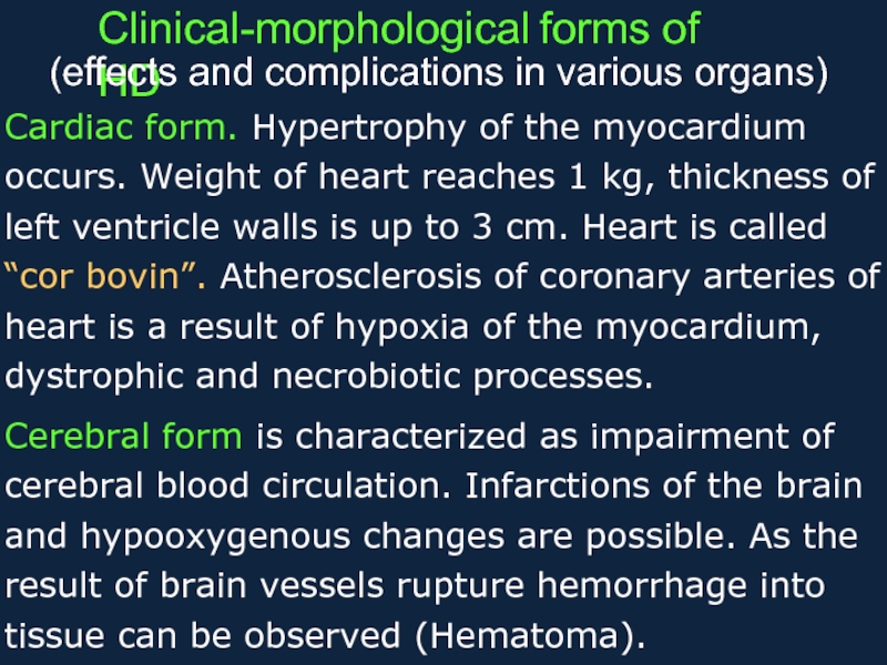
Clinical-morphological forms of HD
(effects and complications in various organs)
Cardiac form. Hypertrophy of the
myocardium occurs. Weight of heart reaches 1 kg, thickness of left ventricle walls is up to 3 cm. Heart is called “cor bovin”. Atherosclerosis of coronary arteries of heart is a result of hypoxia of the myocardium, dystrophic and necrobiotic processes.
Cerebral form is characterized as impairment of cerebral blood circulation. Infarctions of the brain and hypooxygenous changes are possible. As the result of brain vessels rupture hemorrhage into tissue can be observed (Hematoma).
Cerebral form is characterized as impairment of cerebral blood circulation. Infarctions of the brain and hypooxygenous changes are possible. As the result of brain vessels rupture hemorrhage into tissue can be observed (Hematoma).
Слайд 22
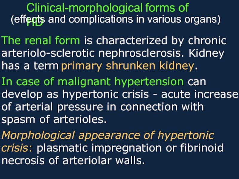
Clinical-morphological forms of HD
(effects and complications in various organs)
The renal form is characterized
by chronic arteriolo-sclerotic nephrosclerosis. Kidney has a term primary shrunken kidney.
In case of malignant hypertension can develop as hypertonic crisis - acute increase of arterial pressure in connection with spasm of arterioles.
Morphological appearance of hypertonic crisis: plasmatic impregnation or fibrinoid necrosis of arteriolar walls.
In case of malignant hypertension can develop as hypertonic crisis - acute increase of arterial pressure in connection with spasm of arterioles.
Morphological appearance of hypertonic crisis: plasmatic impregnation or fibrinoid necrosis of arteriolar walls.
