- Главная
- Разное
- Дизайн
- Бизнес и предпринимательство
- Аналитика
- Образование
- Развлечения
- Красота и здоровье
- Финансы
- Государство
- Путешествия
- Спорт
- Недвижимость
- Армия
- Графика
- Культурология
- Еда и кулинария
- Лингвистика
- Английский язык
- Астрономия
- Алгебра
- Биология
- География
- Детские презентации
- Информатика
- История
- Литература
- Маркетинг
- Математика
- Медицина
- Менеджмент
- Музыка
- МХК
- Немецкий язык
- ОБЖ
- Обществознание
- Окружающий мир
- Педагогика
- Русский язык
- Технология
- Физика
- Философия
- Химия
- Шаблоны, картинки для презентаций
- Экология
- Экономика
- Юриспруденция
Alphabet soup and interstitial lung disease презентация
Содержание
- 1. Alphabet soup and interstitial lung disease
- 2. Overview Classification scheme Individual diseases within the alphabet soup Tables Quiz
- 3. Reminder Pathologic changes in interstitial lung disease
- 4. Classification of ILDs (In total,
- 5. Idiopathic interstitial pneumonias IPF NSIP COP (BOOP)
- 6. Granulomatous lung disease T lymphocytes, macrophages, and
- 7. Inflammation and fibrosis Injury to the epithelial
- 8. IPF Most common idiopathic interstitial pneumonia with
- 9. DIP Only in cigarette smokers Occurs in
- 10. AIP (Hamman-Rich Syndrome) Often in previously healthy
- 11. NSIP Younger set of patients than IPF
- 12. COP/BOOP Presents in 40’s-50’s Bilateral patchy or
- 13. LIP Rarest form, F > M Ground
- 14. PLCH smoking-related Men 20-40y.o. PTX in ~25%,
- 15. LAM Premenopausal women with emphysema, PTX, hemoptysis,
- 16. PAP Not actually and ILD, actually autoimmune
- 19. Case #1
- 20. Answer: IPF CT scan: heterogeneous pattern with
- 21. Case #2
- 22. Answer: DIP CT: Mosaic ground-glass opacity with
- 23. Case #3
- 24. Answer: AIP CT: Bilateral alveolar and interstital
- 25. Case #4
- 26. Answer: NSIP A: Fibrotic variant with reticular
- 27. Case #5
- 28. Answer: COP CT: patchy non-segmental consolidations in
- 29. Case #6
- 30. Answer: LIP CXR: diffuse, fine nodular changes
- 31. Case #7
- 32. Answer: PLCH CT: multiple small, irregularly-shaped, cysts
- 33. Case #8
- 34. Answer: LAM CT: Diffuse parenchymal cysts Path:
- 35. Case #9
- 36. Answer: PAP CT: patchy ground glass opacities
- 37. Sources MKSAP 14, ILD section, p. 18-32


Слайд 3Reminder
Pathologic changes in interstitial lung disease involve cellular infiltration, scarring, and/or
architectural disruption of the pulmonary parenchyma involving the interstitium, alveolar space, airways, and vascular and lymphatic structures as well as pleura.

Слайд 4Classification of ILDs
(In total, there are over 200!)
Unknown cause
(idiopathic)
Systemic causes
Sarcoidosis
Rheumatologic/autoimmune
Lymphoproliverative/neoplastic
Systemic causes
Sarcoidosis
Rheumatologic/autoimmune
Lymphoproliverative/neoplastic
Unknown cause (idiopathic)Systemic causesSarcoidosisRheumatologic/autoimmuneLymphoproliverative/neoplastic")
Слайд 5Idiopathic interstitial pneumonias
IPF
NSIP
COP (BOOP)
DIP/RB-ILD
AIP
LIP
Eosinophilic pneumonia
Pulmonary histiocytosis X
LAM
PAP
Primary amyloidosis
Note: The histology of
ALL of these except histiocytosis X is
Inflammatory and fibrosing; histiocytosis X is granulomatous
Inflammatory and fibrosing; histiocytosis X is granulomatous
DIP/RB-ILDAIPLIPEosinophilic pneumoniaPulmonary histiocytosis XLAMPAPPrimary amyloidosisNote: The histology of ALL of these except")
Слайд 6Granulomatous lung disease
T lymphocytes, macrophages, and epithelioid cells make up the
granuloma
Can progress to fibrosis
Most common forms are sarcoidosis and hypersensitivity pneumonitis
Can progress to fibrosis
Most common forms are sarcoidosis and hypersensitivity pneumonitis

Слайд 7Inflammation and fibrosis
Injury to the epithelial surface causes an inflammatory response
in the air spaces and alveolar walls
In chronic disease, this spreads to adjacent interstitium and vasculature
Progressive fibrosis leads to impairments in ventilation and oxygenation
In chronic disease, this spreads to adjacent interstitium and vasculature
Progressive fibrosis leads to impairments in ventilation and oxygenation

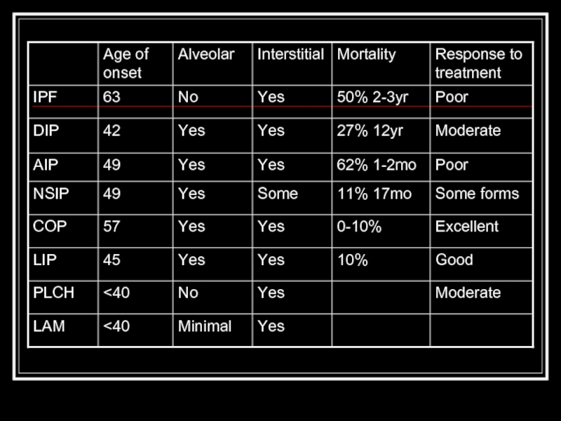
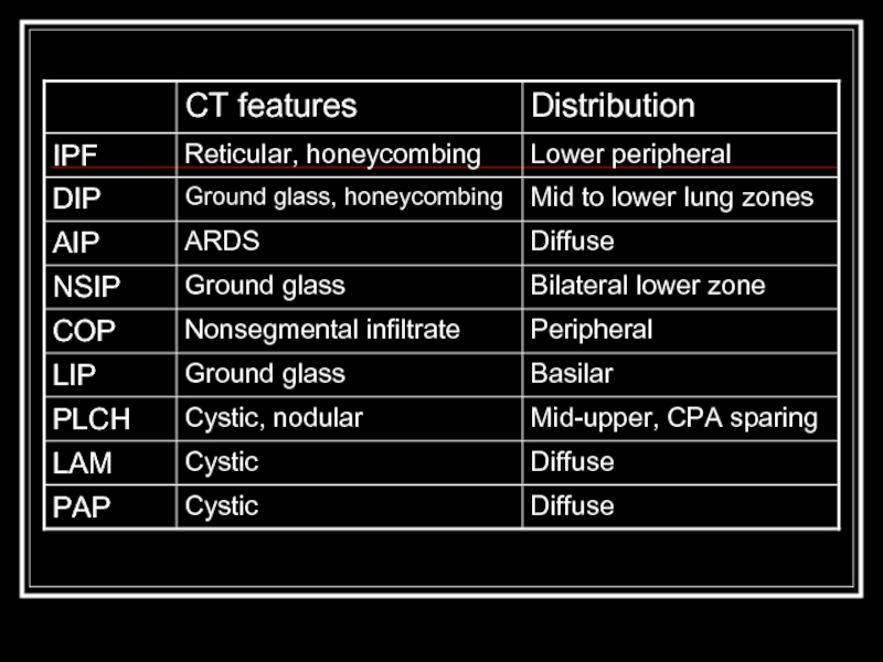
Слайд 8IPF
Most common idiopathic interstitial pneumonia with distinctly poor prognosis
Older age group
(>50y.o.)
Patchy, basilar subpleural reticular opacities with traction bronchiectasis
Temporal and spacial heterogeneity
UIP*—alternating normal lung, interstitial inflammation, foci of proliferating fibroblasts, dense collagen fibrosis, and honeycombing; lymphocytoplasmic infiltrate in alveolar septa; type 2 pneumocyte hyperplasia
*can also be seen in CTDs, pneumoconioses, radiation, drug-induced lung disease,
Chronic aspiration, sarcoidosis, and other conditions
Patchy, basilar subpleural reticular")
Слайд 9DIP
Only in cigarette smokers
Occurs in 30’s-40’s
Diffuse hazy opacities
Intra-alveolar macrophage infiltrate with
minimal interstitial fibrosis
Good response to smoking cessation and glucocorticoids
RB-ILD is a subset in which macrophages accumulate in peribronchial alveoli
Good response to smoking cessation and glucocorticoids
RB-ILD is a subset in which macrophages accumulate in peribronchial alveoli

Слайд 10AIP (Hamman-Rich Syndrome)
Often in previously healthy patients with 7-14 day prodrome
Most
patients >40y.o.
Diffuse, symmetric bilateral ground-glass opacities. May also be subpleural.
Diffuse alveolar damage
ARDS is a subset, but lung biopsy is required to confirm the diagnosis
High requirement for mechanical ventilation and high mortality, but good recovery of lung function in survivors
Diffuse, symmetric bilateral ground-glass opacities. May also be subpleural.
Diffuse alveolar damage
ARDS is a subset, but lung biopsy is required to confirm the diagnosis
High requirement for mechanical ventilation and high mortality, but good recovery of lung function in survivors
Often in previously healthy patients with 7-14 day prodromeMost patients >40y.o.Diffuse, symmetric bilateral")
Слайд 11NSIP
Younger set of patients than IPF present with fevers and without
clubbing
Bilateral, subpleural ground-glass opacities and associated lower lobe volume loss. Honeycombing unusual
Temporally and spacially homogenous
Good response to steroids
Bilateral, subpleural ground-glass opacities and associated lower lobe volume loss. Honeycombing unusual
Temporally and spacially homogenous
Good response to steroids

Слайд 12COP/BOOP
Presents in 40’s-50’s
Bilateral patchy or diffuse alveolar and small nodular opacities
with normal lung volumes and bronchial wall thickening and dilatation; often have recurrent and migratory opacities. Changes most common in periphery and lower lung zones
Granulation tissue within small airways, alveolar ducts, airspaces, with chronic inflammation in the surrounding alveoli
2/3 respond to steroids
Granulation tissue within small airways, alveolar ducts, airspaces, with chronic inflammation in the surrounding alveoli
2/3 respond to steroids
“BOOP pattern” can be present with crypto, Wegener’s lymphoma, hypersensitivity
Pneumonitis, and eosinophilic pneumonia

Слайд 13LIP
Rarest form, F > M
Ground glass, reticular pattern with perivascular cysts
BAL
shows lymphocytosis
Path pattern—cellular interstitial pneumonia with dense lymphoid infiltrate—associated with autoimmune and immunodeficiency disorders
Ddx includes low-grade lymphoma
Path pattern—cellular interstitial pneumonia with dense lymphoid infiltrate—associated with autoimmune and immunodeficiency disorders
Ddx includes low-grade lymphoma

Слайд 14PLCH
smoking-related
Men 20-40y.o.
PTX in ~25%, rarely hemoptysis and DI
Ill-defined or stellate nodules,
reticular or nodular opacities, and bizarre-shaped upper zone cysts, with preserved lung volumes and sparing of the costophrenic angles

Слайд 15LAM
Premenopausal women with emphysema, PTX, hemoptysis, chylous pleural effusion, mostly caucasians
Proliferation
of atypical pulmonary interstitial smooth muscle and cyst formation, react with monoclonal Ab HMB45
Accelerates in pregnancy, abates after oophrectomy
Median survival 8-10 years
Accelerates in pregnancy, abates after oophrectomy
Median survival 8-10 years

Слайд 16PAP
Not actually and ILD, actually autoimmune with and IgG against GM-CSF
Defect
in macrophage processing of surfactant leads to accumulation of PAS-positive lipoproteinaceous material in the distal air spaces with little or no inflammation
Presents in 30’s-50’s, M > F
Labs show polycythemia, hypergammaglobunlinemia, increased LDH
Ground-glass opacities and thickened intralobular strucutres and septa
BAL can be therapeutic
Presents in 30’s-50’s, M > F
Labs show polycythemia, hypergammaglobunlinemia, increased LDH
Ground-glass opacities and thickened intralobular strucutres and septa
BAL can be therapeutic


Слайд 20Answer: IPF
CT scan: heterogeneous pattern with subpleural disease concentrated posteriorly, traction
bronchiectasis/honeycombing, no nodules, little ground glass
Path: heterogeneous paraseptal collagen deposition and fibroblast foci
Path: heterogeneous paraseptal collagen deposition and fibroblast foci


Слайд 22Answer: DIP
CT: Mosaic ground-glass opacity with vascular definition in the areas
of ground-glass opacity and lobular sparing
Path: large numbers of slightly-eosinophilic staining macrophages with interstitial lymphoid aggregates
Path: large numbers of slightly-eosinophilic staining macrophages with interstitial lymphoid aggregates


Слайд 24Answer: AIP
CT: Bilateral alveolar and interstital infiltrates
Path: Early exudative phase showing
vascular congestion, with interstitial and airspace edema and inflammatory cell infiltrates (top left) and fibrinous exudates (top right), organizing phase diffuse alveolar damage (bottom two)


Слайд 26Answer: NSIP
A: Fibrotic variant with reticular subpleural lines with uniform distribution,
bronchiolectasis, and areas of ground glass attenuation
B: Cellular variant with ground glass opacities and traction bronchiectasis
Path: homogeneous expansion of interstitium by inflammatory cells, myofibroblasts, and Type II pneumocytes hyperplasia
B: Cellular variant with ground glass opacities and traction bronchiectasis
Path: homogeneous expansion of interstitium by inflammatory cells, myofibroblasts, and Type II pneumocytes hyperplasia


Слайд 28Answer: COP
CT: patchy non-segmental consolidations in a subpleural and peripheral distribution
Path: diffuse fibrous organization of the airways with obliteration of normal lung architecture


Слайд 30Answer: LIP
CXR: diffuse, fine nodular changes particularly in the lower lobes
Path:
Lymphocytes and plasma cells within interstitial tissue


Слайд 32Answer: PLCH
CT: multiple small, irregularly-shaped, cysts of varying sizes with thin
walls
scattered throughout the lungs (yellow arrows) relatively sparing the bases
Path: eosinophilic granuloma
Path: eosinophilic granuloma


Слайд 34Answer: LAM
CT: Diffuse parenchymal cysts
Path: nodular proliferation of smooth muscle (LAM)
cells replacing the lung parenchyma and jutting into air spaces
 cells replacing the lung")

Слайд 36Answer: PAP
CT: patchy ground glass opacities and septal thickening in a
geographic distribution
Path: intra-alveolar accumulation of surfactant components and cellular debris, with minimal interstitial inflammation or fibrosis
Path: intra-alveolar accumulation of surfactant components and cellular debris, with minimal interstitial inflammation or fibrosis

Слайд 37Sources
MKSAP 14, ILD section, p. 18-32
Gross, T and Gary Hunninghake. “Idiopathic
Pulmonary Fibrosis.” NEJM Vol. 345, No. 7, p. 517-526
Harrisons’s Chapters 237 and 243
Multiple “google images” searches
Harrisons’s Chapters 237 and 243
Multiple “google images” searches