Psychiatrists begin to map genetic architecture of mental disorders презентация

The nucleus accumbens (NAc), the amygdala (Amy), the thalamic and subthalamic nuclei (THAL/SubThal), and hypothalamic nuclei (HT) that have been reported to regulate expression and perception of aggression in

Слайд 1Psychiatrists begin to map genetic architecture of mental disorders
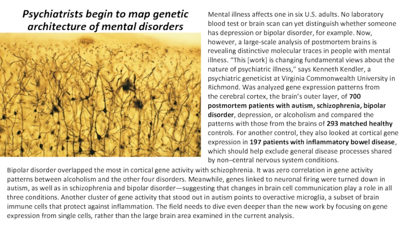
Mental illness affects one

in six U.S. adults. No laboratory blood test or brain scan can yet distinguish whether someone has depression or bipolar disorder, for example. Now, however, a large-scale analysis of postmortem brains is revealing distinctive molecular traces in people with mental
illness. “This [work] is changing fundamental views about the
nature of psychiatric illness,” says Kenneth Kendler, a psychiatric geneticist at Virginia Commonwealth University in Richmond. Was analyzed gene expression patterns from
the cerebral cortex, the brain’s outer layer, of 700 postmortem patients with autism, schizophrenia, bipolar disorder, depression, or alcoholism and compared the patterns with those from the brains of 293 matched healthy controls. For another control, they also looked at cortical gene expression in 197 patients with inflammatory bowel disease, which should help exclude general disease processes shared by non–central nervous system conditions.

Bipolar disorder overlapped the most in cortical gene activity with schizophrenia. It was zero correlation in gene activity patterns between alcoholism and the other four disorders. Meanwhile, genes linked to neuronal firing were turned down in autism, as well as in schizophrenia and bipolar disorder—suggesting that changes in brain cell communication play a role in all three conditions. Another cluster of gene activity that stood out in autism points to overactive microglia, a subset of brain immune cells that protect against inflammation. The field needs to dive even deeper than the new work by focusing on gene expression from single cells, rather than the large brain area examined in the current analysis.
Слайд 2

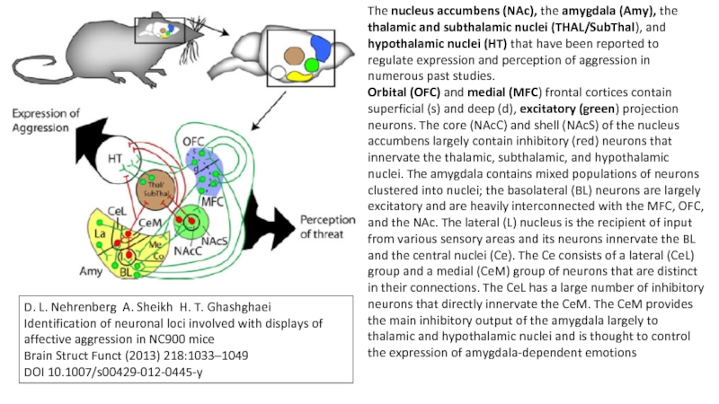
The nucleus accumbens (NAc), the amygdala (Amy), the thalamic and subthalamic

nuclei (THAL/SubThal), and hypothalamic nuclei (HT) that have been reported to regulate expression and perception of aggression in numerous past studies.
Orbital (OFC) and medial (MFC) frontal cortices contain superficial (s) and deep (d), excitatory (green) projection neurons. The core (NAcC) and shell (NAcS) of the nucleus accumbens largely contain inhibitory (red) neurons that innervate the thalamic, subthalamic, and hypothalamic nuclei. The amygdala contains mixed populations of neurons clustered into nuclei; the basolateral (BL) neurons are largely excitatory and are heavily interconnected with the MFC, OFC, and the NAc. The lateral (L) nucleus is the recipient of input from various sensory areas and its neurons innervate the BL and the central nuclei (Ce). The Ce consists of a lateral (CeL) group and a medial (CeM) group of neurons that are distinct in their connections. The CeL has a large number of inhibitory neurons that directly innervate the CeM. The CeM provides the main inhibitory output of the amygdala largely to thalamic and hypothalamic nuclei and is thought to control the expression of amygdala-dependent emotions

D. L. Nehrenberg A. Sheikh H. T. Ghashghaei
Identification of neuronal loci involved with displays of affective aggression in NC900 mice
Brain Struct Funct (2013) 218:1033–1049
DOI 10.1007/s00429-012-0445-y


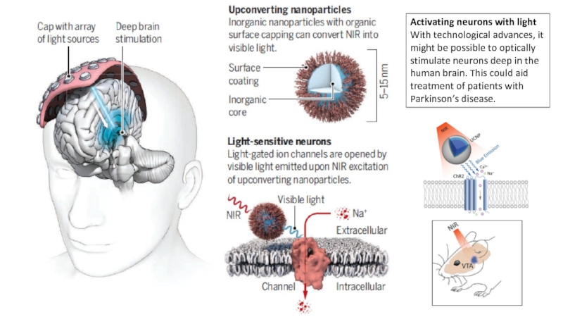
Слайд 3Activating neurons with light
With technological advances, it might be possible to

optically stimulate neurons deep in the human brain. This could aid treatment of patients with
Parkinson’s disease.

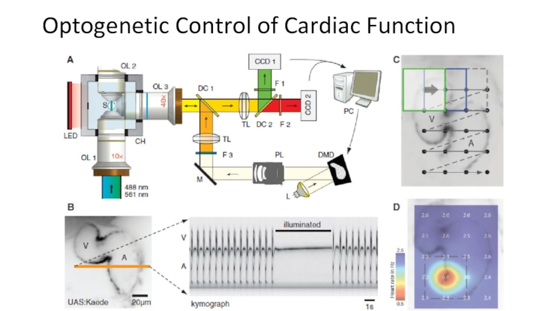
Слайд 4Optogenetic Control of Cardiac Function


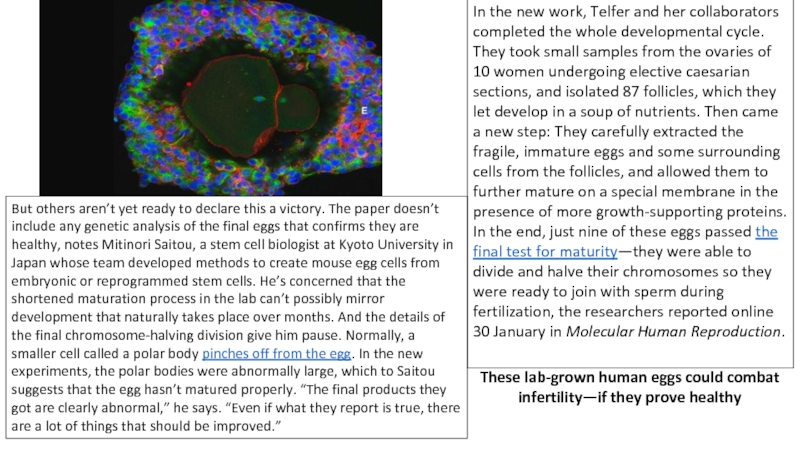
Слайд 5In the new work, Telfer and her collaborators completed the whole

developmental cycle. They took small samples from the ovaries of 10 women undergoing elective caesarian sections, and isolated 87 follicles, which they let develop in a soup of nutrients. Then came a new step: They carefully extracted the fragile, immature eggs and some surrounding cells from the follicles, and allowed them to further mature on a special membrane in the presence of more growth-supporting proteins. In the end, just nine of these eggs passed the final test for maturity—they were able to divide and halve their chromosomes so they were ready to join with sperm during fertilization, the researchers reported online 30 January in Molecular Human Reproduction.

But others aren’t yet ready to declare this a victory. The paper doesn’t include any genetic analysis of the final eggs that confirms they are healthy, notes Mitinori Saitou, a stem cell biologist at Kyoto University in Japan whose team developed methods to create mouse egg cells from embryonic or reprogrammed stem cells. He’s concerned that the shortened maturation process in the lab can’t possibly mirror development that naturally takes place over months. And the details of the final chromosome-halving division give him pause. Normally, a smaller cell called a polar body pinches off from the egg. In the new experiments, the polar bodies were abnormally large, which to Saitou suggests that the egg hasn’t matured properly. “The final products they got are clearly abnormal,” he says. “Even if what they report is true, there are a lot of things that should be improved.”

These lab-grown human eggs could combat infertility—if they prove healthy


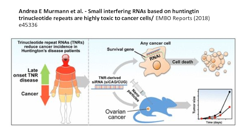
Слайд 6Andrea E Murmann et al. - Small interfering RNAs based on

huntingtin trinucleotide repeats are highly toxic to cancer cells/ EMBO Reports (2018) e45336

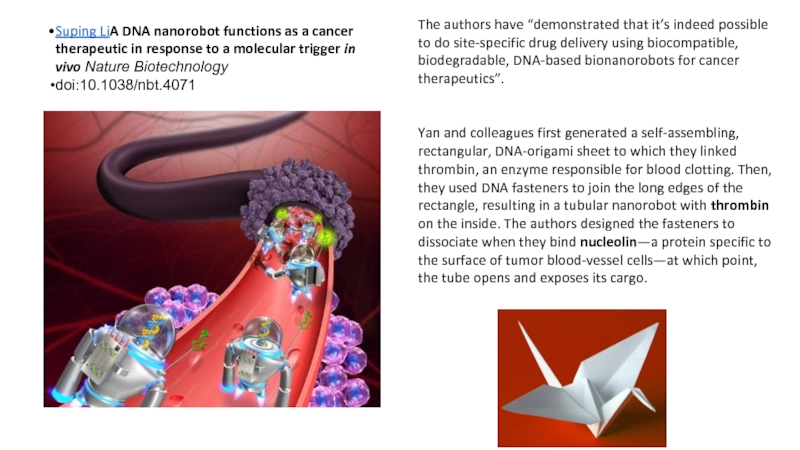
Слайд 7Suping LiA DNA nanorobot functions as a cancer therapeutic in response

to a molecular trigger in vivo Nature Biotechnology
doi:10.1038/nbt.4071


The authors have “demonstrated that it’s indeed possible to do site-specific drug delivery using biocompatible, biodegradable, DNA-based bionanorobots for cancer therapeutics”.


Yan and colleagues first generated a self-assembling, rectangular, DNA-origami sheet to which they linked thrombin, an enzyme responsible for blood clotting. Then, they used DNA fasteners to join the long edges of the rectangle, resulting in a tubular nanorobot with thrombin on the inside. The authors designed the fasteners to dissociate when they bind nucleolin—a protein specific to the surface of tumor blood-vessel cells—at which point, the tube opens and exposes its cargo.


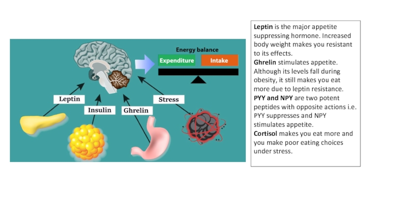
Слайд 18Leptin is the major appetite suppressing hormone. Increased body weight makes

you resistant to its effects.
Ghrelin stimulates appetite. Although its levels fall during obesity, it still makes you eat more due to leptin resistance.
PYY and NPY are two potent peptides with opposite actions i.e. PYY suppresses and NPY stimulates appetite.
Cortisol makes you eat more and you make poor eating choices under stress.


Обратная связь

Если не удалось найти и скачать презентацию, Вы можете заказать его на нашем сайте. Мы постараемся найти нужный Вам материал и отправим по электронной почте. Не стесняйтесь обращаться к нам, если у вас возникли вопросы или пожелания:

Email: Нажмите что бы посмотреть 

Что такое ThePresentation.ru?

Это сайт презентаций, докладов, проектов, шаблонов в формате PowerPoint. Мы помогаем школьникам, студентам, учителям, преподавателям хранить и обмениваться учебными материалами с другими пользователями.


Для правообладателей

Яндекс.Метрика