- Главная
- Разное
- Дизайн
- Бизнес и предпринимательство
- Аналитика
- Образование
- Развлечения
- Красота и здоровье
- Финансы
- Государство
- Путешествия
- Спорт
- Недвижимость
- Армия
- Графика
- Культурология
- Еда и кулинария
- Лингвистика
- Английский язык
- Астрономия
- Алгебра
- Биология
- География
- Детские презентации
- Информатика
- История
- Литература
- Маркетинг
- Математика
- Медицина
- Менеджмент
- Музыка
- МХК
- Немецкий язык
- ОБЖ
- Обществознание
- Окружающий мир
- Педагогика
- Русский язык
- Технология
- Физика
- Философия
- Химия
- Шаблоны, картинки для презентаций
- Экология
- Экономика
- Юриспруденция
What is respiration презентация
Содержание
- 1. What is respiration
- 2. WHAT IS RESPIRATION Respiration is the exchange
- 3. Parts of Human Respiratory System: Nose Pharynx
- 4. The Path of Air From the
- 7. Pharynx is located behind the mouth cavity.
- 8. The lungs are located in the chest
- 9. Left lung: Upper lobe Left
- 10. The lungs consist of many alveoli. The
- 11. Artery Bronchiole
- 13. BREATHING: Movement of air into & out.
- 14. REGULATION OF RESPIRATION Inhalation and exhalation
- 15. HEMOGLOBIN Hemoglobin is red color pigment that
- 16. 98% of oxygen is transported by hemoglobin
- 17. In body tissues HbO2 is divided into
- 18. 70% of CO2 is transported as bicarbonate
- 19. 20% of CO2 is transported by hemoglobin
- 22. Section 4 Breathing— Air In, Air Out
- 23. Breathing When you breathe, air pressure gradients
- 24. Breathing During the cycle, the volume of
- 25. Fig. 11.7, p. 200 INWARD BULK FLOW
- 26. Breathing The basic respiratory cycle. To inhale,
- 27. Breathing Another pressure gradient aids the process.
- 28. Breathing How much air is in a
- 29. inspiratory reserve volume expiratory reserve volume tidal
- 30. Breathing A residual volume of about 1,200

Слайд 2WHAT IS RESPIRATION
Respiration is the exchange of gases between the organism
In HUMAN BODY the respiratory system and circulatory system cooperate in the transport of gases to the cells.


Слайд 4The Path of Air
From the nose, air passes through a muscular
Air then passes into the trachea. The trachea divides into two smaller tubes, the bronchi, which lead to the lungs. Within the lungs, gas exchange occurs in clusters of tiny sacs called alveoli.
Chapter 38

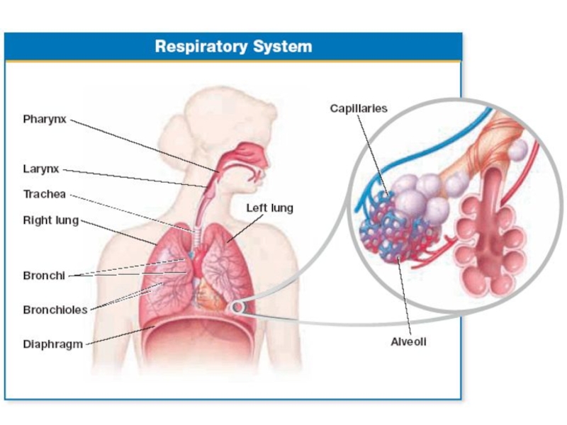
Слайд 7Pharynx is located behind the mouth cavity. (It serves as a
Voice box is located in the larynx.
Vocal cords can be controlled to make sounds.
The trachea is divided into two branches that enter into each lung.
Branches is divided into many smaller bronchioles.

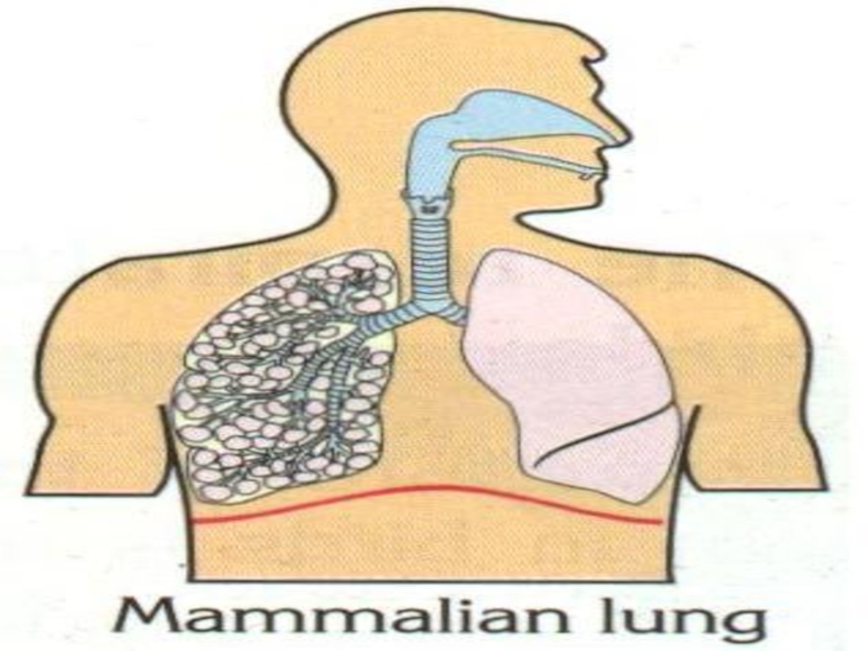
Слайд 8The lungs are located in the chest cavity or thorax.
Lungs are
Both lungs are surrounded by double layered membrane called pleura.
LUNGS

Слайд 9 Left lung: Upper lobe
Left lung: Lower lobe
Right lung: Upper
Right lung: Middle lobe
Right lung: Lower lobe

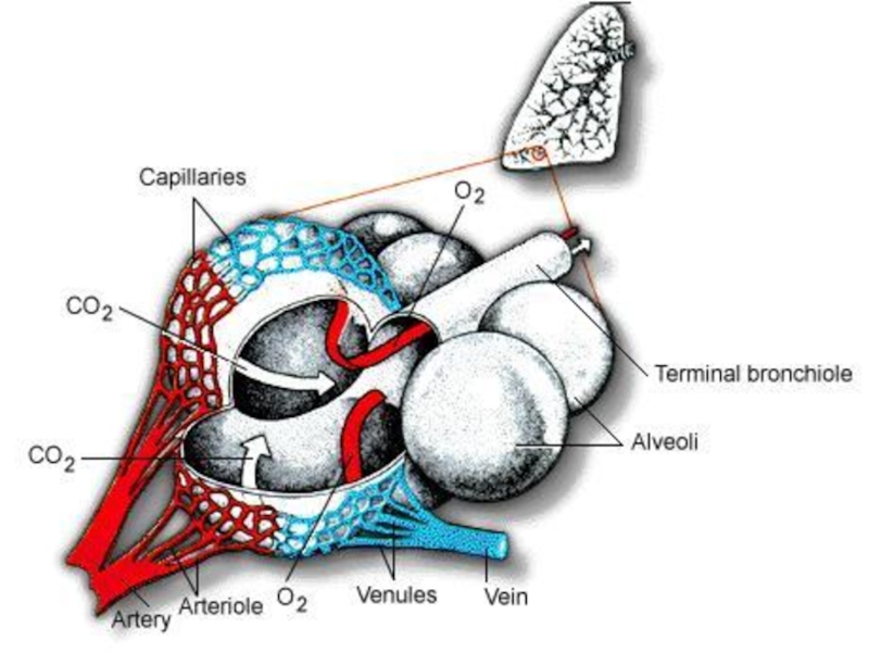
Слайд 10The lungs consist of many alveoli.
The alveoli increases the internal surface
Alveoli are surrounded by a network of capillaries.
Gases are transported by the blood in all land animals except insects.
respiration of Insects is Trachae


Слайд 13BREATHING: Movement of air into & out. Have 2 phases inhalation
EXTERNAL RESPIRATION: Exchange of O2 & CO2 between air & blood in lungs.
CIRCULATION: Carrying dissolved gases.
INTERNAL RESPIRATION: Exchange of O2 & CO2 between blood & body cells.
PHASIS OF HUMAN RESPIRATION

Слайд 14REGULATION OF RESPIRATION
Inhalation and exhalation are under the control of

Слайд 15HEMOGLOBIN
Hemoglobin is red color pigment that found in mammals, birds, amphibia,
There are iron atoms in structure of hemoglobin.

Слайд 1698% of oxygen is transported by hemoglobin in erythrocytes.
2% of oxygen
Oxygen combines with hemoglobine to form oxyhemoglobin (HbO2). HbO2 molecules combines with K ions.
oxygen transport
KHb + O2
Lung capillaries
KHbO2

Слайд 17In body tissues HbO2 is divided into Hb and O2.
Because
And O2 diffuses to body tissue.
KHbO2
KHb + O2
Tissue capillaries

Слайд 18 70% of CO2 is transported as bicarbonate ions.
Carbondioxide Transport
CO2
H2CO3 (Carbonic Acid)
H2CO3
H+ + HCO3- (Bicarbonate ion)
(In the body tissues with action of Carboxylase enzyme)
H+ + HCO3-
H2CO3
H2CO3
CO2 + H2O
In the lungs by
Carboxylase
H2CO3")
Слайд 1920% of CO2 is transported by hemoglobin in erythrocytes
Hb + CO2
HbCO2
plasma.
(Carboxyhemoglobin)


Слайд 23Breathing
When you breathe, air pressure gradients reverse in a cycle.
The respiratory
Inspiration (inhalation) draws breath into the airways.
Expiration (exhalation) moves a breath out of the airways.

Слайд 24Breathing
During the cycle, the volume of the chest cavity increases, then
This works because the air in the airways is the
same pressure as the outside atmosphere.
Pressure in the alveoli (intrapulmonary pressure)
is also the same as the outside air.

Слайд 25Fig. 11.7, p. 200
INWARD BULK
FLOW OF AIR
OUTWARD BULK
FLOW OF AIR
Inhalation
Diaphragm
Exhalation
Diaphragm and external intercostal muscles return to the resting positions. Rib cage moves down. Lungs recoil passively.

Слайд 26Breathing
The basic respiratory cycle.
To inhale, the diaphragm contracts and flattens, muscles
To exhale, the actions listed above are reversed; the elastic lung tissue recoils passively and air flows out of the lungs.
Active exhalation involves contraction of the abdominal muscles to push the diaphragm upward, forcing more air out.

Слайд 27Breathing
Another pressure gradient aids the process.
The lungs are stretched to fill
In a collapsed lung (pneumothorax), air enters the pleural cavity, disrupting the normal expansion and contraction of the lungs.

Слайд 28Breathing
How much air is in a “breath”?
About 500 ml of air
A human can forcibly inhale 3,100 ml of air (inspiratory reserve volume) and forcibly exhale 1,200 ml (expiratory reserve volume).
The maximum volume that can be moved in and out is called the vital capacity (4,800 ml for males, 3,800 ml for females).
 enters and")
Слайд 29inspiratory
reserve volume
expiratory
reserve volume
tidal volume
vital capacity
total lung capacity
Fig. 11.8, p. 201
time
Lung volume
6,000
1,000
2,000
3,000
4,000
5,000
0
residual
volume
6,0001,0002,0003,0004,0005,0000residual volume")
Слайд 30Breathing
A residual volume of about 1,200 ml remains in the lungs
Sometimes food enters the trachea rather than the esophagus; it can be forced out by the Heimlich maneuver, which forces the diaphragm to elevate, pushing air into the trachea to dislodge the obstruction.