- Главная
- Разное
- Дизайн
- Бизнес и предпринимательство
- Аналитика
- Образование
- Развлечения
- Красота и здоровье
- Финансы
- Государство
- Путешествия
- Спорт
- Недвижимость
- Армия
- Графика
- Культурология
- Еда и кулинария
- Лингвистика
- Английский язык
- Астрономия
- Алгебра
- Биология
- География
- Детские презентации
- Информатика
- История
- Литература
- Маркетинг
- Математика
- Медицина
- Менеджмент
- Музыка
- МХК
- Немецкий язык
- ОБЖ
- Обществознание
- Окружающий мир
- Педагогика
- Русский язык
- Технология
- Физика
- Философия
- Химия
- Шаблоны, картинки для презентаций
- Экология
- Экономика
- Юриспруденция
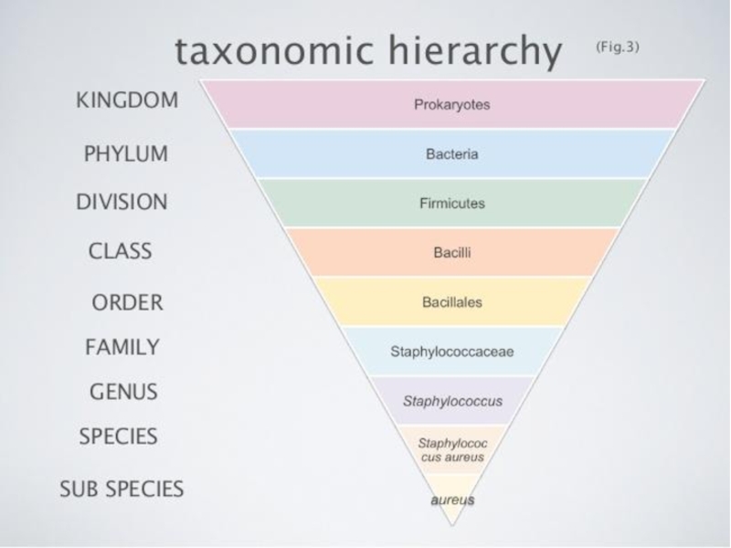
Microbiology. Microbiological laboratory systematics of microorganisms morphology of microorganims презентация
Содержание
- 1. Microbiology. Microbiological laboratory systematics of microorganisms morphology of microorganims
- 2. SAFETY RULES Always wear lab coats and
- 3. IT IS STRICKTLY PROHIBITED to pump fluid
- 4. PURPOSES to get acquainted with principles of
- 5. MICROBIOLOGY Microbiology (from Greek μῑκρος, mīkros, "small"; βίος, bios, "life"; and -λογία, -logia) is the
- 8. MICROBIOLOGICAL LABORATORY
- 9. Laboratory rooms and laminar flow cabinets are designed for specific activities in aseptic conditions
- 10. Room for preparation of nutrient media
- 11. Table automatic boiler for the preparation of small volumes of nutrient media
- 12. Specially equipped rooms for sterilization of nutrient media, laboratory glassware, disinfection of infectious material
- 13. Vivarium for laboratory animals
- 14. LABORATORY EQUIPMENT Biological immersion microscope Instruments: inoculation
- 15. BIOLOGICAL IMMERSION MICROSCOPE TASK 1 (P. 13) NAME PARTS OF LIGHT MICROSCOPE
- 16. IMMERSION MICROSCOPY
- 17. IMMERSION MICROSCOPY TASK 2 (P. 13) DRAW WAY OF LIGHT IN IMMERSION SYSTEM
- 18. INSTRUMENTS inoculation loops spatula tweezers spirit lamp
- 19. LABORATORY GLASSWARE test tubes Petri dish flasks pipettes
- 20. DEVICES FOR STERILIZATION autoclave Pasteur oven
- 21. NUTRIENT MEDIA Blood agar Endo media
- 22. REAGENTS
- 23. pH Meters
- 24. DISTILLERS
- 25. CENTRIFUGES
- 26. BALANCES technical analytical
- 27. FILTRATION EQUIPMENT
- 29. STUDENT’S LABORATORY EQUIPMENT Microscope Immersion oil Inoculating
- 30. MORPHOLOGY OF MICROORGANISMS Size of microbial cells
- 31. SIZE OF MICROORGANISMS Bacteria are of about
- 35. STAINING Because microbial cytoplasm is usually transparent,
- 37. STAINING Simple stain techniques Staining can be
- 39. IMMERSION MICROSCOPY PROCEDURE 1. Work well seated.
- 40. Escherichia coli (simple stain by fuchsine) Escherichia coli (scanning electron microscope)
- 41. Staphylococcus aureus (simple stain by fuchsine) Staphylococcus aureus (scanning electron microscope)
- 42. Bacillus anthracoides (simple stain by fuchsine)
- 43. Vibrio cholerae (simple stain by fuchsine) Vibrio cholerae (scanning electron microscopy)
- 44. TASK 3 DESCRIBE THE MORPHOLOGY OF YOUR
- 45. TASK 3 DESCRIBE THE MORPHOLOGY OF YOUR
- 46. TASK 3 DESCRIBE THE MORPHOLOGY OF YOUR
- 47. TASK 3 DESCRIBE THE MORPHOLOGY OF YOUR
- 48. Recommendations Attend all lectures and lessons
Слайд 1MICROBIOLOGY
MICROBIOLOGICAL LABORATORY
SYSTEMATICS OF MICROORGANISMS
MORPHOLOGY OF MICROORGANIMS
LESSON №1

Слайд 2SAFETY RULES
Always wear lab coats and caps
Don’t put your bags and
personal things on lab table
DON’T EAT, DRINK AND SMOKE IN THE DEPARTMENT
Used reagents and materials or pipettes put in bottle with disinfectant
If dangerous material got on the table, floor or clothes tell about it immediately to professor or lab technician and strictly follow his recommendations
Table must be clean all time
Be careful with the equipment
Wash your hands with soap after lesson

Слайд 3IT IS STRICKTLY PROHIBITED
to pump fluid into the pipette by mouth
to
move a burning spirit lamp
to light up one burner from the other
to light up one burner from the other

Слайд 4PURPOSES
to get acquainted with principles of organization, equipment of microbiology laboratory
and rules of work in it
to get acquainted with main microscopic methods, preparation of bacterial smears, simple staining and immersion microscopy technique and main morphological forms of bacteria
to get acquainted with main microscopic methods, preparation of bacterial smears, simple staining and immersion microscopy technique and main morphological forms of bacteria

Слайд 5MICROBIOLOGY
Microbiology (from Greek μῑκρος, mīkros, "small"; βίος, bios, "life"; and -λογία, -logia) is the study of microorganisms, those being unicellular(single cell), multicellular (cell
colony), or acellular (lacking cells)
Microbiology encompasses numerous sub-disciplines including virology, parasitology,
mycology and bacteriology
Microbiology encompasses numerous sub-disciplines including virology, parasitology,
mycology and bacteriology


Слайд 9Laboratory rooms and laminar flow cabinets are designed for specific activities
in aseptic conditions


Слайд 12Specially equipped rooms for sterilization of nutrient media, laboratory glassware, disinfection
of infectious material


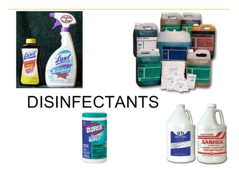
Слайд 14LABORATORY EQUIPMENT
Biological immersion microscope
Instruments: inoculation loops, spatulas, tweezers, spirit lamps, etc
Laboratory
glassware: tubes, Petri dishes, flasks, pipettes, etc
Devices for sterilization of glassware, nutrient media, reagents, pH meters, distillers, centrifuges, technical and analytical balances, filtering equipment, etc
Other fire and chemical safety equipment (fire extinguishers, disinfectants, etc)
Devices for sterilization of glassware, nutrient media, reagents, pH meters, distillers, centrifuges, technical and analytical balances, filtering equipment, etc
Other fire and chemical safety equipment (fire extinguishers, disinfectants, etc)

 NAME PARTS OF LIGHT MICROSCOPE")

 DRAW WAY OF LIGHT IN IMMERSION SYSTEM")


Слайд 29STUDENT’S LABORATORY EQUIPMENT
Microscope
Immersion oil
Inoculating loop
Burner or spirit lamp
Staining kits
Water for washing
smears
Slides
Stands for tubes
Crystallizer and bridge
Tweezers for collecting slides
Filter paper for drying smears
Flask for used slides
Slides
Stands for tubes
Crystallizer and bridge
Tweezers for collecting slides
Filter paper for drying smears
Flask for used slides

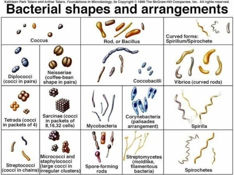
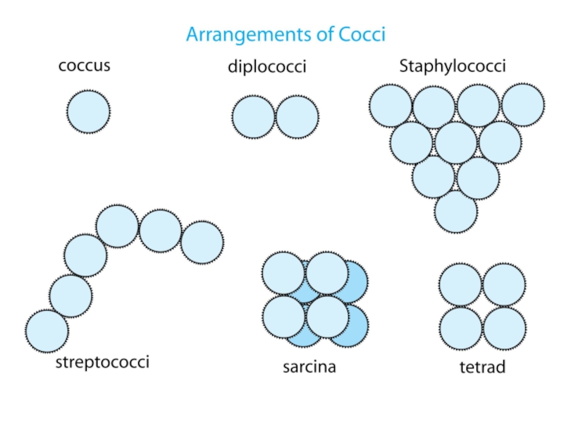
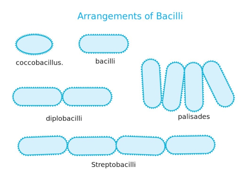
Слайд 30MORPHOLOGY OF MICROORGANISMS
Size of microbial cells
Shape of microbial cells
Arrangement of microbial
cells
Bacteria are of about 0,5—5 µm in size
Bacteria are of about 0,5—5 µm in size

Слайд 31SIZE OF MICROORGANISMS
Bacteria are of about 0,5—5 µm in size
The range
of sizes shown by prokaryotes, relative to those of other organisms and biomolecules

Слайд 35STAINING
Because microbial cytoplasm is usually transparent, it is necessary to stain
microorganisms before they can be viewed with the light microscope. In some cases, staining is unnecessary, for example when microorganisms are very large or when motility is to be studied, and a drop of the microorganisms can be placed directly on the slide and observed
Staining is an auxiliary technique used in microscopy to enhance contrast in the microscopic image
Staining is an auxiliary technique used in microscopy to enhance contrast in the microscopic image

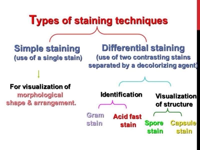
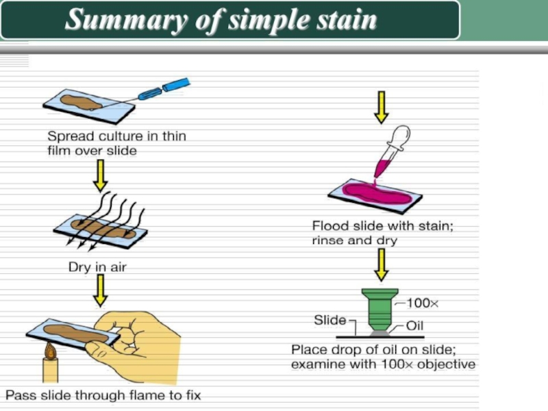
Слайд 37STAINING
Simple stain techniques
Staining can be performed with basic dyes such as
crystal violet or methylene blue, positively charged dyes that are attracted to the negatively charged materials of the microbial cytoplasm. Such a procedure is the simple stain procedure
The differential stain technique distinguishes two kinds of organisms. An example is the Gram stain technique. This differential technique separates bacteria into two groups, Gram‐positive bacteria and Gram‐negative bacteria.
The differential stain technique distinguishes two kinds of organisms. An example is the Gram stain technique. This differential technique separates bacteria into two groups, Gram‐positive bacteria and Gram‐negative bacteria.

Слайд 39IMMERSION MICROSCOPY PROCEDURE
1. Work well seated.
2. Lift up the condenser to
the level of an object table.
3. Set up the lightening looking through an ocular.
4. Place a preparation with a drop of immersion oil on an object table and fix it with clamps.
5. Install an immersion lens (with a magnification of 90 or 100). Work very carefully with an immersion lens. Be careful while immersing the lens into a drop of oil, because it can crush the glass.
6. Lower a tube under the control of the eyes using the macroscrew, immerse the lens into the oil, don't touch the glass surface.
7. Looking into an ocular, slowly raise a tube up with the macro-screw until an image appears (until something flashes in the field of view).
8. After that, turn the micro-screw to receive the clear image of an object. Both eyes should be open, using the left hand move a preparation in such way for general review.
9. After treating the preparation, raise a tube up with the macro-screw. Remove a preparation, lower a condenser, wipe the oil from an oil immersion lens with a soft napkin, then return a drawtube to its initial position.

Escherichia coli (scanning electron microscope)")
Слайд 41Staphylococcus aureus
(simple stain by fuchsine)
Staphylococcus aureus
(scanning electron microscope)
Staphylococcus aureus(scanning electron microscope)")
")
Vibrio cholerae (scanning electron microscopy)")
Слайд 44TASK 3
DESCRIBE THE MORPHOLOGY OF YOUR PREPARATION
Species Escherichia coli
Size small
Cell form
rods
Cell location single
Cell arrangement chaotically
Form of cell edge rounded edges
Stain simple stain by fuchsine
Magnification 1000х
Cell location single
Cell arrangement chaotically
Form of cell edge rounded edges
Stain simple stain by fuchsine
Magnification 1000х

Слайд 45TASK 3
DESCRIBE THE MORPHOLOGY OF YOUR PREPARATION
Species Staphylococcus aureus
Size large
Cell form
coccus (spherical)
Cell location ”grapes”-like clusters
Form of cell edge –
Stain simple stain by fuchsine
Magnification 1000х
Cell location ”grapes”-like clusters
Form of cell edge –
Stain simple stain by fuchsine
Magnification 1000х
Cell location")
Слайд 46TASK 3
DESCRIBE THE MORPHOLOGY OF YOUR PREPARATION
Species Bacillus anthracoides
Size large
Cell
form bacillus
Cell location chains
Form of cell edge chopped edges
Stain simple stain by fuchsine
Magnification 1000х
Cell location chains
Form of cell edge chopped edges
Stain simple stain by fuchsine
Magnification 1000х

Слайд 47TASK 3
DESCRIBE THE MORPHOLOGY OF YOUR PREPARATION
Species Vibrio cholerae
Size small
Cell
form vibrio (curved comma-like rods)
Cell location single
Cell arrangement chaotically
Form of cell edge rounded edges
Stain simple stain by fuchsine
Magnification 1000х
Cell location single
Cell arrangement chaotically
Form of cell edge rounded edges
Stain simple stain by fuchsine
Magnification 1000х

Слайд 48Recommendations
Attend all lectures and lessons
Prepare home task for each lesson
At
the end of each lesson show results in workbook and answer questions
Books, slides and other useful materials will be published in this public: https://vk.com/pmedpharm_mb
Our official web-page:
https://www.pmedpharm.ru/departments/kafedra_biologicheskoy_himii_i_mikrobiologii/
Books, slides and other useful materials will be published in this public: https://vk.com/pmedpharm_mb
Our official web-page:
https://www.pmedpharm.ru/departments/kafedra_biologicheskoy_himii_i_mikrobiologii/