- Главная
- Разное
- Дизайн
- Бизнес и предпринимательство
- Аналитика
- Образование
- Развлечения
- Красота и здоровье
- Финансы
- Государство
- Путешествия
- Спорт
- Недвижимость
- Армия
- Графика
- Культурология
- Еда и кулинария
- Лингвистика
- Английский язык
- Астрономия
- Алгебра
- Биология
- География
- Детские презентации
- Информатика
- История
- Литература
- Маркетинг
- Математика
- Медицина
- Менеджмент
- Музыка
- МХК
- Немецкий язык
- ОБЖ
- Обществознание
- Окружающий мир
- Педагогика
- Русский язык
- Технология
- Физика
- Философия
- Химия
- Шаблоны, картинки для презентаций
- Экология
- Экономика
- Юриспруденция
Информационная связь между днк, рнк и белками презентация
Содержание
- 1. Информационная связь между днк, рнк и белками
- 2. 19 РЕПЛИКАЦИЯ ДНК а - полуконсервативная б
- 5. 20 РЕПЛИКАЦИЯ ДНК общая схема
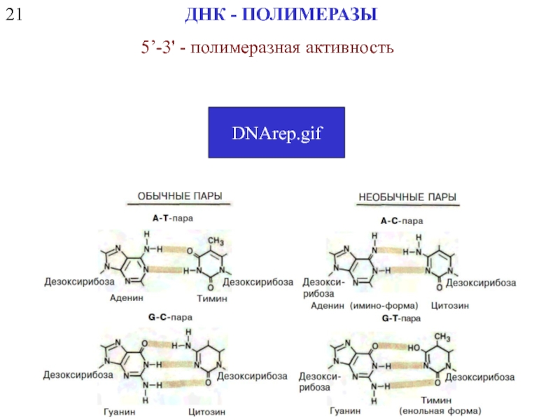
- 6. 21 ДНК - ПОЛИМЕРАЗЫ 5’-3' - полимеразная активность DNArep.gif
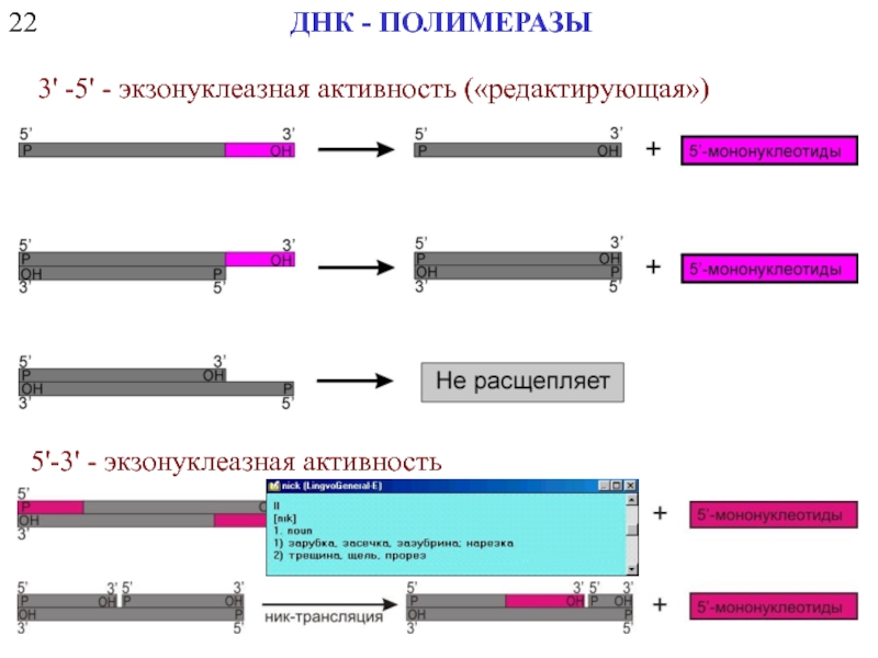
- 7. 22 ДНК - ПОЛИМЕРАЗЫ 3' -5' - экзонуклеазная активность («редактирующая») 5'-3' - экзонуклеазная активность
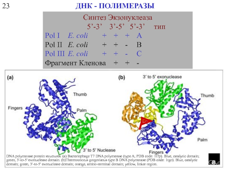
- 8. 23 ДНК - ПОЛИМЕРАЗЫ
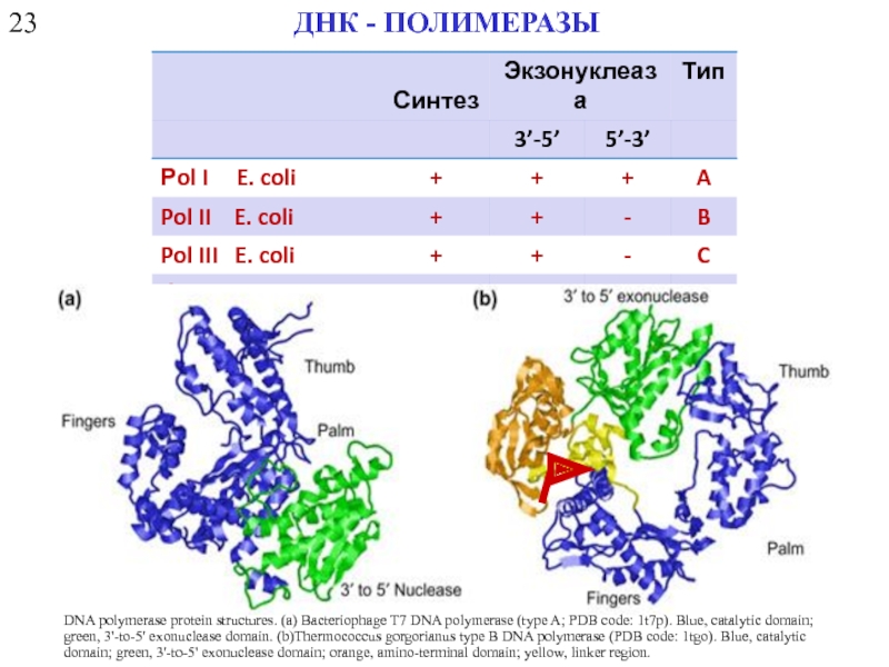
- 9. 23 ДНК - ПОЛИМЕРАЗЫ
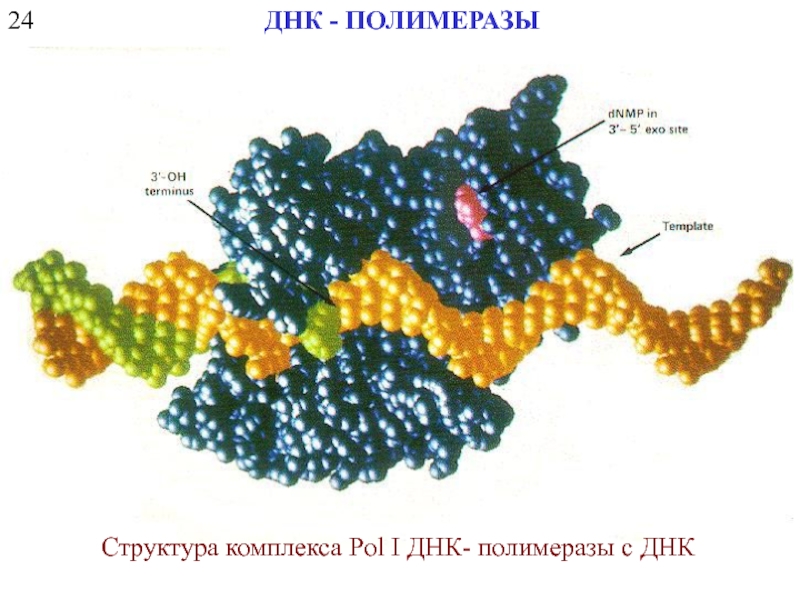
- 10. 24 ДНК - ПОЛИМЕРАЗЫ Структура комплекса Pol I ДНК- полимеразы с ДНК
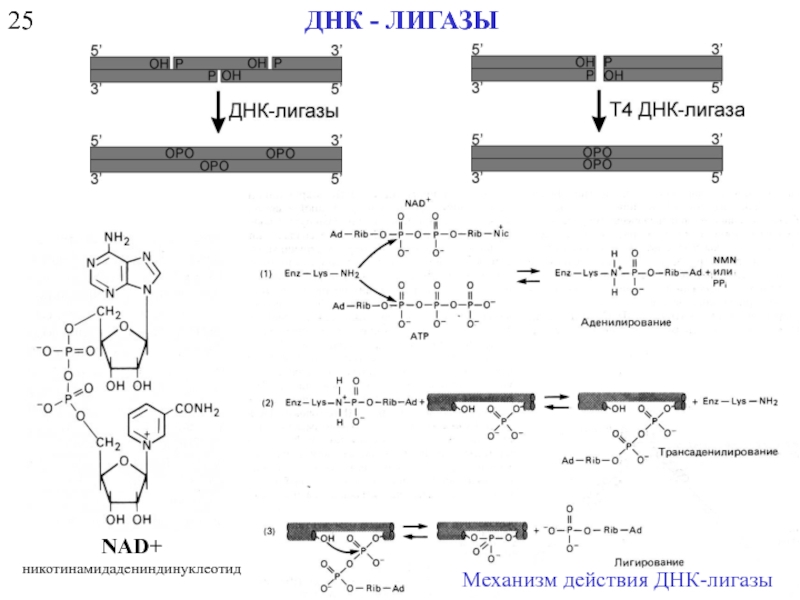
- 11. 25 ДНК - ЛИГАЗЫ NAD+ никотинамидадениндинуклеотид Механизм действия ДНК-лигазы
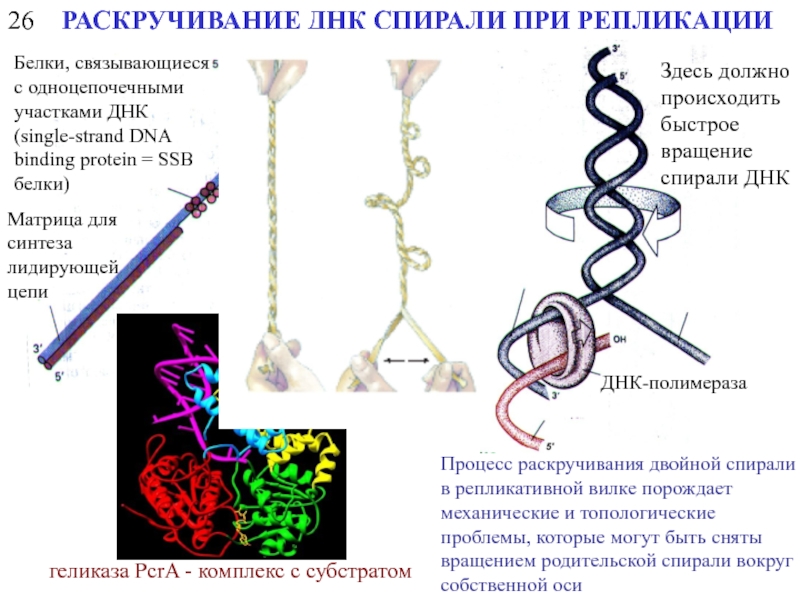
- 12. 26 РАСКРУЧИВАНИЕ ДНК СПИРАЛИ ПРИ РЕПЛИКАЦИИ геликаза
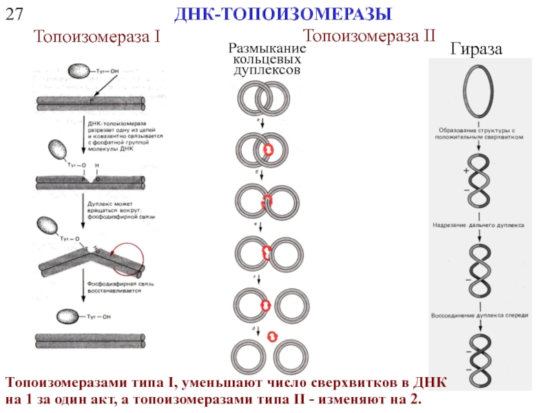
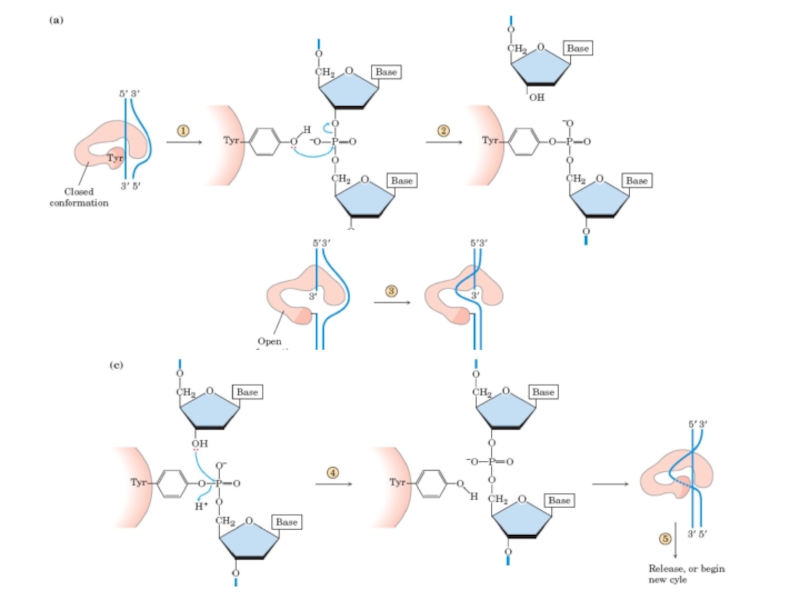
- 13. 27 ДНК-ТОПОИЗОМЕРАЗЫ Топоизомераза I Топоизомераза II Размыкание
- 15. 28 ТОЧКИ НАЧАЛА РЕПЛИКАЦИИ (ORIGIN) репликационный «пузырек»
- 16. A.C. Leonard and M. Mechali DNA Replication
- 17. A.C. Leonard and M. Mechali DNA Replication
- 18. 29 ИНИЦИАЦИЯ ОБРАЗОВАНИЯ НОВЫХ ЦЕПЕЙ ДНК Инициация
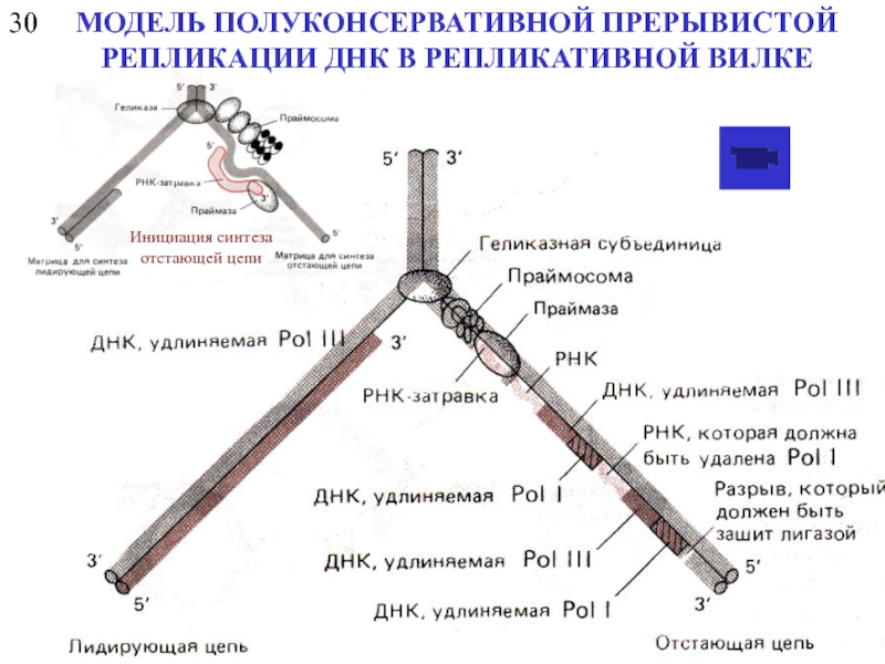
- 19. 30 Инициация синтеза отстающей цепи МОДЕЛЬ ПОЛУКОНСЕРВАТИВНОЙ ПРЕРЫВИСТОЙ РЕПЛИКАЦИИ ДНК В РЕПЛИКАТИВНОЙ ВИЛКЕ
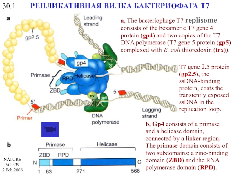
- 20. 30.1 РЕПЛИКАТИВНАЯ ВИЛКА БАКТЕРИОФАГА Т7 a, The
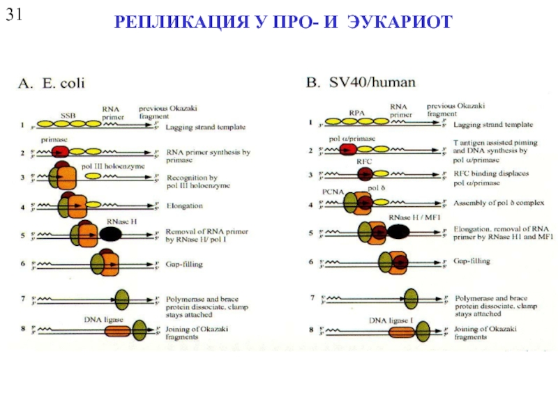
- 21. 31 РЕПЛИКАЦИЯ У ПРО- И ЭУКАРИОТ
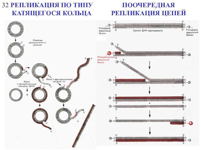
- 22. 32 РЕПЛИКАЦИЯ ПО ТИПУ КАТЯЩЕГОСЯ КОЛЬЦА ПООЧЕРЕДНАЯ РЕПЛИКАЦИЯ ЦЕПЕЙ
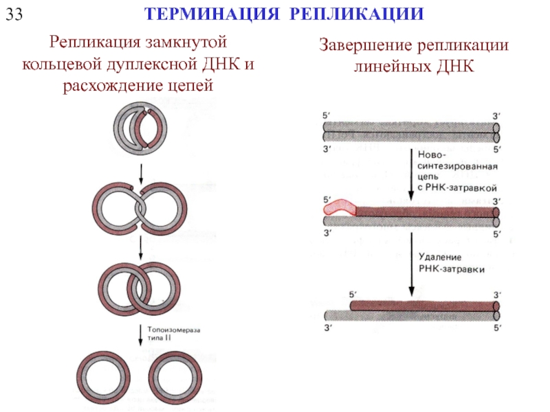
- 23. 33 ТЕРМИНАЦИЯ РЕПЛИКАЦИИ Репликация замкнутой кольцевой дуплексной ДНК и расхождение цепей Завершение репликации линейных ДНК
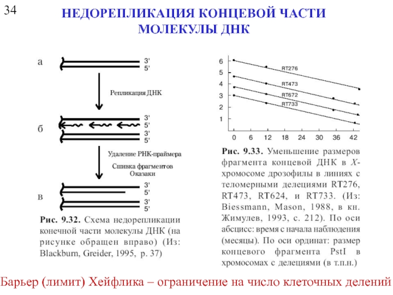
- 24. 34 НЕДОРЕПЛИКАЦИЯ КОНЦЕВОЙ ЧАСТИ МОЛЕКУЛЫ ДНК Барьер (лимит) Хейфлика – ограничение на число клеточных делений
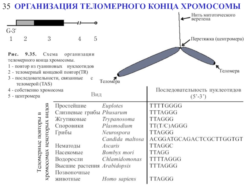
- 25. 35 ОРГАНИЗАЦИЯ ТЕЛОМЕРНОГО КОНЦА ХРОМОСОМЫ Вид Теломерные повторы в хромосомах некоторых видов
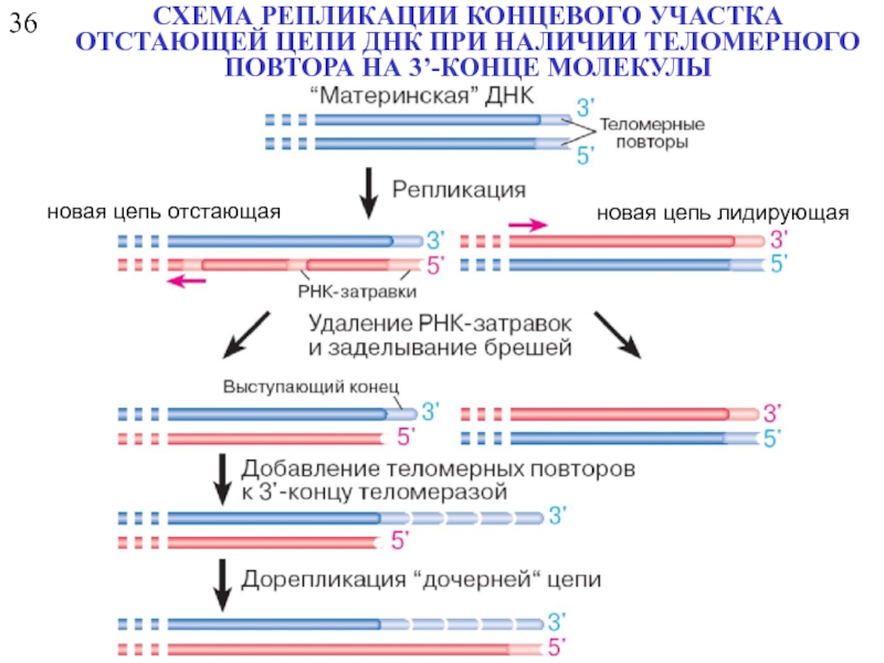
- 26. 36 СХЕМА РЕПЛИКАЦИИ КОНЦЕВОГО УЧАСТКА ОТСТАЮЩЕЙ ЦЕПИ
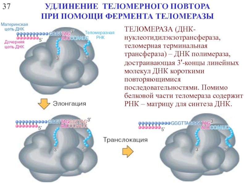
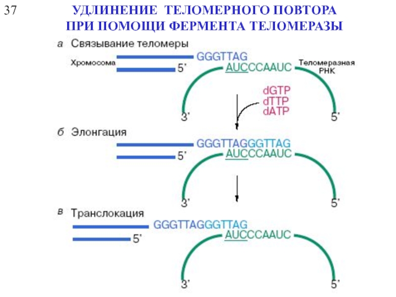
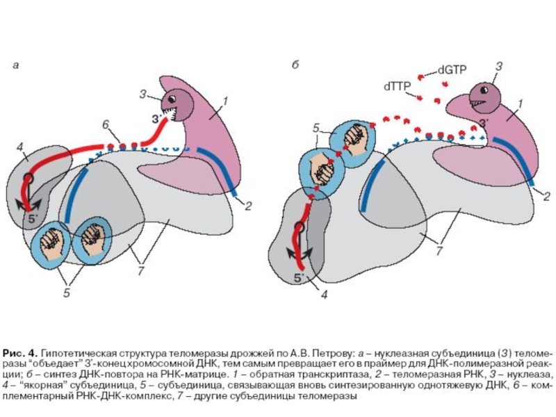
- 27. 37 УДЛИНЕНИЕ ТЕЛОМЕРНОГО ПОВТОРА ПРИ ПОМОЩИ ФЕРМЕНТА
- 28. 37 УДЛИНЕНИЕ ТЕЛОМЕРНОГО ПОВТОРА ПРИ ПОМОЩИ ФЕРМЕНТА ТЕЛОМЕРАЗЫ
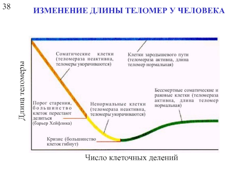
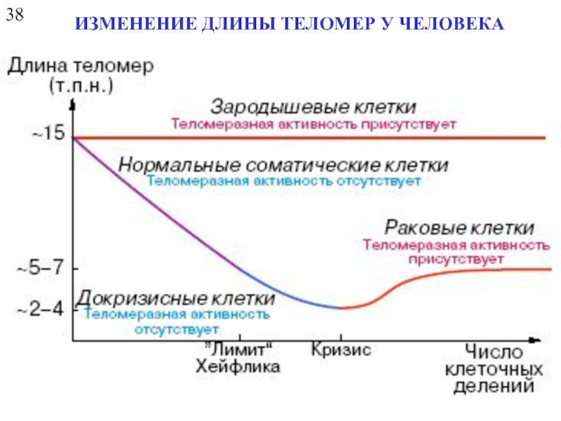
- 30. 38 ИЗМЕНЕНИЕ ДЛИНЫ ТЕЛОМЕР У ЧЕЛОВЕКА Число клеточных делений Длина теломеры (барьер Хейфлика)
- 31. 38 ИЗМЕНЕНИЕ ДЛИНЫ ТЕЛОМЕР У ЧЕЛОВЕКА
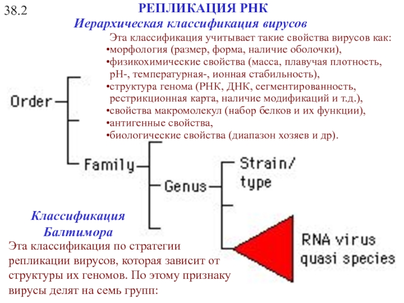
- 32. 38.2 РЕПЛИКАЦИЯ РНК Иерархическая классификация вирусов Эта
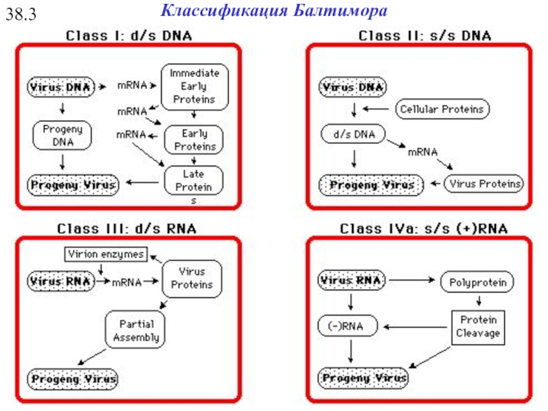
- 33. 38.3 Классификация Балтимора
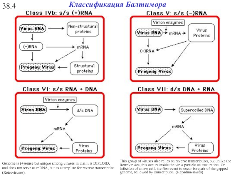
- 34. 38.4 Genome is (+)sense but unique among
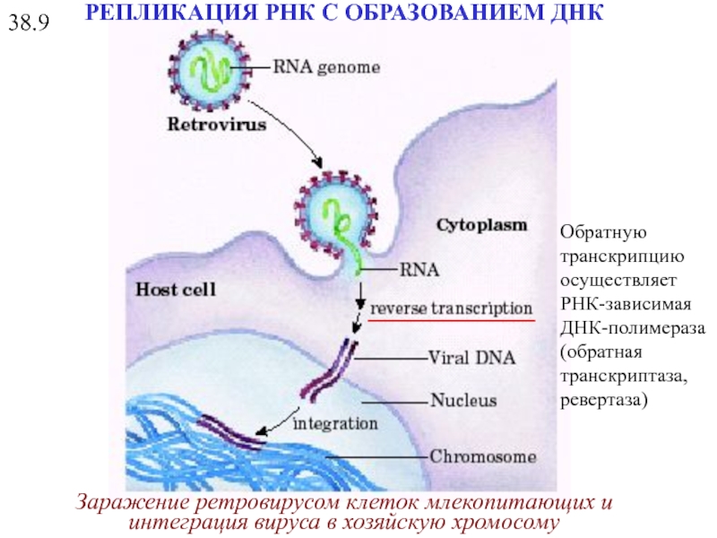
- 35. Заражение ретровирусом клеток млекопитающих и интеграция вируса
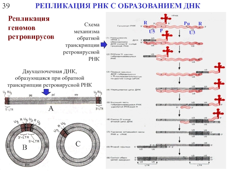
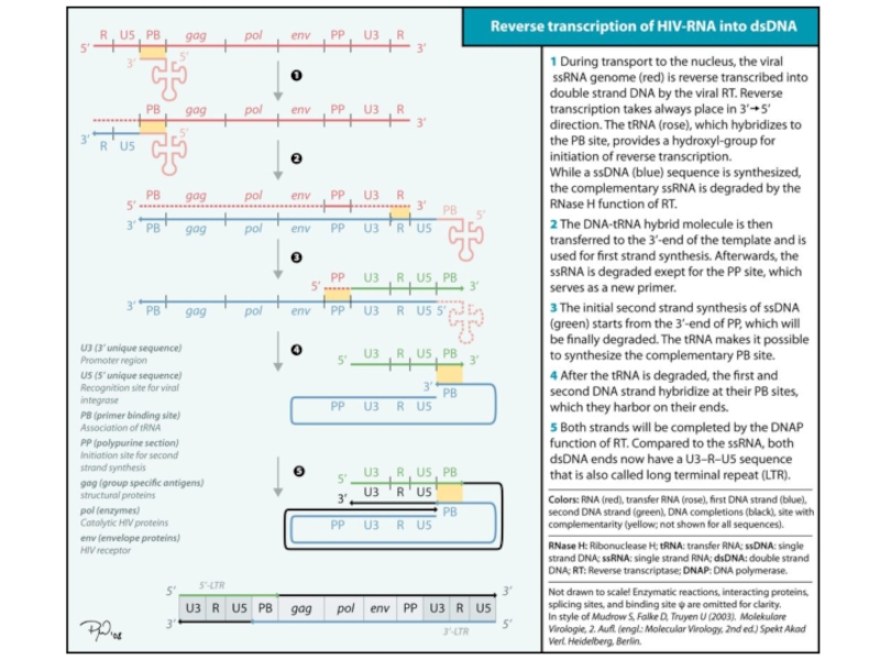
- 36. 39 РЕПЛИКАЦИЯ РНК С ОБРАЗОВАНИЕМ ДНК Репликация
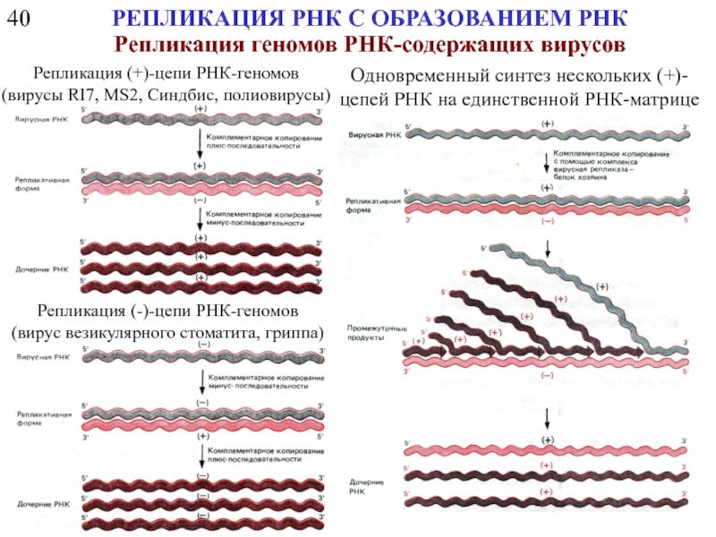
- 39. 40 РЕПЛИКАЦИЯ РНК С ОБРАЗОВАНИЕМ РНК Репликация
Слайд 118
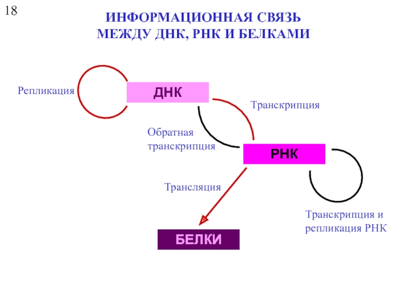
ИНФОРМАЦИОННАЯ СВЯЗЬ
МЕЖДУ ДНК, РНК И БЕЛКАМИ
ДНК
РНК
БЕЛКИ
Репликация
Транскрипция
Обратная
транскрипция
Трансляция
Транскрипция и
репликация РНК
Слайд 219
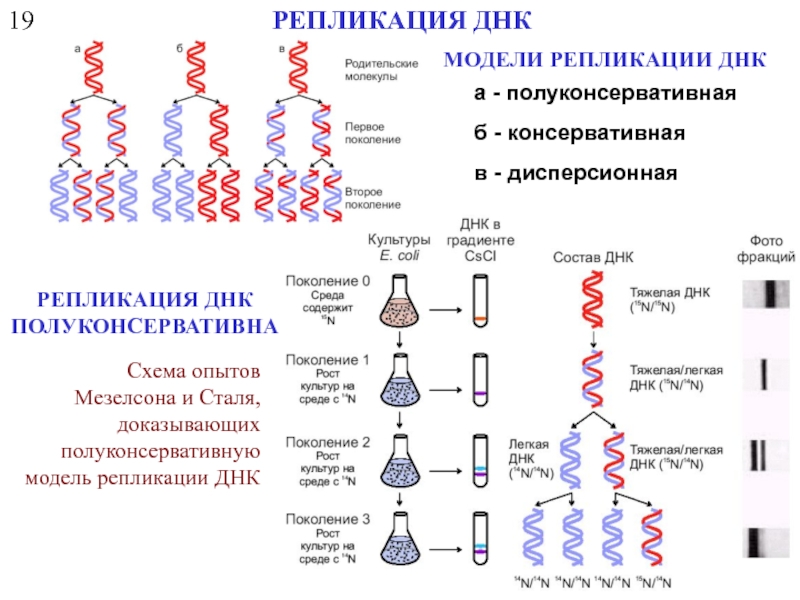
РЕПЛИКАЦИЯ ДНК
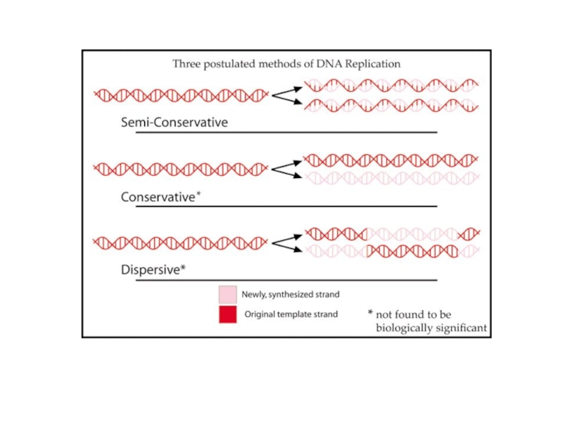
а - полуконсервативная
б - консервативная
в - дисперсионная
МОДЕЛИ РЕПЛИКАЦИИ ДНК
РЕПЛИКАЦИЯ ДНК
ПОЛУКОНСЕРВАТИВНА
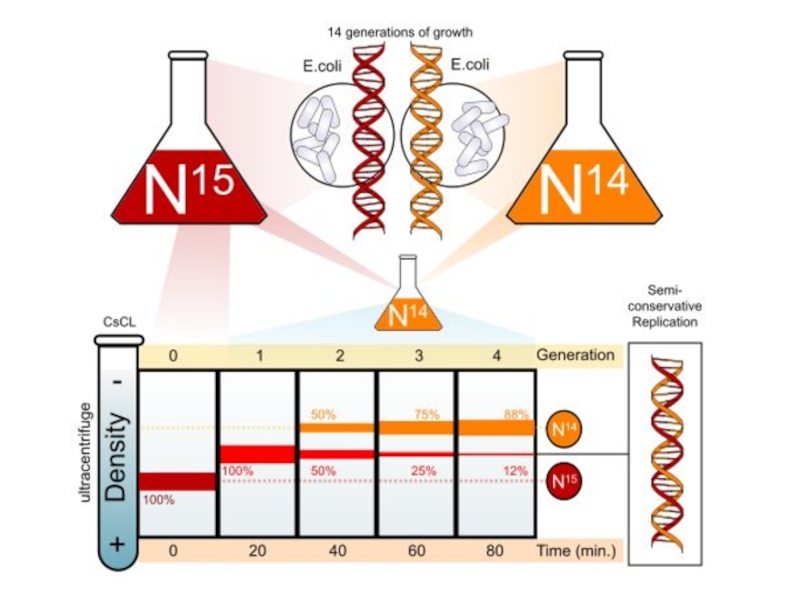
Схема опытов Мезелсона и Сталя, доказывающих полуконсервативную модель репликации ДНК
Слайд 722
ДНК - ПОЛИМЕРАЗЫ
3' -5' - экзонуклеазная активность («редактирующая»)
5'-3' - экзонуклеазная активность
Слайд 823
ДНК - ПОЛИМЕРАЗЫ
Синтез Экзонуклеаза
Рol I E. coli + + + A
Pol II E. coli + + - B
Pol III E. coli + + - C
Фрагмент Кленова + + -
DNA polymerase protein structures. (a) Bacteriophage T7 DNA polymerase (type A; PDB code: 1t7p). Blue, catalytic domain; green, 3'-to-5' exonuclease domain. (b)Thermococcus gorgorianus type B DNA polymerase (PDB code: 1tgo). Blue, catalytic domain; green, 3'-to-5' exonuclease domain; orange, amino-terminal domain; yellow, linker region.
Слайд 1226
РАСКРУЧИВАНИЕ ДНК СПИРАЛИ ПРИ РЕПЛИКАЦИИ
геликаза PcrA - комплекс с субстратом
ДНК-геликаза
Белки, связывающиеся
Матрица для синтеза отстающей цепи
Матрица для синтеза лидирующей цепи
Процесс раскручивания двойной спирали в репликативной вилке порождает механические и топологические проблемы, которые могут быть сняты вращением родительской спирали вокруг собственной оси
ДНК-полимераза
Здесь должно происходить быстрое вращение спирали ДНК
Слайд 1327
ДНК-ТОПОИЗОМЕРАЗЫ
Топоизомераза I
Топоизомераза II
Размыкание
кольцевых
дуплексов
Гираза
Топоизомеразами типа I, уменьшают число сверхвитков в ДНК на
Слайд 1528
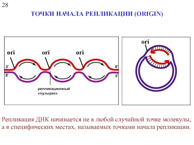
ТОЧКИ НАЧАЛА РЕПЛИКАЦИИ (ORIGIN)
репликационный
«пузырек»
ori
ori
Репликация ДНК начинается не в любой случайной точке
Слайд 16A.C. Leonard and M. Mechali DNA Replication Origins. // Cold Spring
Revised versions of the replicon model for all domains of life
For cells of each domain type, trans-acting initiators recognize replication origins to assemble prereplicative complexes required to unwind the DNA and load DNA helicase. Eukaryotic initiators are preassembled into hexameric origin recognition complexes (ORCs) before interacting with DNA. In prokaryotes, single initiators (archaeal Orc1/Cdc6 or bacterial DnaA) bind to recognition sites and assemble into complexes on DNA. In all cases, the DNA helicases (MCMs or DnaB) are recruited to the origin and loaded onto single DNA strands. In bacteria, DNA-bending proteins, such as Fis or IHF, may modulate the assembly of pre-RC by bending the origin DNA. Two activities of DnaA are described in the figure. The larger version binds to recognition sites, and the smaller version represents DnaA required to assist DnaC in loading DnaB helicase on single-stranded DNA.
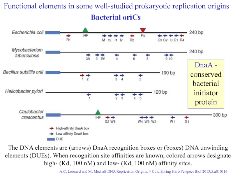
Слайд 17A.C. Leonard and M. Mechali DNA Replication Origins. // Cold Spring
The DNA elements are (arrows) DnaA recognition boxes or (boxes) DNA unwinding elements (DUEs). When recognition site affinities are known, colored arrows designate high- (Kd, 100 nM) and low- (Kd, 100 nM) affinity sites.
Functional elements in some well-studied prokaryotic replication origins
Bacterial oriCs
DnaA - conserved bacterial initiator protein
Слайд 1829
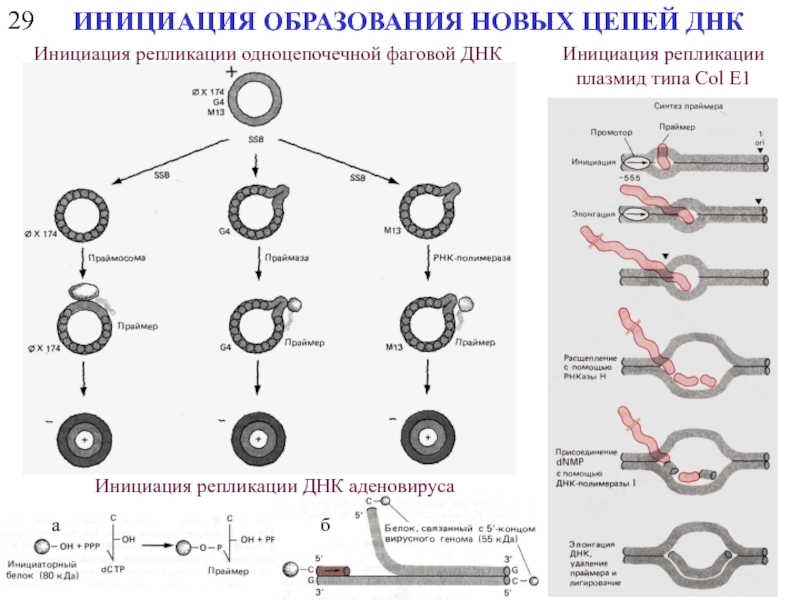
ИНИЦИАЦИЯ ОБРАЗОВАНИЯ НОВЫХ ЦЕПЕЙ ДНК
Инициация репликации одноцепочечной фаговой ДНК
Инициация репликации плазмид
Инициация репликации ДНК аденовируса
а
б
Слайд 1930
Инициация синтеза отстающей цепи
МОДЕЛЬ ПОЛУКОНСЕРВАТИВНОЙ ПРЕРЫВИСТОЙ РЕПЛИКАЦИИ ДНК В РЕПЛИКАТИВНОЙ ВИЛКЕ
Слайд 2030.1
РЕПЛИКАТИВНАЯ ВИЛКА БАКТЕРИОФАГА Т7
a, The bacteriophage T7 replisome consists of the
b, Gp4 consists of a primase and a helicase domain, connected by a linker region. The primase domain consists of two subdomains: a zinc-binding domain (ZBD) and the RNA polymerase domain (RPD).
T7 gene 2.5 protein (gp2.5), the ssDNA-binding protein, coats the transiently exposed ssDNA in the replication loop.
NATURE
Vol 439
2 Feb 2006
Слайд 2333
ТЕРМИНАЦИЯ РЕПЛИКАЦИИ
Репликация замкнутой кольцевой дуплексной ДНК и расхождение цепей
Завершение репликации линейных
Слайд 2434
НЕДОРЕПЛИКАЦИЯ КОНЦЕВОЙ ЧАСТИ МОЛЕКУЛЫ ДНК
Барьер (лимит) Хейфлика – ограничение на число
Слайд 2636
СХЕМА РЕПЛИКАЦИИ КОНЦЕВОГО УЧАСТКА ОТСТАЮЩЕЙ ЦЕПИ ДНК ПРИ НАЛИЧИИ ТЕЛОМЕРНОГО ПОВТОРА
новая цепь лидирующая
новая цепь отстающая
Слайд 2737
УДЛИНЕНИЕ ТЕЛОМЕРНОГО ПОВТОРА
ПРИ ПОМОЩИ ФЕРМЕНТА ТЕЛОМЕРАЗЫ
ТЕЛОМЕРАЗА (ДНК-нуклеотидилэкзотрансфераза, теломерная терминальная трансфераза) –
Слайд 3238.2
РЕПЛИКАЦИЯ РНК
Иерархическая классификация вирусов
Эта классификация по стратегии репликации вирусов, которая зависит
Классификация
Балтимора
Эта классификация учитывает такие свойства вирусов как:
морфология (размер, форма, наличие оболочки),
физикохимические свойства (масса, плавучая плотность, pH-, температурная-, ионная стабильность),
структура генома (РНК, ДНК, сегментированность, рестрикционная карта, наличие модификаций и т.д.),
свойства макромолекул (набор белков и их функции),
антигенные свойства,
биологические свойства (диапазон хозяев и др).
Слайд 3438.4
Genome is (+)sense but unique among viruses in that it is
This group of viruses also relies on reverse transcription, but unlike the Retroviruses, this occurs inside the virus particle on maturation. On infection of a new cell, the first event to occur is repair of the gapped genome, followed by transcription. (Hepadnaviruses)
Классификация Балтимора
Слайд 35Заражение ретровирусом клеток млекопитающих и интеграция вируса в хозяйскую хромосому
38.9
РЕПЛИКАЦИЯ РНК
Обратную транскрипцию осуществляет РНК-зависимая ДНК-полимераза (обратная транскриптаза, ревертаза)
Слайд 3639
РЕПЛИКАЦИЯ РНК С ОБРАЗОВАНИЕМ ДНК
Репликация геномов ретровирусов
Двухцепочечная ДНК, образующаяся при обратной
Схема механизма обратной транскрипции ретровирусной РНК
R
U3
Pu
U5
R
P
Слайд 3940
РЕПЛИКАЦИЯ РНК С ОБРАЗОВАНИЕМ РНК
Репликация геномов РНК-содержащих вирусов
Одновременный синтез нескольких (+)-цепей
Репликация (+)-цепи РНК-геномов
(вирусы RI7, MS2, Синдбис, полиовирусы)
Репликация (-)-цепи РНК-геномов
(вирус везикулярного стоматита, гриппа)
