- Главная
- Разное
- Дизайн
- Бизнес и предпринимательство
- Аналитика
- Образование
- Развлечения
- Красота и здоровье
- Финансы
- Государство
- Путешествия
- Спорт
- Недвижимость
- Армия
- Графика
- Культурология
- Еда и кулинария
- Лингвистика
- Английский язык
- Астрономия
- Алгебра
- Биология
- География
- Детские презентации
- Информатика
- История
- Литература
- Маркетинг
- Математика
- Медицина
- Менеджмент
- Музыка
- МХК
- Немецкий язык
- ОБЖ
- Обществознание
- Окружающий мир
- Педагогика
- Русский язык
- Технология
- Физика
- Философия
- Химия
- Шаблоны, картинки для презентаций
- Экология
- Экономика
- Юриспруденция
Esophagus. Esophageal Structure презентация
Содержание
- 1. Esophagus. Esophageal Structure
- 2. Esophagus Esophageal anatomy and physiology Esophageal symptoms Diagnostic procedures GERD Dysphagia
- 3. Esophageal Structure
- 4. Esophagus Endoscopic View GEJ Columnar epithelium Squamous epithelium
- 5. Physiology Upper esophageal sphincter Lower esophageal
- 7. Physiology- Deglutitive Inhibition The swallow-evoked peristaltic
- 9. Physiology Primary peristalsis esophageal peristaltic contraction
- 10. Transient Lower Esophageal Sphincter Relaxations LES relaxation
- 12. Physiology The esophagus is innervated by both
- 13. Symptoms Heartburn (pyrosis)- the most common esophageal
- 14. Symptoms Regurgitation - effortless return of food
- 15. Symptoms Chest pain - common esophageal symptom
- 16. Symptoms Dysphagia - feeling of food "sticking"
- 17. Symptoms Odynophagia - pain caused by swallowing
- 18. Diagnostic Studies Endoscopy Radiography Endoscopic Ultrasound Esophageal Manometry Video swallow study Reflux Testing
- 19. ENDOSCOPY Endoscopy
- 20. Radiography- Barium Swallow Normal barium swallow Esophageal spasm Cork screw esophagus Hiatal hernia
- 21. Esophageal manometry
- 22. Motility Testng High Resolution Esophageal Manometry
- 23. 24-hour transnasally positioned wire electrode with the
- 24. pH study: intranasal wire electrode with the sensor in the distal esophagus.
- 25. Wireless Bravo pH Capsule for acid reflux detection
- 28. Acid and non-acid acid reflux detection Gold
- 29. Gastroesophageal Reflux Disease (GERD)
- 30. GERD- definitions Physiologic reflux episodes typically occur
- 31. Pathophysiology of GERD Castell Do
- 32. Pathophysiology of GERD Hiatal hernia
- 33. GERD Epidemiology Prevalence : 10 -20
- 34. GERD Symptoms Common: Heartburn and regurgitation
- 35. GERD- Ds Ds is usually based
- 37. GERD Differential Diagnosis Infectious, pill, or
- 38. GERD Treatment Lifestyle modifications Avoidance
- 39. GERD Treatment Inhibitors of gastric acid secretion
- 40. bv
- 42. GERD Treatment- surgical Nissen fundoplication the proximal
- 43. GERD Complications Chronic esophagitis (bleeding and
- 44. Barrett’s esophagus Endoscopy: Tongues of reddish mucosa extending proximally from GE junction
- 45. Barrett’s esophagus Histology: columnar metaplasia with Goblet cells
- 46. GERD Complications- Barrett’s Obese white males in
- 47. Dysphagia
- 48. Approach to Dysphagia Dysphagia Oropharyngeal Esophageal
- 49. Oropharyngeal Dysphagia Etiology Neurogenic -
- 50. Zenker's diverticulum Elderly Prevalence
- 51. Esophageal Dysphagia Solid food dysphagia appears when the lumen is
- 52. Esophageal Dysphagia Structural causes Schatzki's rings Eosinophilic
- 53. Esophageal Dysphagia Upper endoscopy Dysphagia is
- 54. Esophageal Dysphagia- Schatzki's ring Distal esophagus
- 55. Achalasia Incidence 1-3:100,000 Age - 25 to
- 56. Achalasia Etiology: Loss of ganglion
- 57. Achalasia Differential diagnosis
- 58. Achalasia Diagnosis Endoscopy -
- 59. Achalasia Conventional manometry - Impaired LES relaxation - Absent peristalsis of esophageal body
- 60. Achalasia Normal High Resolution Manometry
- 61. Three Subtypes of Achalasia on High Resolution
- 62. Three Subtypes of Achalasia on High Resolution
- 63. Achalasia Barium swallow x-ray dilated esophagus with
- 64. Achalasia Treatment Therapy is directed at reducing
- 65. Pneumatic balloon dilation of LES
- 67. Achalasia- Complications Squamous cell carcinoma risk increased
- 68. Diffuse Esophageal Spasm (DES) Episodes of
- 69. Diffuse Esophageal Spasm Corkscrew esophagus
- 70. Dysphagia Diffuse Esophageal Spasm (DES) Diffferntial
- 71. Scleroderma - Dilated esophagus - Ineffective peristalsis - Low LES pressure - Severe GERD
- 72. Eosinophilic Esophagitis Prevalence 1:1000 with a
- 73. Eosinophilic Esophagitis Endoscopy: multiple esophageal rings, linear
- 74. Eosinophilic Esophagitis Complications: food impaction and esophageal
- 75. Infectious Esophagitis Common infections in Immunocompromized
- 76. Infectious Esophagitis Candida Esophagitis C.
- 77. Infectious Esophagitis Herpetic Esophagitis Herpes simplex
- 78. Other Types of Esopahgitis Radiation esopahgitis
- 79. Esophageal Cancer Squamous cell carcinoma Adenocarcinoma
- 80. Esophageal Cancer Squamous cell carcinoma risk factors:
- 82. Esophageal Cancer incidence of squamous cell cancer
- 83. Esophageal Cancer Adenocarcinomas arise in the distal
- 84. Esophageal Cancer Location 10% upper third of
- 85. Clinical features Progressive dysphagia (solids) Weight loss
- 86. Esophageal carcinoma Endoscopic and cytologic screening for
- 87. Thank you! !תודה רבה Спасибо за внимание!

Слайд 2Esophagus
Esophageal anatomy and physiology
Esophageal symptoms
Diagnostic procedures
GERD
Dysphagia


Слайд 5Physiology
Upper esophageal sphincter
Lower esophageal sphincter
Diaphragmatic sphincter
Esophageal body
Function
Esophageal bolus transport

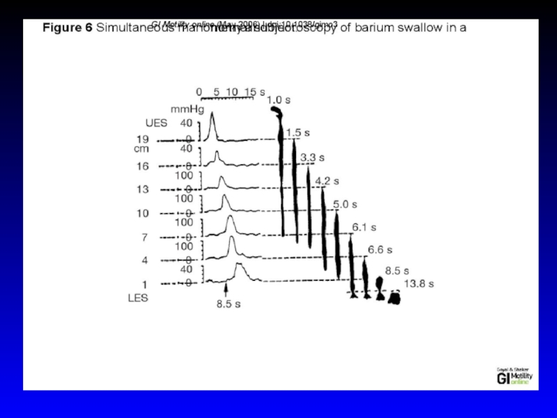
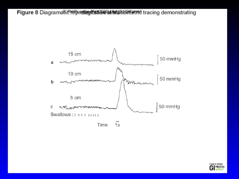
Слайд 7Physiology- Deglutitive Inhibition
The swallow-evoked peristaltic contraction consist of wave of
The wave of inhibition that precedes peristaltic contraction is deglutitive inhibition
Esophageal contraction in response to a single swallow lasts 8 to 10 seconds, and this will obstruct the bolus of a second swallow taken less than 8 second afterward.
The phenomenon of deglutitive inhibition is essential for drinking of fluids (rate of swallows faster than one swallow every 10 seconds)
During the usual drinking of water, swallows can be every 1 to 2 seconds, possible by the phenomenon of deglutitive inhibition in which a swallow abruptly inhibits any ongoing contraction in the esophagus.

Слайд 9Physiology
Primary peristalsis
esophageal peristaltic contraction wave associated with swallowing
Secondary peristalsis
It
Not associated with swallowing and does not involve full swallowing reflex
Residual food in the esophagus can be cleared by what is called secondary peristalsis

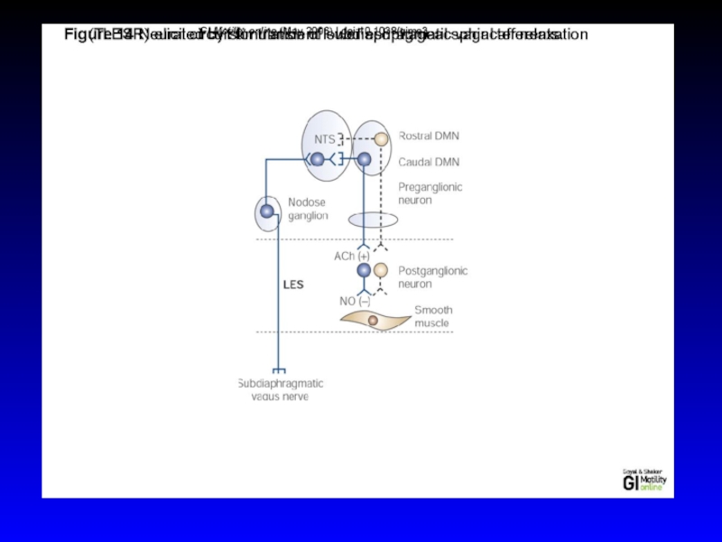
Слайд 10Transient Lower Esophageal Sphincter Relaxations
LES relaxation during belching, retching, vomiting, and
TLESR are not associated with swallowing
TLESR are increased after gastric distention or in the presence of a nasogastric tube.
Vagal afferents in the stomach cause reflex LES relaxation via a vasovagal pathway that involves inhibitory vagal pathway neurons in the caudal part of the DMN and nNOS-containing neurons in the LES
GERD and TELSR:
Most esophageal reflux episodes occurring during TLESR
TLESR are increased in patients with reflux esophagitis
TELSR associated with reflux of gas, and belch
Not all TLESRs were associated with reflux events

Слайд 12Physiology
The esophagus is innervated by both parasympathetic and sympathetic nerves
The

Слайд 13Symptoms
Heartburn (pyrosis)- the most common esophageal symptom
Discomfort or burning sensation behind
Appears after eating, during exercise, and while lying recumbent
Relieved with drinking water or antacid
- the most common esophageal symptomDiscomfort or burning sensation behind the sternum that arises")
Слайд 14Symptoms
Regurgitation - effortless return of food or fluid into the pharynx
Fluid - a sour or burning in the throat or mouth, may also contain undigested food particles
Bending, belching, or maneuvers increasing intraabdominal pressure can provoke regurgitation (not vomiting or rumination)

Слайд 15Symptoms
Chest pain - common esophageal symptom with characteristics similar to cardiac
pressure type sensation in the mid chest, radiating to the mid back, arms, or jaws
GE reflux is the most common cause of esophageal chest pain

Слайд 16Symptoms
Dysphagia - feeling of food "sticking" or lodging in the chest
Solid
Episodic /constant dysphagia
Progressive /static dysphagia
Oropharyngeal /esophageal
A patient's localization of food hang-up in the esophagus is very imprecise!
Oropharyngeal dysphagia is often associated with aspiration, nasopharyngeal regurgitation, cough, drooling, or history of CVA

Слайд 17Symptoms
Odynophagia - pain caused by swallowing
common with pill or infectious esophagitis,
Globus sensation - perception of a lump or fullness in the throat that is felt irrespective of swallowing
anxiety, GERD
Water brash – unpleasant sensation of the mouth rapidly filling with salty thin fluid
excessive salivation resulting from a vagal reflex triggered by acidification of the esophageal mucosa

Слайд 18Diagnostic Studies
Endoscopy
Radiography
Endoscopic Ultrasound
Esophageal Manometry
Video swallow study
Reflux Testing


Слайд 20Radiography- Barium Swallow
Normal barium swallow
Esophageal spasm
Cork screw esophagus
Hiatal hernia


Слайд 2324-hour transnasally positioned wire electrode with the tip stationed in the
48-hour esophageal pH recording using a wireless pH-sensitive transmitter (capsule)
Intraluminal impedance monitoring to detect reflux events irrespective of their pH


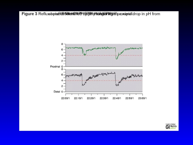
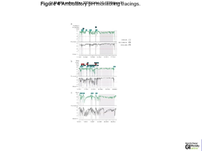
Слайд 28Acid and non-acid acid reflux detection
Gold standard of reflux testing
PH-MII detects
The method is based on measuring the resistance to alternating current (i.e., impedance) of the content of the esophageal lumen
Pairs of electrodes, separated by an isolator (i.e., catheter), are placed inside the esophagus
Reflux Monitoring: pH- MII
Multichannel Intraluminal Impedance
Esophageal Reflux Monitoring

")
Слайд 30GERD- definitions
Physiologic reflux episodes typically occur postprandially, are short-lived, asymptomatic, and
Pathologic reflux is associated with symptoms or mucosal injury, often including nocturnal episodes
Gastroesophageal reflux disease (GERD) - a condition that develops when the reflux of stomach contents causes troublesome symptoms and/or complications
Reflux esophagitis - endoscopic or histopathologic evidence of esophageal inflammation in a subset of patients with GERD
.

Слайд 31
Pathophysiology of GERD
Castell Do et al. Aliment Pharmacol Ther 2004; 20
Lower esophageal sphincter (LES)
Decreased salivation
Impaired esophageal acid clearance
Impaired tissue resistance
Decreasing resting tone of LES
Delayed gastric emptying
Transient LES relaxation
Duodenum
Hiatal hernia
:14Lower esophageal sphincter")

Слайд 33GERD
Epidemiology
Prevalence : 10 -20 % in the Western world ,
Incidence : 5 per 1000 person-years

Слайд 34GERD Symptoms
Common: Heartburn and regurgitation
Less common: dysphagia and chest pain
Extraesophageal manifestations
chronic cough
laryngitis
hoarsness
asthma
dental erosions

Слайд 35GERD- Ds
Ds is usually based on clinical symptoms
Utilization of
Upper endoscopy
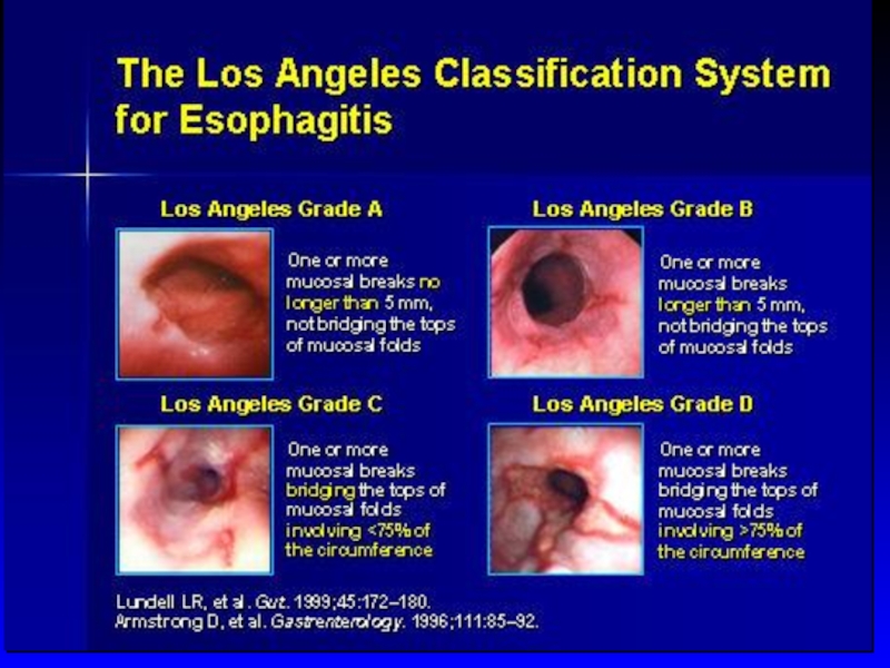
Los Angeles classification of esophagitis
pH metry
Manometry

Слайд 37GERD Differential Diagnosis
Infectious, pill, or eosinophilic esophagitis
Peptic ulcer disease
Dyspepsia
Biliary colic
Coronary artery
Esophageal motility disorders

Слайд 38GERD Treatment
Lifestyle modifications
Avoidance of
Foods that reduce LES pressure -"refluxogenic" (fatty
Acidic foods
Smoking
Carbohydrated beverages
elevated head of the bed
avoidance of eating before lying down
weight reduction

Слайд 39GERD Treatment
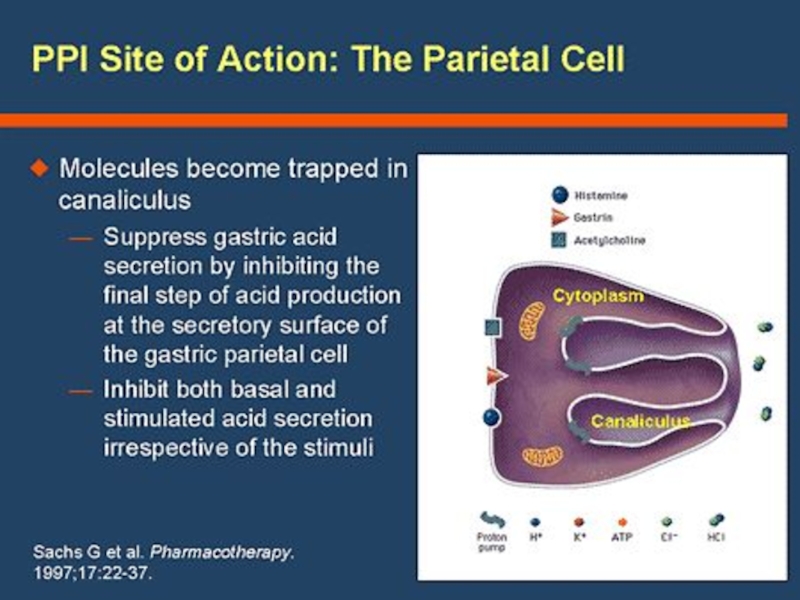
Inhibitors of gastric acid secretion
Reducing the acidity of gastric juice
Proton pump inhibitors (PPI) /omeprazole/
PPI is given 20- 30 min before meal for maximal efficacy
Histamine2 receptor antagonists (H2RAs) /famotidine/
PPIs are more efficacious than H2RAs; and both are superior to placebo
Anti- acid /Maalox- aluminium hydrocide and magnesium hydroxide, neutralizes gastric acid/. Symptomatic treatment.


Слайд 42GERD Treatment- surgical
Nissen fundoplication
the proximal stomach is wrapped around the distal
Potential side effects:
- temporary solution in majority of cases (5-10y)
- surgical morbidity and mortality
- postoperative dysphagia
- failure or breakdown requiring reoperation
- an inability to belch (increased bloating)

Слайд 43GERD Complications
Chronic esophagitis (bleeding and stricture)
increasingly rare due to potent
Esophageal adenocarcinoma
Barrett's metaplasia
 increasingly rare due to potent antisecretory medicationsEsophageal adenocarcinoma")
Слайд 44Barrett’s esophagus
Endoscopy: Tongues of reddish mucosa extending proximally from GE junction


Слайд 46GERD Complications- Barrett’s
Obese white males in 6th decade of lie are
Barrett's metaplasia can progress to adenocarcinoma through the intermediate stages of low- and high-grade dysplasia
The rate of cancer development - 0.5% per year
No evidence that aggressive antisecretory therapy or antireflux surgery causes regression of Barrett's esophagus or prevents adenocarcinoma
Management of Barrett's esophagus remains controversial
High-grade dysplasia in Barrett’s mandates further intervention
Esophagectomy
Mucosal ablation
Endoscopic Mucosal Resection


Слайд 48Approach to Dysphagia
Dysphagia
Oropharyngeal
Esophageal
Video swallow study
Type of Bolus
Abnormal
Address specific cause
Normal
other causes
(e.g.
Solids only
Solids and Liquids
Character
Character
Progressive
Intermittent
Progressive
Intermittent
No weight loss
Age > 50 or weight loss
Caustic stricture
Diverticula
Peptic stricture
Carcinoma
EoE
Esophageal ring
Achalasia
Chagas’ disease
Scleroderma
Non specific motility disorder
Sleisenger et al., 9th edition
 Solids onlySolids")
Слайд 49Oropharyngeal Dysphagia
Etiology
Neurogenic - major source of morbidity related to aspiration and
CVA
Parkinson's disease
ALS
Structural lesions
Zenker's diverticulum
cricopharyngeal bar
neoplasia
Iatrogenic causes
surgery and radiation (head and neck cancer)
Striated muscle pathology
usually involves both the oropharynx and the cervical esophagus

Слайд 50Zenker's diverticulum
Elderly
Prevalence 1:1000 - 1:10,000
Symptoms: dysphagia, regurgitation of
Pathogenesis: stenosis of the cricopharyngeus, causing diminished UES opening and increased hypopharyngeal pressure during swallowing with development of a pulsion diverticulum immediately above the cricopharyngeus
Zenker's diverticulum

Слайд 51Esophageal Dysphagia
Solid food dysphagia appears when the lumen is

Слайд 52Esophageal Dysphagia
Structural causes
Schatzki's rings
Eosinophilic esophagitis
Peptic strictures
Neoplasia
GERD without a stricture, perhaps on
Propulsive disorders
Abnormalities of peristalsis and/or deglutitive inhibition (achalasia)
Diseases affecting smooth muscle

Слайд 53Esophageal Dysphagia
Upper endoscopy
Dysphagia is an alarm symptom
Esophageal manometry
Barium swallow

Слайд 54Esophageal Dysphagia- Schatzki's ring
Distal esophagus
Mucosal ring
Intermittent dysphagia
Treatment ( if symptomatic): dilatation
: dilatation +/- acid supression")
Слайд 55Achalasia
Incidence 1-3:100,000
Age - 25 to 60 yo
Symptoms
Dysphagia: solid and liquid
Regurgitation: food, fluid, and secretions are retained in the dilated esophagus (risk for bronchitis, pneumonia, or lung abscess from chronic regurgitation and aspiration)
Chest pain: a squeezing, pressure-like retrosternal pain, sometimes radiating to the neck, arms, jaw, and back.
Weight loss

Слайд 56Achalasia
Etiology:
Loss of ganglion cells- inhibitory (nitric oxide) ganglionic neurons
Excitatory (cholinergic) ganglionic neurons are variably affected
Impaired deglutitive LES relaxation and absent peristalsis
Progressive dilatation and sigmoid deformity of the esophagus with hypertrophy of the LES
 ganglionic neurons within the esophageal myenteric")
Слайд 57Achalasia
Differential diagnosis
Diffuse esopghageal spasm (DES)
Chagas' disease (Trypanosoma cruzi)
-The
Pseudoachalasia
- Tumor infiltration - up to 5% of suspected acalasia cases (more likely with advanced age, abrupt onset of symptoms, and weight loss).
- Paraneoplastic syndrome with circulating antineuronal antibodies- rare.
Chagas' disease (Trypanosoma cruzi)-The chronic phase of the disease")
Слайд 58Achalasia Diagnosis
Endoscopy
- rarely diagnostic, to exclude pseudo-achalasia
Manometry
- most
Barium swallow x-ray

Слайд 59Achalasia
Conventional manometry
- Impaired LES relaxation
- Absent peristalsis of esophageal body


Слайд 61Three Subtypes of Achalasia on
High Resolution Manometry
Alexander J. Eckardt & Volker
Nature Reviews Gastroenterology & Hepatology 8, 311-319 (June 2011)

Слайд 62Three Subtypes of Achalasia on
High Resolution Manometry
Peter J Kahrilas, The Am

Слайд 63Achalasia
Barium swallow x-ray
dilated esophagus with poor emptying
air-fluid level
tapering at the LES
“bird’s beak”

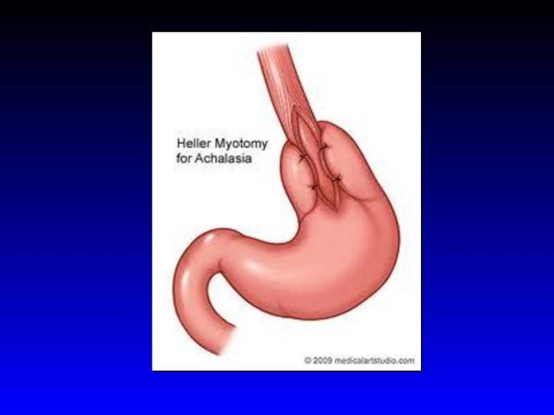
Слайд 64Achalasia Treatment
Therapy is directed at reducing LES pressure
Pharmacologicals therapies are
Botulinum toxin, injected into the LES
Pneumatic balloon dilatation
Surgical: Heller myotomy, good to excellent results are reported in 62–100% of cases


Слайд 67Achalasia- Complications
Squamous cell carcinoma risk increased 17-fold in inadequately treated achalasia
Malnutrition
There is no known way of preventing or reversing achalasia

Слайд 68Diffuse Esophageal Spasm (DES)
Episodes of dysphagia and chest pain attributable to
Diagnosis
Barium swallow: tertiary contractions or a "corkscrew esophagus" , "rosary bead esophagus," pseudodiverticula”
Manometry: simultaneous contractions in the distal esophagus, but normal deglutitive LES relaxation
 Episodes of dysphagia and chest pain attributable to abnormal esophageal contractions.Diagnosis")

Слайд 70Dysphagia
Diffuse Esophageal Spasm (DES)
Diffferntial diagnosis:
angina pectoris
peptic or infectious esophagitis
Achalasia
Treatment
- Partial
Diffferntial diagnosis:angina pectorispeptic or infectious esophagitisAchalasiaTreatment- Partial response to nitrates, calcium")

Слайд 72Eosinophilic Esophagitis
Prevalence 1:1000 with a predilection for white males, incidence is
Symptoms: dysphagia, food impaction, atypical chest pain, heartburn, particularly heartburn that is refractory to PPI therapy.
An atopic history of food allergy, asthma, eczema, or allergic rhinitis is present in the majority of patients
EoE is an allergic disorder induced by antigen sensitization in susceptible individuals.
dietary allergens
aeroallergens
The natural history of the disorder is uncertain

Слайд 73Eosinophilic Esophagitis
Endoscopy: multiple esophageal rings, linear furrows, and punctate exudates
Histology:

Слайд 74Eosinophilic Esophagitis
Complications: food impaction and esophageal perforation
Treatment:
Dietary restrictions
PPIs
Systemic or topical
Montelukast
Immunomodulators
Endoscopic dilatation of strictures (increased risk of esophageal mural disruption and perforation!)
 glucocorticoids MontelukastImmunomodulatorsEndoscopic dilatation")
Слайд 75Infectious Esophagitis
Common infections in Immunocompromized pts (organ transplantation, chronic inflammatory diseases,
Candida species
Herpesvirus
CMV
Nonimmunocompromised pts: herpes simplex and Candida albicans are the most common pathogens
Odynophagia is characteristic
Dysphagia, chest pain, and hemorrhage are also common
Candida speciesHerpesvirusCMVNonimmunocompromised")
Слайд 76Infectious Esophagitis
Candida Esophagitis
C. albicans is the most common.
Endoscopy with biopsy
Endoscopic appearance of white plaques with friability
If oral thrush is present, empirical therapy is appropriate
Oral fluconazole (200 mg on the first day, followed by 100 mg daily) for 7–14 days is the preferred treatment.
IV echinocandin or Amphotericin B in severe cases

Слайд 77Infectious Esophagitis
Herpetic Esophagitis
Herpes simplex virus type 1 or 2 may cause
Endoscopy: vesicles and small, punched-out ulcerations
Biopsies from the ulcer margins
Treatment: Acyclovir
CMV esophagitis
Only in immunocompromised patients, particularly transplant recipients
Endoscopy: serpiginous ulcers in an otherwise normal mucosa Biopsies of the ulcer bases
Treatment: Ganciclovir
CMV esophagitis

Слайд 78Other Types of Esopahgitis
Radiation esopahgitis
Pill- induced esophagitis
doxyclin, tertacyclin, minocycline, peniciliin, clindamycin,
Corrosive esophagitis


Слайд 80Esophageal Cancer
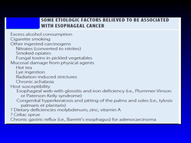
Squamous cell carcinoma risk factors:
excess alcohol consumption and/or cigarette smoking
ingestion
smoked opiates
fungal toxins in pickled vegetables
chronic mucosal injury as extremely hot tea, the ingestion of lye, radiation induced strictures, and chronic achalasia
esophageal web in association with glossitis and iron deficiency (Plummer-Vinson syn)

Слайд 82Esophageal Cancer
incidence of squamous cell cancer decreases over the past 30
incidence of adenocarcinoma has risen dramatically, particularly in white males.
Adenocarcinomas arise in the distal esophagus in the presence of chronic gastric reflux and gastric metaplasia of the epithelium (Barrett’s esophagus)
Adenocarcinomas arise within dysplastic columnar epithelium in the distal esophagus.
Adenocarcinomas are >60% of esophageal cancers.

Слайд 83Esophageal Cancer
Adenocarcinomas arise in the distal esophagus in the presence of
Adenocarcinomas are now >60% of esophageal cancers

Слайд 84Esophageal Cancer
Location
10% upper third of the sophagus
35% in the middle
55% in the lower third
Squamous cell and adenocarcinomas cannot be distinguished radiographically or endoscopically

Слайд 85Clinical features
Progressive dysphagia (solids)
Weight loss
When these symptoms develop, the disease is
The disease most commonly spreads to adjacent and supraclavicular lymph nodes, liver, lungs, pleura, and bone
Weight lossWhen these symptoms develop, the disease is usually incurableThe disease most")
Слайд 86Esophageal carcinoma
Endoscopic and cytologic screening for carcinoma in patients with Barrett’s
Prognosis is poor: < 5% 5 yrs survival
Treatment: surgery
radiotherapy
Chemotherapy
Palliation with esophageal stents or endoscopic dilatation