- Главная
- Разное
- Дизайн
- Бизнес и предпринимательство
- Аналитика
- Образование
- Развлечения
- Красота и здоровье
- Финансы
- Государство
- Путешествия
- Спорт
- Недвижимость
- Армия
- Графика
- Культурология
- Еда и кулинария
- Лингвистика
- Английский язык
- Астрономия
- Алгебра
- Биология
- География
- Детские презентации
- Информатика
- История
- Литература
- Маркетинг
- Математика
- Медицина
- Менеджмент
- Музыка
- МХК
- Немецкий язык
- ОБЖ
- Обществознание
- Окружающий мир
- Педагогика
- Русский язык
- Технология
- Физика
- Философия
- Химия
- Шаблоны, картинки для презентаций
- Экология
- Экономика
- Юриспруденция
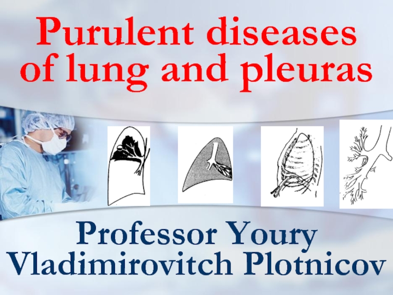
pus01en презентация
Содержание
- 1. pus01en
- 2. Classification: on I. Pathogenesis II. Character
- 3. I. Pathogenesis 1. Bronchogenic (in-cluding aspirational
- 4. II. Pathological process character (abscess and gangrene
- 5. III. Condition gravity easy middle heavy
- 6. IV. Complications 1. Not complicated
- 7. lung abscess classification Pathogenesis Localization Patient
- 8. pathogenesis postpneumonic aspirational hematogenic- embolic traumatic
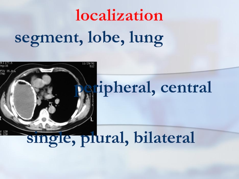
- 9. localization segment, lobe, lung peripheral, central single, plural, bilateral
- 10. Condition gravity easy middle heavy
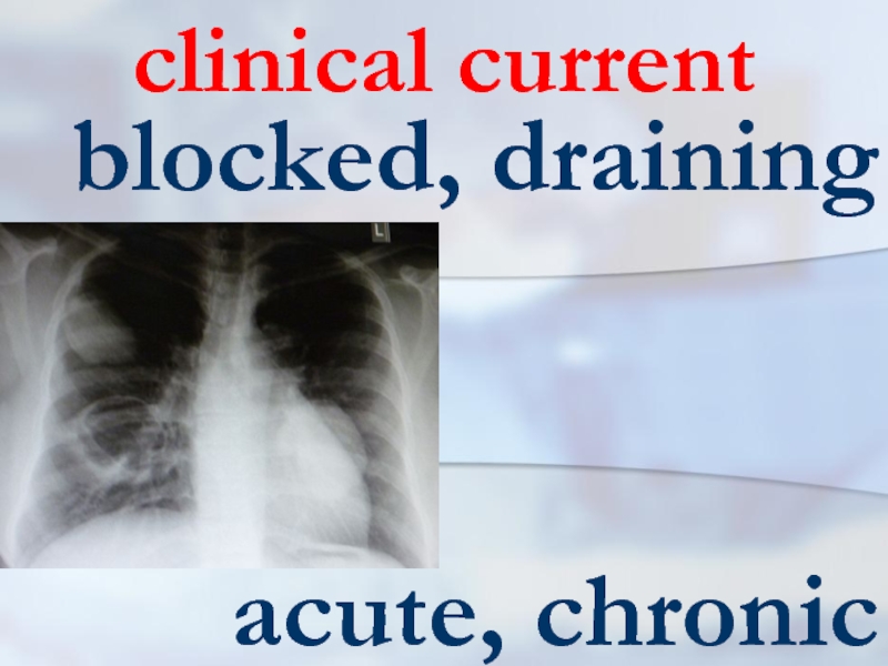
- 11. clinical current blocked, draining acute, chronic
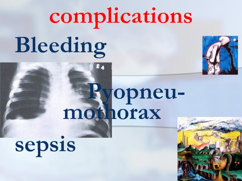
- 12. complications Bleeding Pyopneu- mothorax sepsis
- 13. definition The abscess of lung (a suppuration,
- 14. exciting cause More often activators of an
- 15. Infections ways More often the pyogenic infection
- 16. Infections ways Direct infection of pulmonary tissue
- 17. Infections ways It is necessary to note,
- 18. Infections ways More often it arises at
- 19. Infections ways Aspiration, as a rule, is
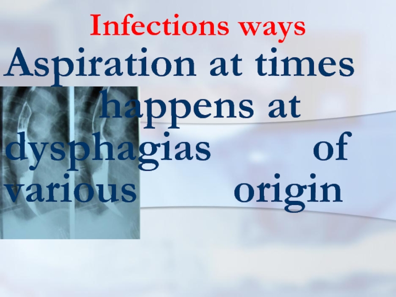
- 20. Infections ways Aspiration at times
- 21. Infections ways After aspiration deve- lops atelectasis
- 22. Infections ways Indirect confirmation of the aspi- ration
- 23. drainage function Infringements of drainage function lung
- 24. background disease Therefore, at the certain situations,
- 25. drainage function Thus, owing to acute obstruc- tion
- 26. sepsis At a sepsis are marked metas-tatic
- 27. causes Hence, the reasons of pulmonary abscesses
- 28. 60 and more 30-59 29 and younger
- 29. Clinical picture First of all it is
- 30. Adverse factors Besides adverse production factors matter
- 31. clinical picture In a clinical picture of
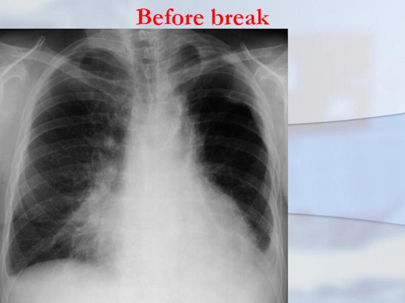
- 32. Before break For the first period is
- 33. Before break There may be pains in
- 34. Before break Infringements of the
- 35. Before break The clinic purulent-resorptive fevers is
- 36. Before break On the average, this clinic
- 37. Before break
- 38. after break In the second period when
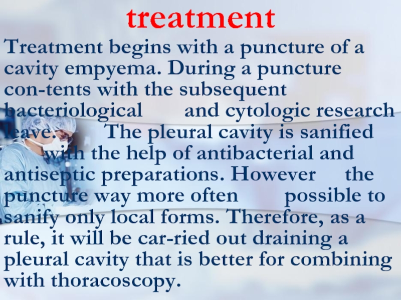
- 39. after break In other cases discharge of
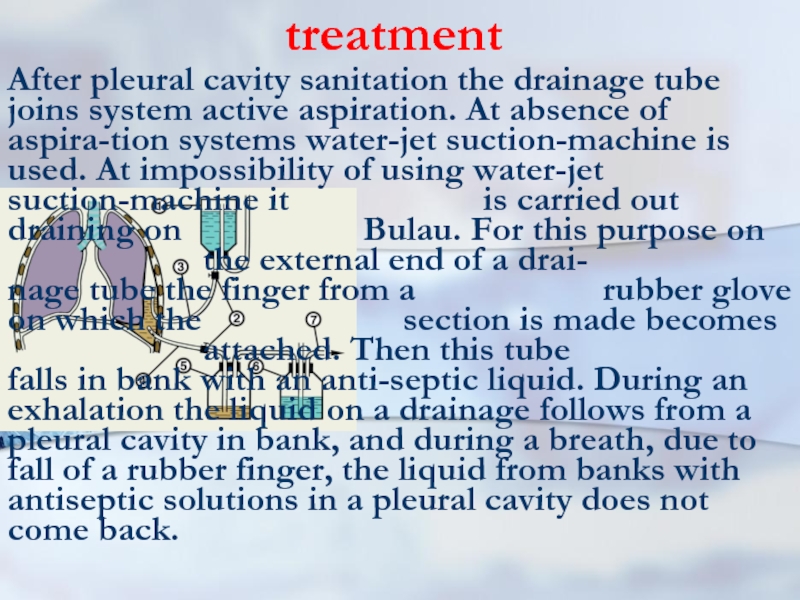
- 40. after break The x-ray picture becomes typical
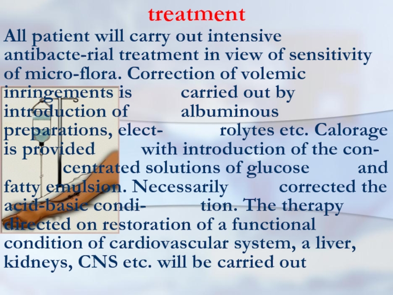
- 41. after break The cavity of an
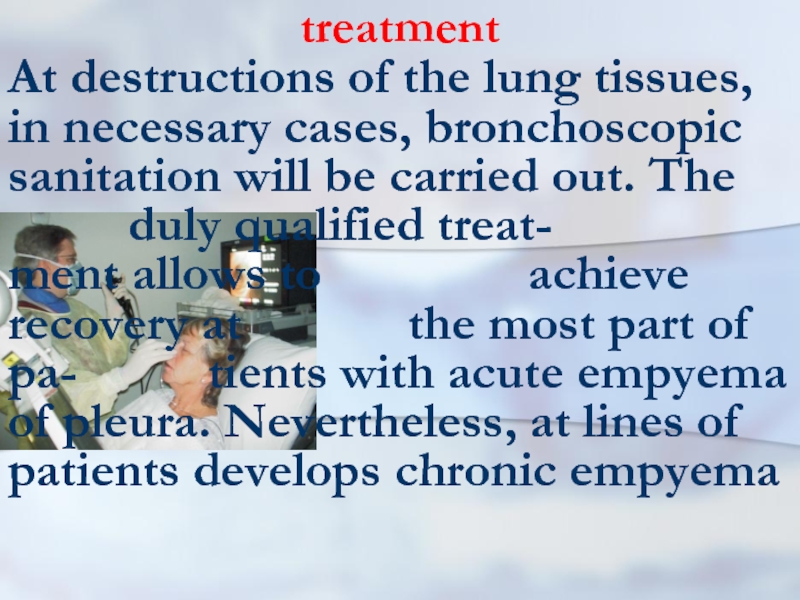
- 42. after break In some situations it is
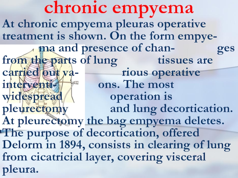
- 43. bad draining In some cases, when it
- 44. bad draining Clinically the constant disharge of
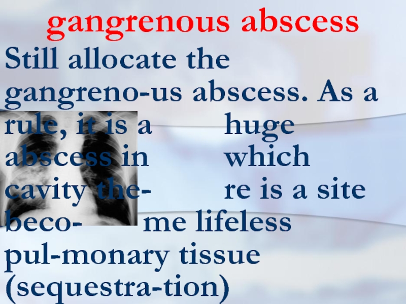
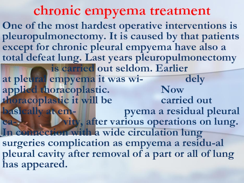
- 45. gangrenous abscess Still allocate the gangreno-us abscess.
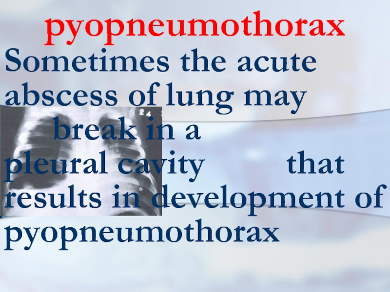
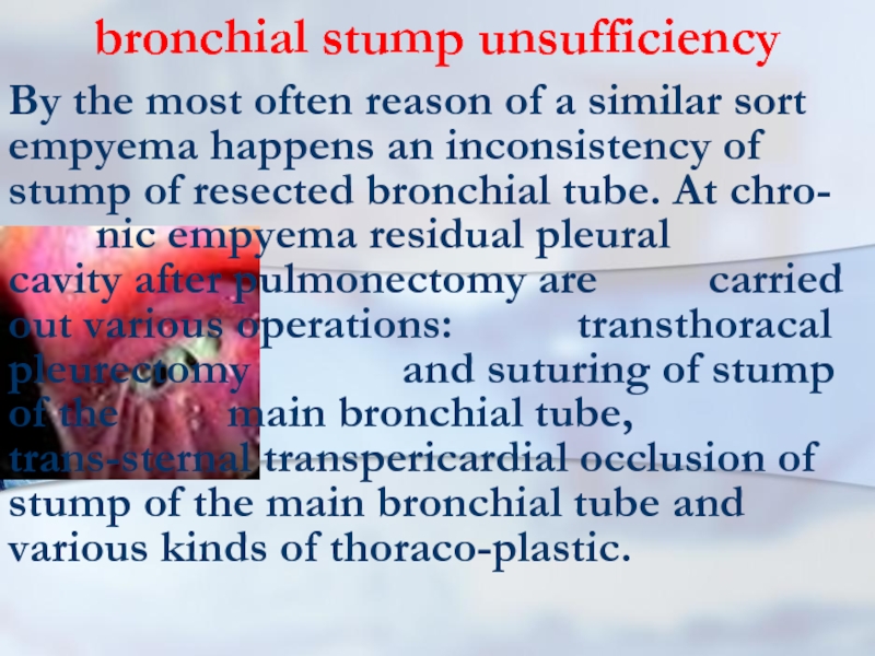
- 46. pyopneumothorax Sometimes the acute abscess of lung
- 47. Radial methods In diagnosis of pulmonary abscesses
- 48. Conservative treatment Conservative treatment of an acute
- 49. draining Sometimes bronchoscopy is car-ried out with
- 50. draining In case of insufficient sanitation with
- 51. antibacterial therapy Sometimes these preparations are entered
- 52. general improving health therapy treatment The pharmacotherapy
- 53. acute abscesses Hence, acute abscesses, as
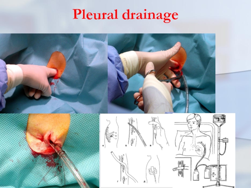
- 54. Pleural drainage
- 55. Pleural drainage rules (K.Mattox) 1. NEVER just
- 56. Pleural drainage rules (K.Mattox) 6. In teenage
- 57. Pleural drainage rules (K.Mattox) 8. ALWAYS connect
- 58. Pleural drainage rules (K.Mattox) 11. ALWAYS have
- 59. sequestration in an abscess At the sequestration
- 60. emergency operation In the extremely rare cases
- 61. chronic abscess The basic indication to operation
- 62. PLEURAL EMPYEMA Empyema - a congestion of
- 63. Pleural empyema The purulent pleurisy is the
- 64. Pleural empyema Pleural empyema in 90% of
- 65. Pleural empyema To outpulmonary diseases resulting in
- 66. Classification of the pleural empyema 1. On
- 67. Classification of the pleural empyema 1. On
- 68. Classification of the pleural empyema 4. On
- 69. Classification of the pleural empyema For the
- 70. Classification of the pleural empyema Limited empyema
- 71. Classification of the pleural empyema To I
- 72. Classification of the pleural empyema Introduction in
- 73. Classification of the pleural empyema It is
- 74. pathogeny As a rule, the purulent inflammation
- 75. Pneumonia and pleurisy Pneumonias may divide on
- 76. clinic Clinical picture. At pleural empyema occur
- 77. clinic The typical answer of an organism
- 78. clinic As it is marked above, frequently
- 79. clinic In other cases the clinical picture
- 80. clinic At the acute form it is
- 81. clinic At the soft form, as a
- 82. clinic The raised body temperature is one
- 83. clinic Frequently pains amplify at breath, there-fore
- 84. clinic Restriction of respiratory excursions of a
- 85. diagnosis One of the important methods of
- 86. diagnosis Sometimes x-ray research will be carried
- 87. treatment Treatment begins with a puncture of
- 88. treatment After pleural cavity sanitation the drainage
- 89. treatment All patient will carry out intensive
- 90. treatment At destructions of the lung tissues,
- 91. chronic empyema At chronic empyema pleuras operative
- 92. chronic empyema treatment As a
- 93. chronic empyema treatment One of the most
- 94. bronchial stump unsufficiency By the most often
- 95. chronic empyema treatment Concluding this section, it
- 96. lung gangrene Purulent-putrefactive necrosis of lobe or
- 97. lung gangrene As a rule, the gangrene
- 98. lung gangrene Etiopathogen moments of a gangrene
- 99. lung gangrene It is frequently marked aspira-tion
- 100. lung gangrene The significant role is played
- 101. Clinic As a rule, the gangrene of
- 102. Clinic Sometimes cough out small slices lifeless
- 103. Clinic Frequently current of a gangrene of
- 104. Clinic At percussion zones of dullness above
- 105. x-ray At x-ray comes to light diffuse
- 106. prognosis The prognosis at a lung gangrene
- 107. gangrene lung treatment It should be started
- 108. gangrene lung treatment The main thing
- 109. In 21 century illiterate the one
Слайд 2Classification: on
I. Pathogenesis
II. Character of pathological process
III. Condition gravity
IV.
Complications
Слайд 3I. Pathogenesis
1. Bronchogenic (in-cluding aspirational and obturatio- nal)
2.Hematogenic (including embolic)
3.
Posttraumatic
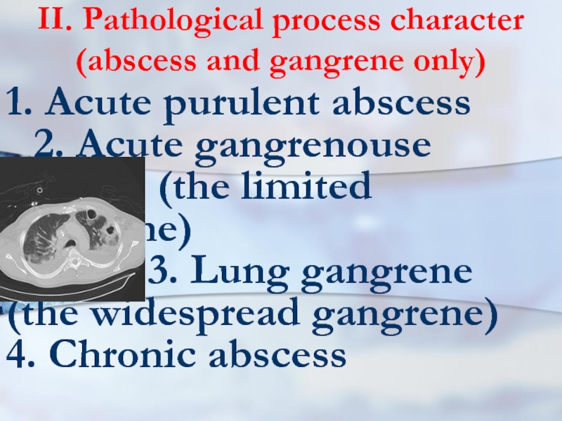
Слайд 4II. Pathological process character (abscess and gangrene only)
1. Acute purulent abscess
2.
Acute gangrenouse abscess (the limited gangrene)
3. Lung gangrene (the widespread gangrene)
4. Chronic abscess
3. Lung gangrene (the widespread gangrene)
4. Chronic abscess
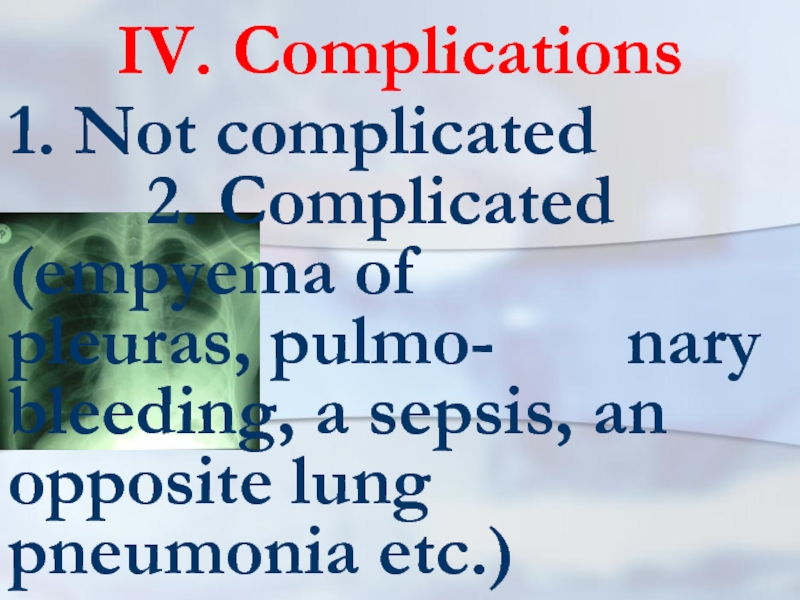
Слайд 6IV. Complications
1. Not complicated
2. Complicated (empyema of
pleuras, pulmo- nary bleeding, a sepsis, an opposite lung pneumonia etc.)
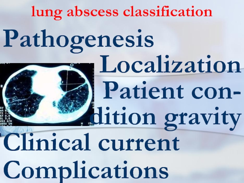
Слайд 7lung abscess classification
Pathogenesis
Localization
Patient con- dition gravity
Clinical current
Complications
Слайд 13definition
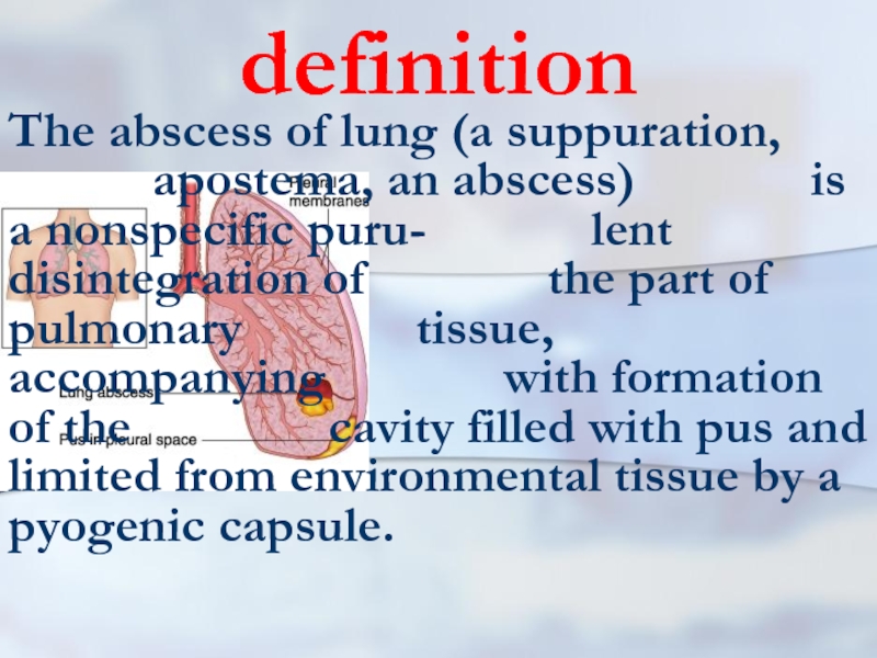
The abscess of lung (a suppuration, apostema, an abscess)
is a nonspecific puru- lent disintegration of the part of pulmonary tissue, accompanying with formation of the cavity filled with pus and limited from environmental tissue by a pyogenic capsule.
Слайд 14exciting cause
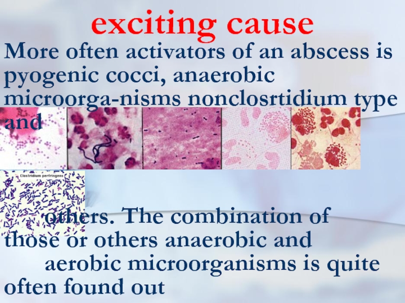
More often activators of an abscess is pyogenic cocci, anaerobic
microorga-nisms nonclosrtidium type and
others. The combination of those or others anaerobic and
aerobic microorganisms is quite often found out
others. The combination of those or others anaerobic and
aerobic microorganisms is quite often found out
Слайд 15Infections ways
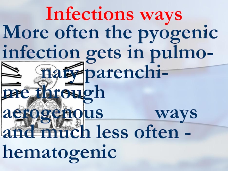
More often the pyogenic infection gets in pulmo- nary
parenchi- me through aerogenous ways and much less often - hematogenic
Слайд 16Infections ways
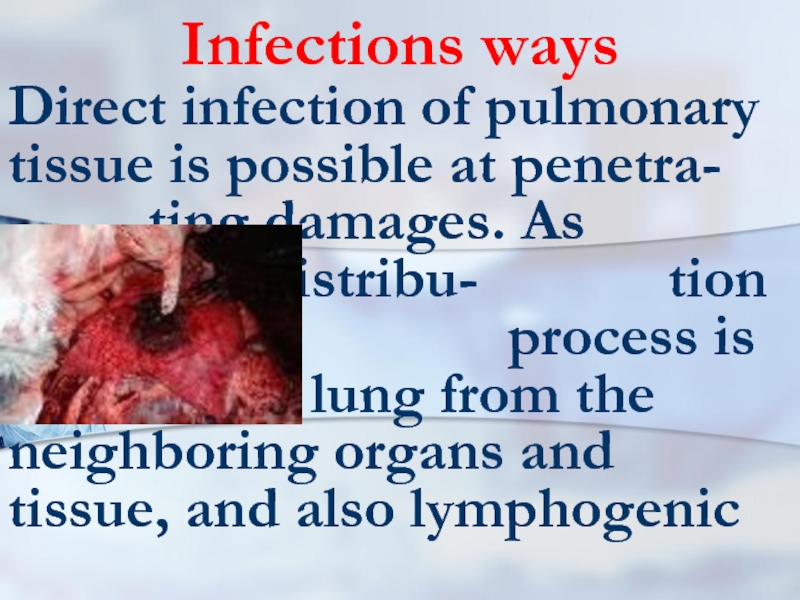
Direct infection of pulmonary tissue is possible at penetra-
ting damages. As casuality, distribu- tion of purulent process is marked in lung from the neighboring organs and tissue, and also lymphogenic
Слайд 17Infections ways
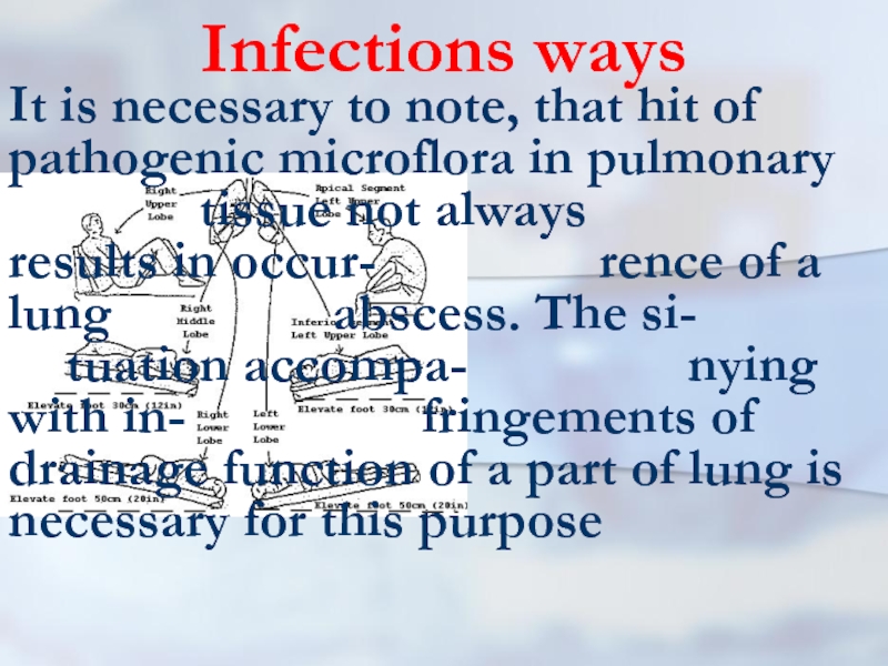
It is necessary to note, that hit of pathogenic microflora
in pulmonary tissue not always results in occur- rence of a lung abscess. The si- tuation accompa- nying with in- fringements of drainage function of a part of lung is necessary for this purpose
Слайд 18Infections ways
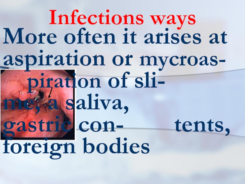
More often it arises at aspiration or mycroas- piration of sli- me,
a saliva, gastric con- tents, foreign bodies
Слайд 19Infections ways
Aspiration, as a rule, is marked at infringements of
consciousness owing to intoxi- cation, epileptic attack, head traumas, and also during a narcosis
Слайд 21Infections ways
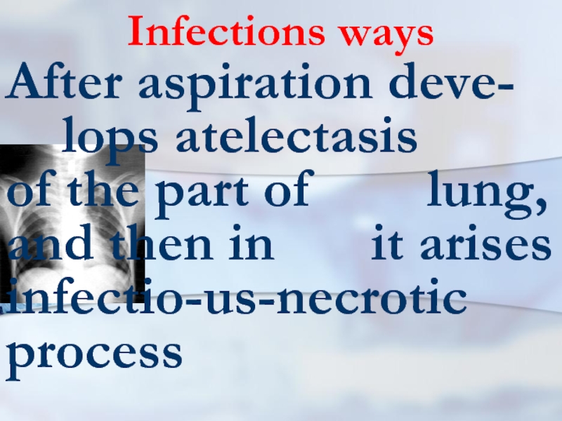
After aspiration deve- lops atelectasis of the part of
lung, and then in it arises infectio-us-necrotic process
Слайд 22Infections ways
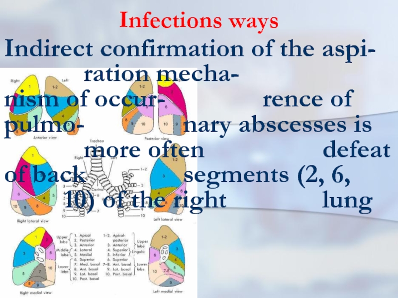
Indirect confirmation of the aspi- ration mecha- nism of occur- rence of pulmo- nary
abscesses is more often defeat of back segments (2, 6, 10) of the right lung
Слайд 23drainage function
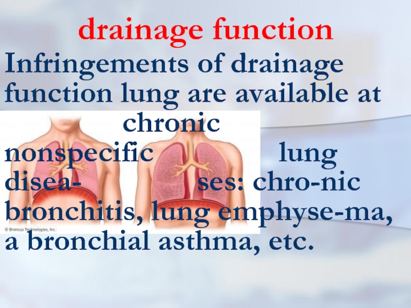
Infringements of drainage function lung are available at
chronic nonspecific lung disea- ses: chro-nic bronchitis, lung emphyse-ma, a bronchial asthma, etc.
Слайд 24background disease
Therefore, at the certain situations, some diseases
promote occur- rence of pulmo- nary abscesses. To a lung abs-cess a grippe and a diabe-tes contribute
Слайд 25drainage function
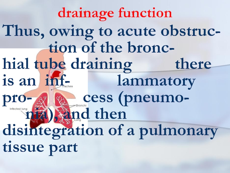
Thus, owing to acute obstruc- tion of the bronc- hial tube draining
there is an inf- lammatory pro- cess (pneumo- nia), and then disintegration of a pulmonary tissue part
Слайд 26sepsis
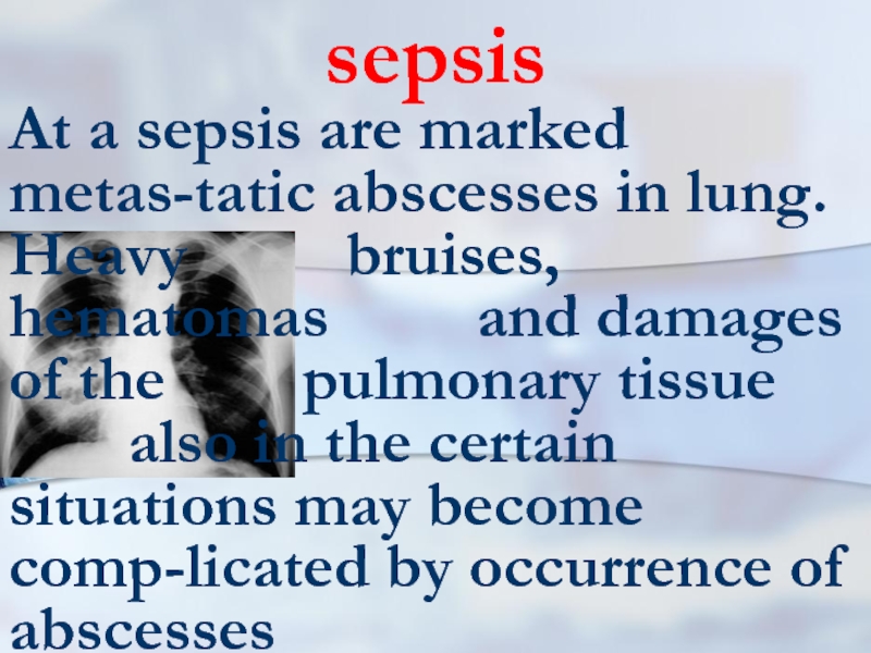
At a sepsis are marked metas-tatic abscesses in lung. Heavy
bruises, hematomas and damages of the pulmonary tissue also in the certain situations may become comp-licated by occurrence of abscesses
Слайд 27causes
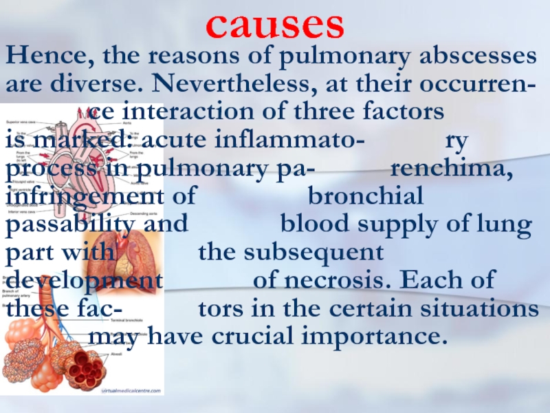
Hence, the reasons of pulmonary abscesses are diverse. Nevertheless, at their
occurren- ce interaction of three factors is marked: acute inflammato- ry process in pulmonary pa- renchima, infringement of bronchial passability and blood supply of lung part with the subsequent development of necrosis. Each of these fac- tors in the certain situations may have crucial importance.
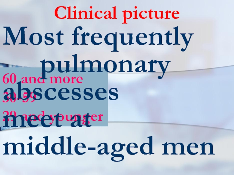
Слайд 2860 and more
30-59
29 and younger
Clinical picture
Most frequently pulmonary
abscesses meet at middle-aged men
Слайд 29Clinical picture
First of all it is caused by that among them
more of- ten there are the per- sons abusing alco- holic drinks and smokers, suffering a chronic bronchitis
Слайд 30Adverse factors
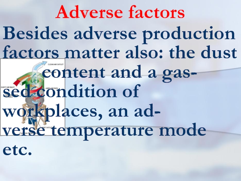
Besides adverse production factors matter also: the dust
content and a gas- sed condition of workplaces, an ad- verse temperature mode etc.
Слайд 31clinical picture
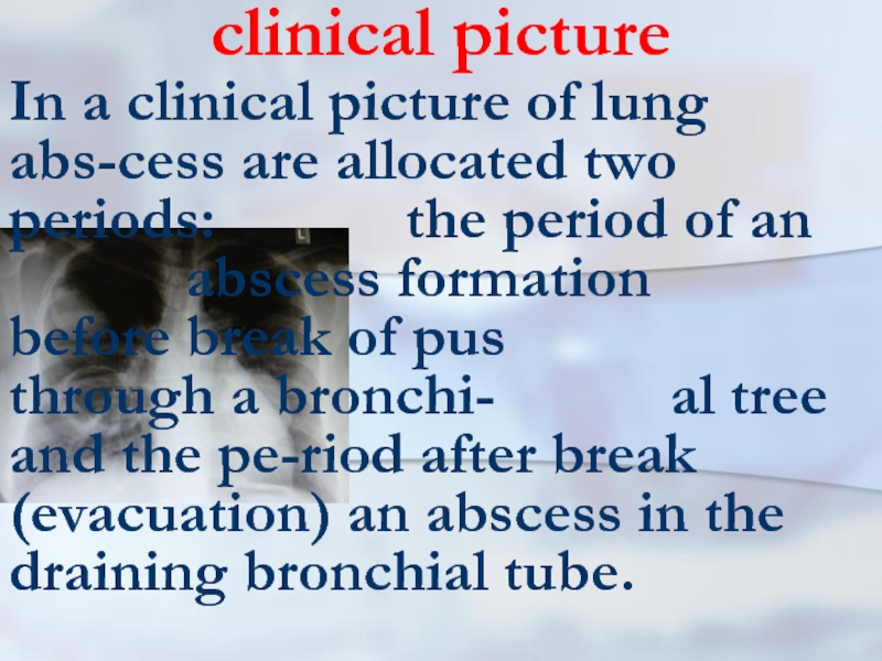
In a clinical picture of lung abs-cess are allocated two
periods: the period of an abscess formation before break of pus through a bronchi- al tree and the pe-riod after break (evacuation) an abscess in the draining bronchial tube.
Слайд 32Before break
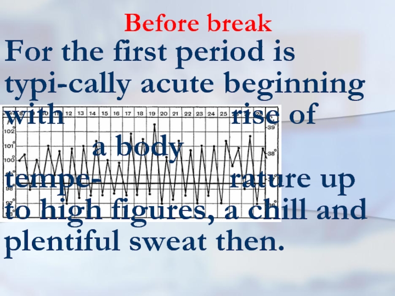
For the first period is typi-cally acute beginning with
rise of a body tempe- rature up to high figures, a chill and plentiful sweat then.
Слайд 33Before break
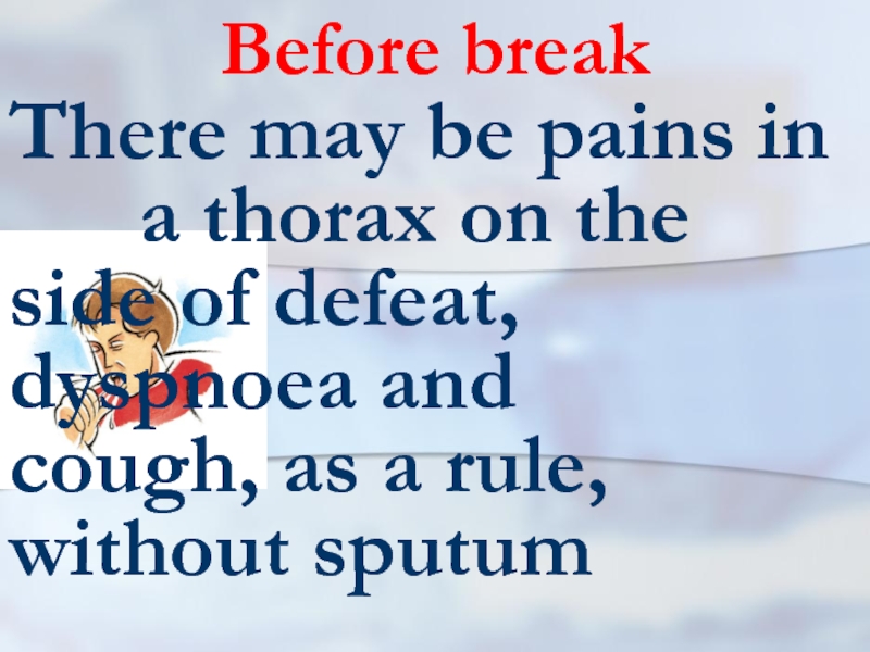
There may be pains in a thorax on the side
of defeat, dyspnoea and cough, as a rule, without sputum
Слайд 34Before break
Infringements of the common condi- tion as
a head- ache, indisposi- tions and weak-ness are marked also
Слайд 35Before break
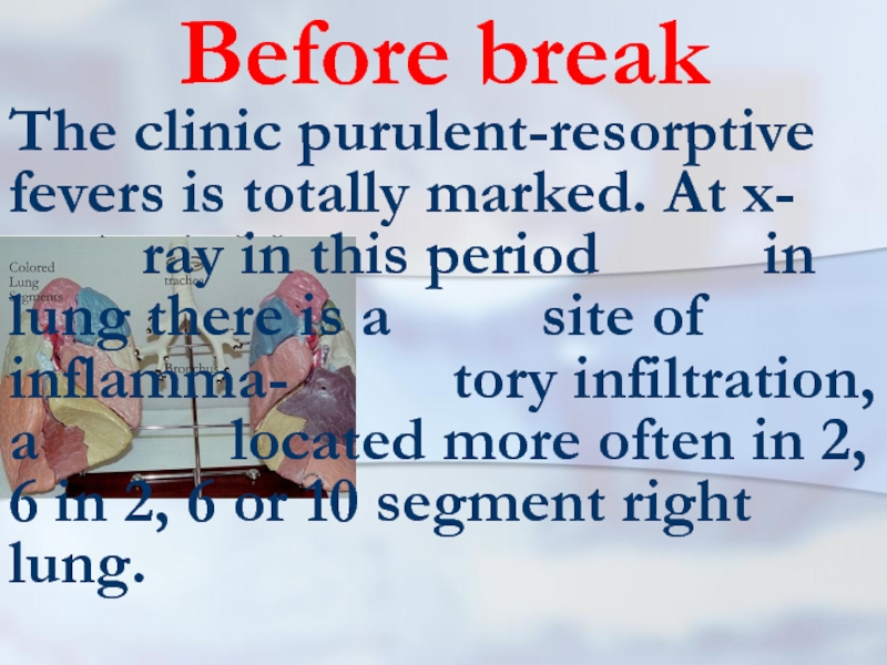
The clinic purulent-resorptive fevers is totally marked. At x- ray in
this period in lung there is a site of inflamma- tory infiltration, a located more often in 2, 6 in 2, 6 or 10 segment right lung.
Слайд 36Before break
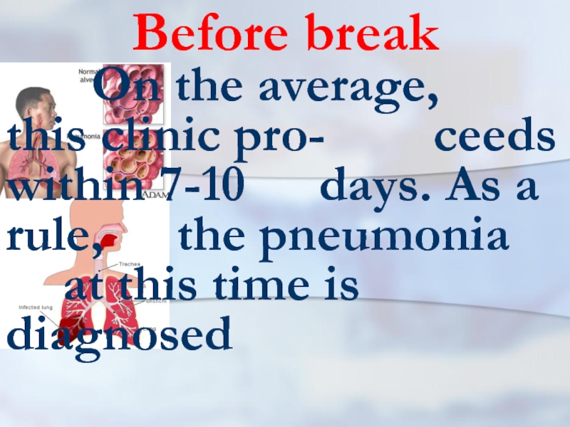
On the average, this clinic pro- ceeds within 7-10 days. As
a rule, the pneumonia at this time is diagnosed
Слайд 38after break
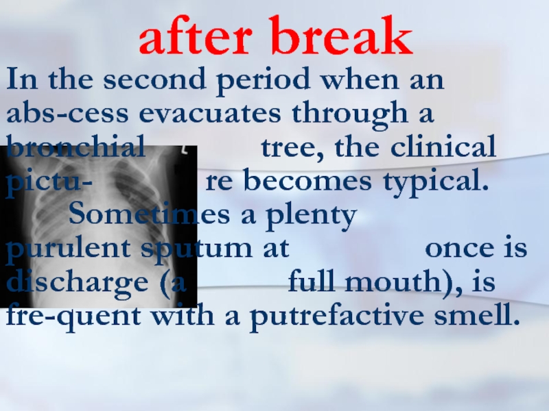
In the second period when an abs-cess evacuates through a
bronchial tree, the clinical pictu- re becomes typical. Sometimes a plenty purulent sputum at once is discharge (a full mouth), is fre-quent with a putrefactive smell.
Слайд 39after break
In other cases discharge of sputum occurs gradually. At once
after discharge of purulent sputum, the condition of the patient is conside- rably improved. The phenomena of an intoxication are acutely reduced
Слайд 40after break
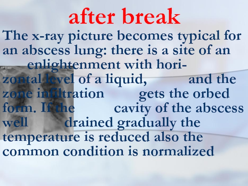
The x-ray picture becomes typical for an abscess lung: there
is a site of an enlightenment with hori- zontal level of a liquid, and the zone infiltration gets the orbed form. If the cavity of the abscess well drained gradually the temperature is reduced also the common condition is normalized
Слайд 41after break
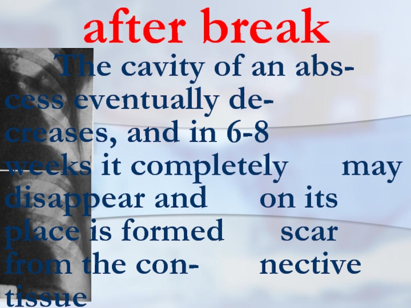
The cavity of an abs- cess eventually de- creases,
and in 6-8 weeks it completely may disappear and on its place is formed scar from the con- nective tissue
Слайд 42after break
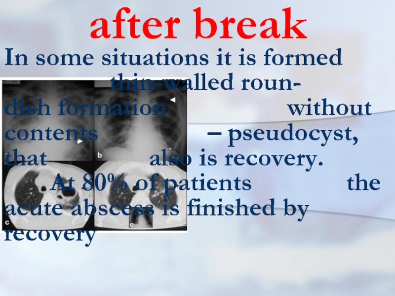
In some situations it is formed
thin-walled roun- dish formation without contents – pseudocyst, that also is recovery. At 80% of patients the acute abscess is finished by recovery
Слайд 43bad draining
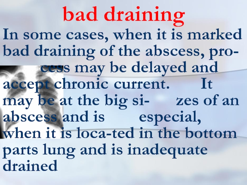
In some cases, when it is marked bad draining of
the abscess, pro- cess may be delayed and accept chronic current. It may be at the big si- zes of an abscess and is especial, when it is loca-ted in the bottom parts lung and is inadequate drained
Слайд 44bad draining
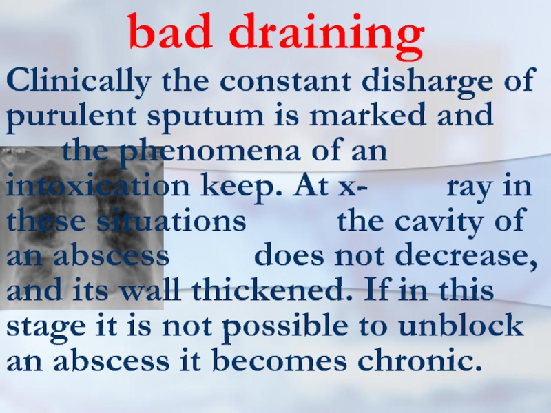
Clinically the constant disharge of purulent sputum is marked and
the phenomena of an intoxication keep. At x- ray in these situations the cavity of an abscess does not decrease, and its wall thickened. If in this stage it is not possible to unblock an abscess it becomes chronic.
Слайд 45gangrenous abscess
Still allocate the gangreno-us abscess. As a rule, it is
a huge abscess in which cavity the- re is a site beco- me lifeless pul-monary tissue (sequestra-tion)
Слайд 46pyopneumothorax
Sometimes the acute abscess of lung may break in
a pleural cavity that results in development of pyopneumothorax
Слайд 47Radial methods
In diagnosis of pulmonary abscesses it is used roentgenography
and tomography of lung. Also it is applied computer tomogra-phy and ultrasonic investigation.
Слайд 48Conservative treatment
Conservative treatment of an acute abscess of lung includes three
obligatory components: optimum draining a purulent cavity and its sanitation, antibacterial therapy, general improving health therapy treatment and the actions directed on restoration of broken homeostasis
Слайд 49draining
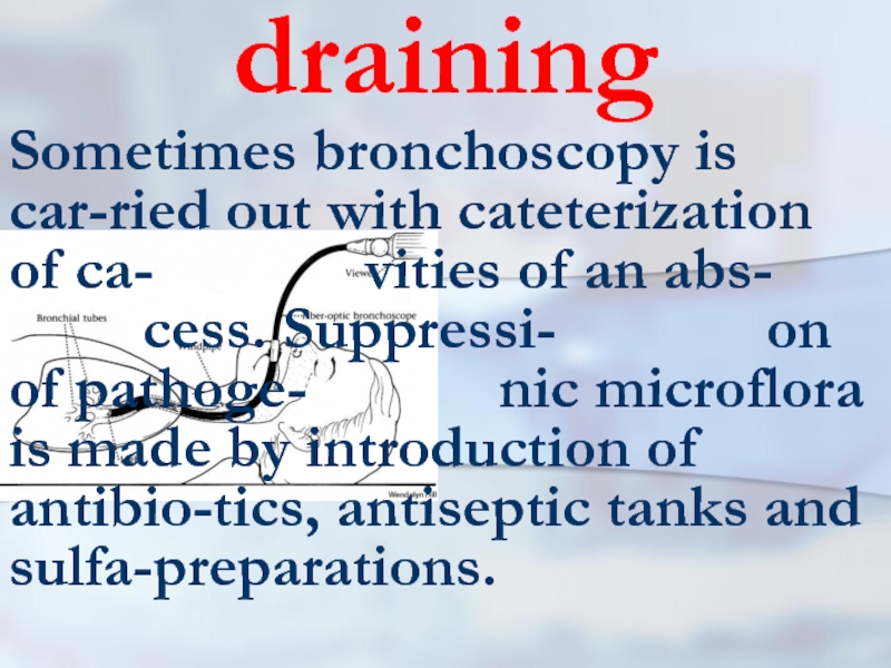
Sometimes bronchoscopy is car-ried out with cateterization of ca- vities of an abs- cess.
Suppressi- on of pathoge- nic microflora is made by introduction of antibio-tics, antiseptic tanks and sulfa-preparations.
Слайд 50draining
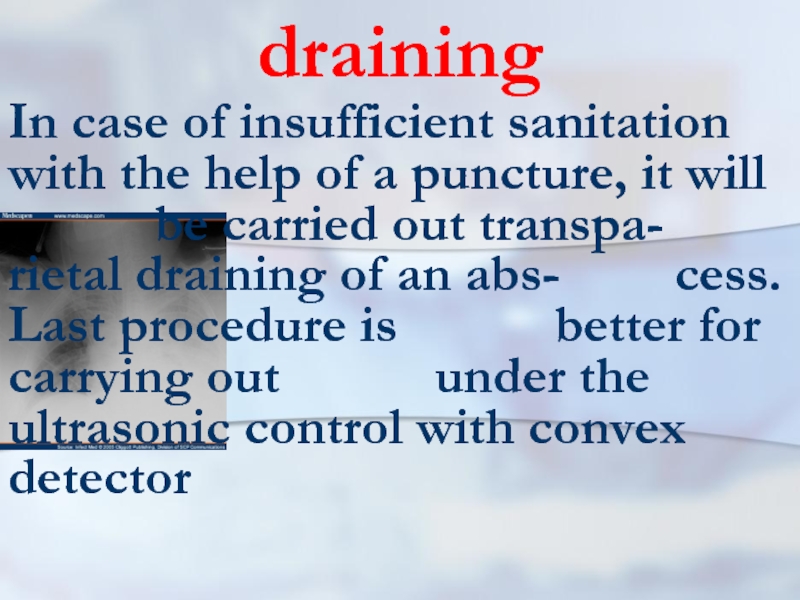
In case of insufficient sanitation with the help of a puncture,
it will be carried out transpa- rietal draining of an abs- cess. Last procedure is better for carrying out under the ultrasonic control with convex detector
Слайд 51antibacterial therapy
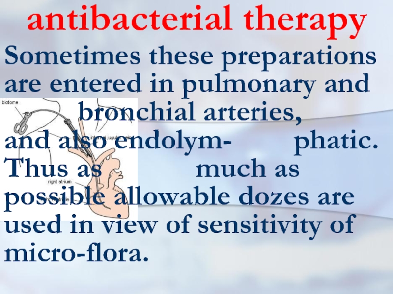
Sometimes these preparations are entered in pulmonary and
bronchial arteries, and also endolym- phatic. Thus as much as possible allowable dozes are used in view of sensitivity of micro-flora.
Слайд 52general improving health therapy treatment
The pharmacotherapy is directed also on stimulation
secretolysis and ex-pectorations, struggle with broncho- spasm and an edema of a mucous membra- ne of a bronchial tube, normalization and improvement of ex-change processes, replacement of immunologic defects etc.
Слайд 53acute abscesses
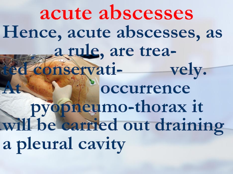
Hence, acute abscesses, as a rule, are trea- ted
conservati- vely. At occurrence pyopneumo-thorax it will be carried out draining a pleural cavity
Слайд 55Pleural drainage rules (K.Mattox)
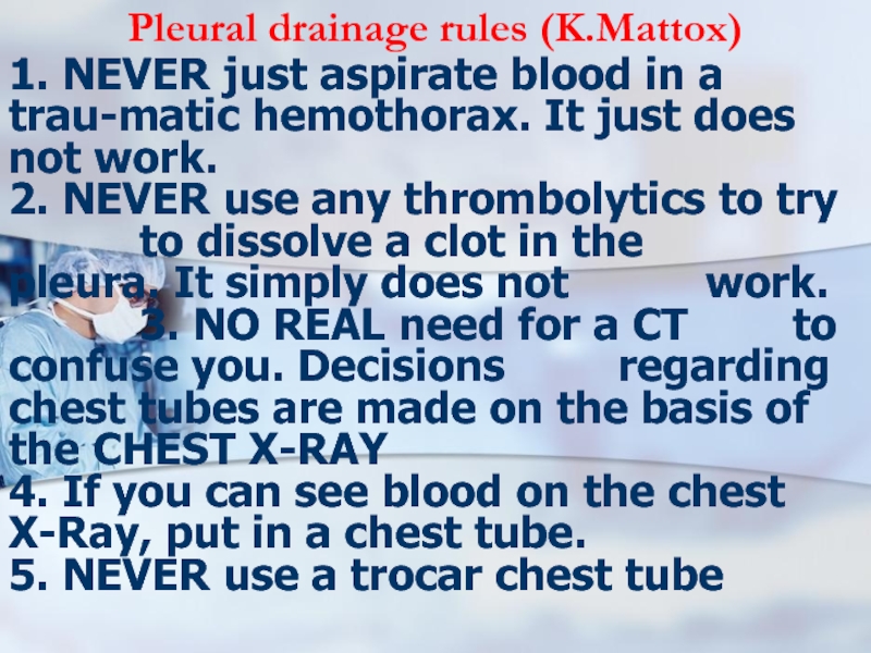
1. NEVER just aspirate blood in a trau-matic
hemothorax. It just does not work.
2. NEVER use any thrombolytics to try to dissolve a clot in the pleura. It simply does not work.
3. NO REAL need for a CT to confuse you. Decisions regarding chest tubes are made on the basis of the CHEST X-RAY
4. If you can see blood on the chest X-Ray, put in a chest tube.
5. NEVER use a trocar chest tube
Слайд 56Pleural drainage rules (K.Mattox)
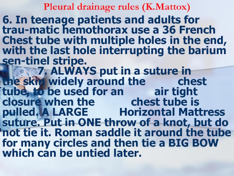
6. In teenage patients and adults for trau-matic
hemothorax use a 36 French Chest tube with multiple holes in the end, with the last hole interrupting the barium sen-tinel stripe.
7. ALWAYS put in a suture in the skin widely around the chest tube, to be used for an air tight closure when the chest tube is pulled. A LARGE Horizontal Mattress suture. Put in ONE throw of a knot, but do not tie it. Roman saddle it around the tube for many circles and then tie a BIG BOW which can be untied later.
7. ALWAYS put in a suture in the skin widely around the chest tube, to be used for an air tight closure when the chest tube is pulled. A LARGE Horizontal Mattress suture. Put in ONE throw of a knot, but do not tie it. Roman saddle it around the tube for many circles and then tie a BIG BOW which can be untied later.
Слайд 57Pleural drainage rules (K.Mattox)
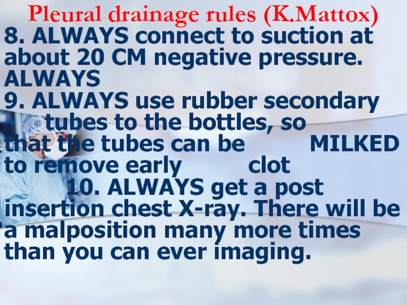
8. ALWAYS connect to suction at about 20
CM negative pressure. ALWAYS
9. ALWAYS use rubber secondary tubes to the bottles, so that the tubes can be MILKED to remove early clot
10. ALWAYS get a post insertion chest X-ray. There will be a malposition many more times than you can ever imaging.
9. ALWAYS use rubber secondary tubes to the bottles, so that the tubes can be MILKED to remove early clot
10. ALWAYS get a post insertion chest X-ray. There will be a malposition many more times than you can ever imaging.
Слайд 58Pleural drainage rules (K.Mattox)
11. ALWAYS have the best person available to
insert the tube who is in the hospital at the time either insert it, or personally and physically supervise the lesser person. Chest tubes in acute hemothorax are NEVER a place for a beginning physician, be they surgeon, emergency physician, etc. to learn.
12. NEVER make your decisions based on an acute CT of the chest in acute chest trauma.
12. NEVER make your decisions based on an acute CT of the chest in acute chest trauma.
Слайд 59sequestration in an abscess
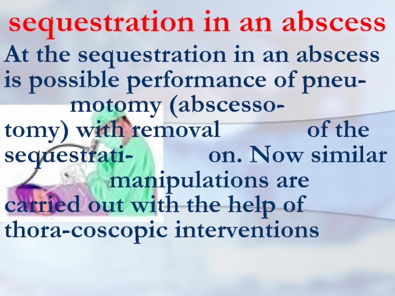
At the sequestration in an abscess is possible
performance of pneu- motomy (abscesso- tomy) with removal of the sequestrati- on. Now similar manipulations are carried out with the help of thora-coscopic interventions
Слайд 60emergency operation
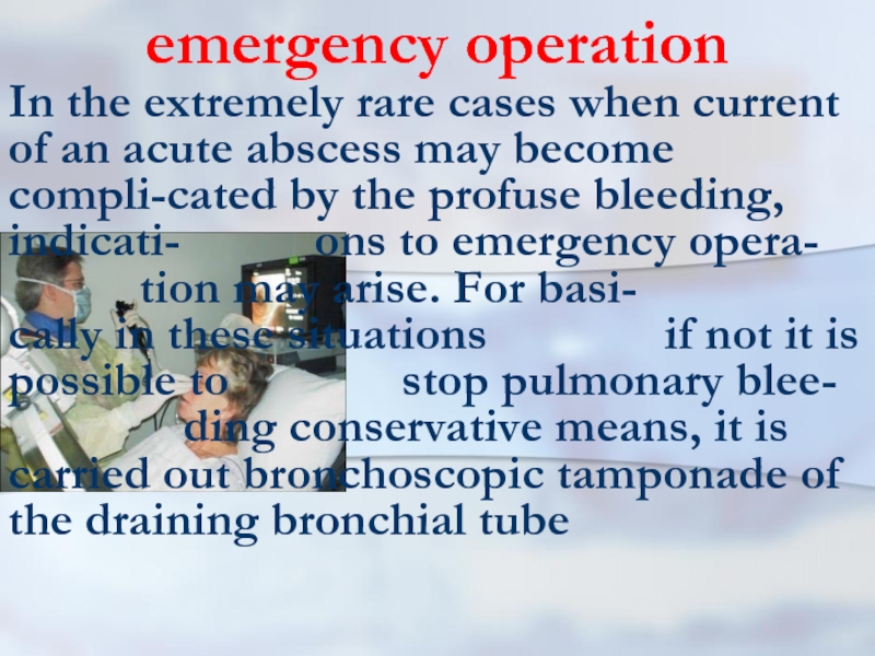
In the extremely rare cases when current of an acute
abscess may become compli-cated by the profuse bleeding, indicati- ons to emergency opera- tion may arise. For basi- cally in these situations if not it is possible to stop pulmonary blee- ding conservative means, it is carried out bronchoscopic tamponade of the draining bronchial tube
Слайд 61chronic abscess
The basic indication to operation is the chronic abscess. The
choice of a method of ope- ration depends on volume of defeat pulmonary tissue. It is carried out segmentec- tomy, lobectomy and in the extremely rare cases bylob-ectomy.
Слайд 62PLEURAL EMPYEMA
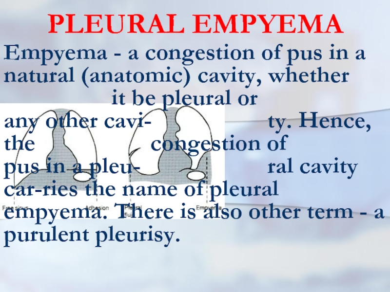
Empyema - a congestion of pus in a natural (anatomic)
cavity, whether it be pleural or any other cavi- ty. Hence, the congestion of pus in a pleu- ral cavity car-ries the name of pleural empyema. There is also other term - a purulent pleurisy.
Слайд 63Pleural empyema
The purulent pleurisy is the inflam-mation of pleural lists accompanying
exudating in a pleural cavity of the purulent exudate. Hence, terms "a purulent pleurisy" and "pleuras empye- ma" are synonyms. Though at times and till now doctors of various specialities confuse these conditions.
Слайд 64Pleural empyema
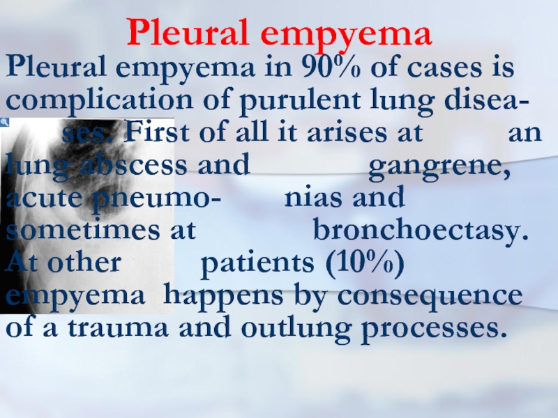
Pleural empyema in 90% of cases is complication of purulent
lung disea- ses. First of all it arises at an lung abscess and gangrene, acute pneumo- nias and sometimes at bronchoectasy. At other patients (10%) empyema happens by consequence of a trauma and outlung processes.
Слайд 65Pleural empyema
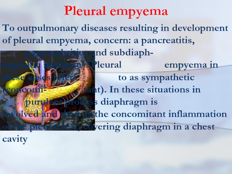
To outpulmonary diseases resulting in development of pleural empyema, concern:
a pancreatitis, paranephrities and subdiaph- ragmatic abscesses. Pleural empyema in these cases refers to as sympathetic (concomi- tant). In these situations in purulent process diaphragm is involved and there is the concomitant inflammation of the pleural leaf, covering diaphragm in a chest cavity
Слайд 66Classification of the pleural empyema
1. On clinical current
2. By the form
3.
On pathogenesis
4. On extent
5. A degree of lung compression
6. Acute and chronic
4. On extent
5. A degree of lung compression
6. Acute and chronic
Слайд 67Classification of the pleural empyema
1. On clinical current: the purulent-resorptive fever
and exhaustion.
2. By the form: empyema without destruction of the pulmonary tissue or with destruction of the pulmo-nary tissue.
3. On pathogenesis: meta- and parapneumonic, posttraumatic, metastatic and sympathetic.
2. By the form: empyema without destruction of the pulmonary tissue or with destruction of the pulmo-nary tissue.
3. On pathogenesis: meta- and parapneumonic, posttraumatic, metastatic and sympathetic.
Слайд 68Classification of the pleural empyema
4. On extent: limited, widespread, total.
5.
A degree of lung compressi- on: 1, 2, 3.
6. Acute and chronic
6. Acute and chronic
Слайд 69Classification of the pleural empyema
For the characteristic of intensity of purulent
process both in lung, and in a pleura, in classification the common typical syndromes deter- mining purulent-resorp- tive fever and very dange- rous condition - the puru- lent-resorptive exhaustion
Слайд 70Classification of the pleural empyema
Limited empyema are in cases of involving
in purulent process only one wall of a pleural cavity. At defeat of two or more walls of a pleu-ral cavity empyema is designa-ted widespread
Слайд 71Classification of the pleural empyema
To I degrees are referred those cases,
when lung compressed within the limits of one third.
II degree means, that lung compressed within the limits of two third.
At III degree lung compressed within the limits of full lung.
Total refers to an empyema at which all pleural cavity from diaphragm up to a dome is amazed.
II degree means, that lung compressed within the limits of two third.
At III degree lung compressed within the limits of full lung.
Total refers to an empyema at which all pleural cavity from diaphragm up to a dome is amazed.
Слайд 72Classification of the pleural empyema
Introduction in classification of empyema with destruction
and without destruction pulmonary tissue is made to show, what exactly destruction of the pulmonary tissue aggravates current of sup- purative process and renders dominant influence on a condition of the internal environment of an organism
Слайд 73Classification of the pleural empyema
It is separately allocated empyema necessitas (perfo- rans)
at which pus acts through intercos- tal intervals in soft tissue of a chest wall. Clinically the phlegmon of a chest wall is defined.
Слайд 74pathogeny
As a rule, the purulent inflammation of pleura begins from fibrinous
pleurisy and arises in two ways: first, owing to direct transition of exudative inflammations with lung on pleura and, second, at break in a pleural cavity of a subpleural lung abscess. The second way of development pleural empyema more often takes place.
Слайд 75Pneumonia and pleurisy
Pneumonias may divide on two groups: exudative type with
insignificant defeat of bronchial tubes and necrotic or absceding type. Thus necrotic sites, single and plural, are frequently located subpleural and consequently, as a rule, are complicated a fibrinous-purulent pleurisy. At absceding pneumonias with plural abscesses of polysegmentary localization and their subpleural arrangement, break of an abscess in a pleura cavity is possible with development of empyema.
Слайд 76clinic
Clinical picture. At pleural empyema occur pains in a thorax on
the side of defeat, the dyspnea is amplifies. Cough may be dry and with purulent sputum. Are marked the raised body temperature and chills. At percussion marked distinct dull sound, is more often behind on the scapular line. Thus, there are clinic purulent-resorptive fevers and attributes of a collecting liquid in a pleural cavity. Nevertheless, the clinical picture is various. It depends on many reasons.
Слайд 77clinic
The typical answer of an organism to any form of a
suppuration including pleural cavity is the purulent-resorptive fever. In its basis three factors lay: suppuration, resorption (absorbing of products of disintegration of tissue and products of ability to live of microorganisms) and the factor of loss. Last factor is caused by losses, which are born with an organism at a purulent inflammation. Clearly, that the degree of purulent-resorptive fevers, no less than intoxications, may be various - beginning from easy and finishing the hardest.
Слайд 78clinic
As it is marked above, frequently by the beginning empyema happens
the absceding pneumonia, therefore in some days after its crisis, again there is rigor, a pain in a side, dyspnoea and high temperature. After 3-5 days comes to light dull sound at percussion sound, weakens vocal fremitus and breath in the field of the struck site
Слайд 79clinic
In other cases the clinical picture of deve-lopment pleural empyema proceeds
latent-ly. It would seem, safely transferred inflam-mation of lung does not bring expected re- covery and, on the contrary, the dyspnea, fever, pains in a side gradually amplify. Probably paral- lel development of a pneumonia and purulent exudate in a cavity of a pleura. At break of a subpleu-ral abscess in a pleural cavity distinguish three clinical forms: acute, soft and erased.
Слайд 80clinic
At the acute form it is observed con-dition as a shock.
Suddenly at per-cussion there is a box sound above a place former dulling. At- tributes of the increasing pneumothorax with total collapsing of the lung are not excluded. The acute form of break of an abscess in a free pleural cavity meets seldom.
Слайд 81clinic
At the soft form, as a rule, an abscess evacuate in
closed incapsulated spa-ce. This form is shown by a moderate pain and change of percus- sion and auscultative attribu- tes. At the erased form which meets most frequently, the moment of the beginning of penetration of pus in a pleura is diffi-cultly perceptible.
Слайд 82clinic
The raised body temperature is one of the major attributes of
empyema of pleura. Temperature reactions may proceed on remitting type, as wrong waves with the tendency to morning downturn. However, the temperature, as a rule, is not reduced up to normal or even subnormal figures. Pains in a breast more often are caused by involving in process parietal pleuras. In the same time a pain may be caused by destruction of lung tissues.
Слайд 83clinic
Frequently pains amplify at breath, there-fore patients avoid deep breath. Trying
to spare the struck half of breast, patient quite often borrow the compelled posi- tion. Thus they are bent aside pathological process. It should be taken into account at diagnostics. Complaints to headaches are quite often marked. Early there are fun-ctional changes on the part of cardiovascu-lar system, a liver and kidneys. Infringe-ments of clotting systems of blood are possible.
Слайд 84clinic
Restriction of respiratory excursions of a chest is marked on the
side of defeat. At widespread and total pleural empyema smoothing intercostal intervals is quite often observed. Thus scapula on the side of defeat rises up slightly and lags behind at breath in comparison with another scapula. At palpation sometimes is marked resistence of soft tissues of chest wall. A characteristic attribute of a con- gestion of a liquid in a pleural cavity is easing vocal fremitus and dullness of percussion sound. At auscultation is marked sharp easing vesicular or bronchial breath. Variegrated moister rattles are listened at empyema, accompanying by destruction of lung tissues more often.
Слайд 85diagnosis
One of the important methods of diag-nosis of the pleural empyema
is the x-ray inspection. Thus it is established, whether there is a liquid in a pleu- ral cavity. A classical x-ray at- tribute pleural empyema slan- ting line of Damuaso. There may be a total and subtotal congestion of a liquid with dis-placement of mediastinum in the heal-thy side. In some cases it is defined li-mited (incapsulated) liquid.
Слайд 86diagnosis
Sometimes x-ray research will be carried out in lateroposition (on one
side). Also are applied computer tomography and USI. At chronic pleural empyema it is applied bronchography which estimates a condition of a bronchial tree and a degree of com- pressing of lung tissues. With the pur- pose of specification of the sizes and a configuration of a cavity of chronic empyema is sometimes used pleurography. At external fistulas it will be carried out fistulography. The big value at last years is given to thoracoscopy, which will be carried out also with the medical purpose.
Слайд 87treatment
Treatment begins with a puncture of a cavity empyema. During a
puncture con-tents with the subsequent bacteriological and cytologic research leave. The pleural cavity is sanified with the help of antibacterial and antiseptic preparations. However the puncture way more often possible to sanify only local forms. Therefore, as a rule, it will be car-ried out draining a pleural cavity that is better for combining with thoracoscopy.
Слайд 88treatment
After pleural cavity sanitation the drainage tube joins system active aspiration.
At absence of aspira-tion systems water-jet suction-machine is used. At impossibility of using water-jet suction-machine it is carried out draining on Bulau. For this purpose on the external end of a drai- nage tube the finger from a rubber glove on which the section is made becomes attached. Then this tube falls in bank with an anti-septic liquid. During an exhalation the liquid on a drainage follows from a pleural cavity in bank, and during a breath, due to fall of a rubber finger, the liquid from banks with antiseptic solutions in a pleural cavity does not come back.
Слайд 89treatment
All patient will carry out intensive antibacte-rial treatment in view of
sensitivity of micro-flora. Correction of volemic inringements is carried out by introduction of albuminous preparations, elect- rolytes etc. Calorage is provided with introduction of the con- centrated solutions of glucose and fatty emulsion. Necessarily corrected the acid-basic condi- tion. The therapy directed on restoration of a functional condition of cardiovascular system, a liver, kidneys, CNS etc. will be carried out
Слайд 90treatment
At destructions of the lung tissues, in necessary cases, bronchoscopic sanitation
will be carried out. The duly qualified treat- ment allows to achieve recovery at the most part of pa- tients with acute empyema of pleura. Nevertheless, at lines of patients develops chronic empyema
Слайд 91chronic empyema
At chronic empyema pleuras operative treatment is shown. On the
form empye- ma and presence of chan- ges from the parts of lung tissues are carried out va- rious operative interventi- ons. The most widespread operation is pleurectomy and lung decortication. At pleurectomy the bag empyema deletes. The purpose of decortication, offered Delorm in 1894, consists in clearing of lung from cicatricial layer, covering visceral pleura.
Слайд 92chronic empyema treatment
As a rule, both operations
(pleurectomy and decortica- tion) are united. Sometimes pleurectomy is combined with removal of a site struck lung tissues. In such cases of operation refer to as: pleurosegmentectomy, pleu-rolobectomy, pleurobilobectomy or pleuropulmonectomy
Слайд 93chronic empyema treatment
One of the most hardest operative interventions is pleuropulmonectomy.
It is caused by that patients except for chronic pleural empyema have also a total defeat lung. Last years pleuropulmonectomy is carried out seldom. Earlier at pleural empyema it was wi- dely applied thoracoplastic. Now thoracoplastic it will be carried out basically at em- pyema a residual pleural ca- vity, after various operations on lung. In connection with a wide circulation lung surgeries complication as empyema a residu-al pleural cavity after removal of a part or all of lung has appeared.
Слайд 94bronchial stump unsufficiency
By the most often reason of a similar sort
empyema happens an inconsistency of stump of resected bronchial tube. At chro- nic empyema residual pleural cavity after pulmonectomy are carried out various operations: transthoracal pleurectomy and suturing of stump of the main bronchial tube, trans-sternal transpericardial occlusion of stump of the main bronchial tube and various kinds of thoraco-plastic.
Слайд 95chronic empyema treatment
Concluding this section, it is necessary to note, that
ade- quate treatment of acute empye- ma with applica- tion in necessary cases thoraco-scopic interventions frequently results pleuras in recovery.
Слайд 96lung gangrene
Purulent-putrefactive necrosis of lobe or all of lung, with ab- sence
of a zone of demar- cation from the healthy lung tissues, having the tendency to the further distribution and shown by the heaviest common con-dition of the patient
Слайд 97lung gangrene
As a rule, the gangrene is formed owing to putrid
disintegration of the massive, beco- me lifeless sites of lung tissues (a lo-be, two lobes or all lung)
Слайд 98lung gangrene
Etiopathogen moments of a gangrene in many re- spects are similar
to those at an abscess of lung. However, at de- velopment of a gang-rene they are expressed in an extreme degree.
Слайд 99lung gangrene
It is frequently marked aspira-tion on a background of alco-
holic intoxication. The big value has the common condition of the patient with reduction of resis- tence (immunity), and al-so heavy accompanying disea-ses (a diabetes etc.).
Слайд 100lung gangrene
The significant role is played with previous chronic non- specific diseases
of lung. More often at a gangrene of lung the microflora in various combinations anaerobic is sowed with aerobic.
Слайд 101Clinic
As a rule, the gangrene of lung begins shar-ply, with significant
rise of a body tempera-ture, a dyspnea, be sick in a chest on the si- de of defeat, weakness and sharp dete- rioration of the common conditi- on. Right at the beginning cough may be dry, and then occurs putre- factive fetid sputum. The conditi- on of the patient the heaviest be-comes very fast. At cough it is increased discharge purulent sputum which has dirty-grey, greenish or (from an impurity of blood) chocolate color.
Слайд 102Clinic
Sometimes cough out small slices lifeless lung tissues. Even being on
significant distance from the patient, it is possible to feel an intolerable fetidity coughed out sputum and exhaled air. It is quite of- ten marked hemoptysis, and at times and fatal pulmonary blee-dings
Слайд 103Clinic
Frequently current of a gangrene of lung is complicated by development
of empyema pleuras. In connection with sharp intoxication, the septic shock with polyorgan insufficiency develops. Quite often at patients euphoria or confusion of consciousness is marked. Integuments of pale-grayish color with expressed acrocyanosis.
Слайд 104Clinic
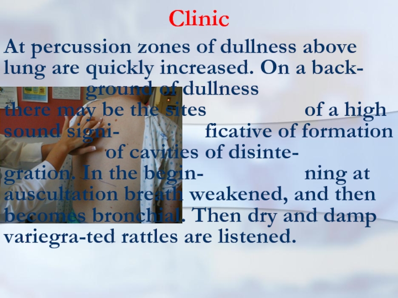
At percussion zones of dullness above lung are quickly increased. On
a back- ground of dullness there may be the sites of a high sound signi- ficative of formation of cavities of disinte- gration. In the begin- ning at auscultation breath weakened, and then becomes bronchial. Then dry and damp variegra-ted rattles are listened.
Слайд 105x-ray
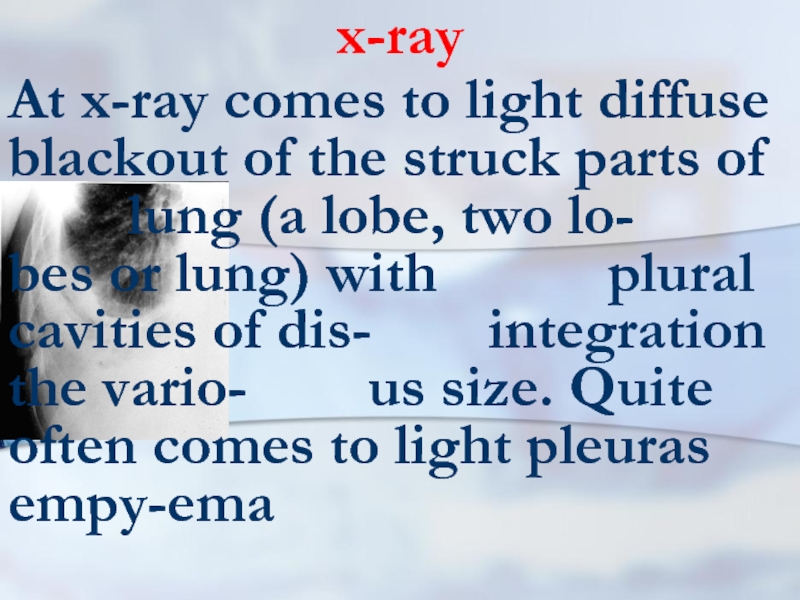
At x-ray comes to light diffuse blackout of the struck parts
of lung (a lobe, two lo- bes or lung) with plural cavities of dis- integration the vario- us size. Quite often comes to light pleuras empy-ema
Слайд 106prognosis
The prognosis at a lung gangrene frequently adverse. Especially it
concerns cases when all lung is struck and there is an inflammatory process in other lung (contralate- ral pneumonia). At a gan- grene of one lobe of lung the prognosis is more often more favorable.
Слайд 107gangrene lung treatment
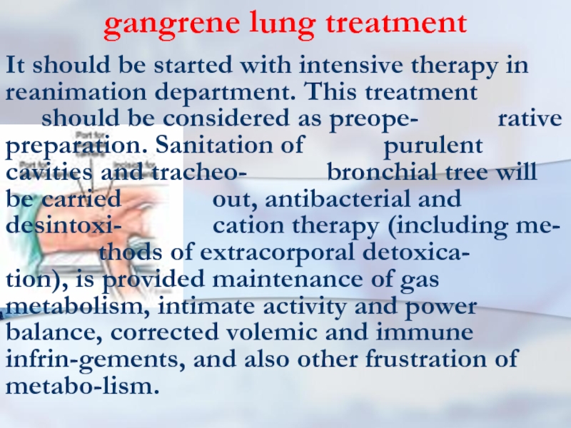
It should be started with intensive therapy in reanimation
department. This treatment should be considered as preope- rative preparation. Sanitation of purulent cavities and tracheo- bronchial tree will be carried out, antibacterial and desintoxi- cation therapy (including me- thods of extracorporal detoxica- tion), is provided maintenance of gas metabolism, intimate activity and power balance, corrected volemic and immune infrin-gements, and also other frustration of metabo-lism.
Слайд 108gangrene lung treatment
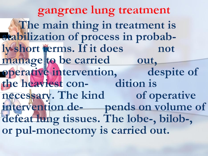
The main thing in treatment is stabilization
of process in probab- ly short terms. If it does not manage to be carried out, operative intervention, despite of the heaviest con- dition is necessary. The kind of operative intervention de- pends on volume of defeat lung tissues. The lobe-, bilob-, or pul-monectomy is carried out.
Слайд 109 In 21 century illiterate the one who is not able
to read and write is considered any more, and the one who is not able to study, to study up and to be retrained. Elwin Toffler
