markers in
primary human fibroblasts
- Главная
- Разное
- Дизайн
- Бизнес и предпринимательство
- Аналитика
- Образование
- Развлечения
- Красота и здоровье
- Финансы
- Государство
- Путешествия
- Спорт
- Недвижимость
- Армия
- Графика
- Культурология
- Еда и кулинария
- Лингвистика
- Английский язык
- Астрономия
- Алгебра
- Биология
- География
- Детские презентации
- Информатика
- История
- Литература
- Маркетинг
- Математика
- Медицина
- Менеджмент
- Музыка
- МХК
- Немецкий язык
- ОБЖ
- Обществознание
- Окружающий мир
- Педагогика
- Русский язык
- Технология
- Физика
- Философия
- Химия
- Шаблоны, картинки для презентаций
- Экология
- Экономика
- Юриспруденция
Accelerated aging diseases and their genetic causes Complex pattern of aging markers in primary human fibroblasts презентация
Содержание
- 1. Accelerated aging diseases and their genetic causes Complex pattern of aging markers in primary human fibroblasts
- 2. Primary non-transformed cell cultures tend to change
- 3. Cells in Culture (5, 10, 15 and 40 p)
- 4. Cell lines from patients AT8SP HGS1SP WS(1609)
- 5. The most frequently used marker of aging
- 6. Primary fibroblasts of skin from donors of
- 7. Hutchinson-Gilford syndrome Autosomal dominant, emerging “de novo”,
- 8. Lamine A
- 9. Nuclear lamina Structural transformations of the nuclear
- 10. Scaffidi P., Misteli T. 2006. Lamin
- 11. Lamin A and progerin processing (Ramirez et al., 2007)
- 12. Chen et al., 2003. LMNA
- 13. ZMPSTE24 gene Ramirez et al., 2006
- 14. Nuclear lamina (lamin A/C detection) The nuclear
- 15. Accumulation of foci of γ-Н2АХ is observed
- 16. Scaffidi P., Misteli T. 2006. Lamin A-dependent nuclear defects in human aging. Science. 312:1059-1063
- 17. Cells change their epigenetic status with age.
- 18. Scaffidi P., Misteli T. 2006. Lamin A-dependent nuclear defects in human aging. Science. 312:1059-1063
- 19. Werner syndrome Autosomal recessive 53
- 20. Werner syndrome gene
- 21. LMNA mutations (Chen et al., 2003. Ramirez et al., 2006)
- 22. Autosomal recessive 20 (40-50) BLM-helicase (RecQ-homologue)
- 23. RecQ family helicases (Hickson 2003)
- 24. RecQ-helicases in DNA repair (Nakoyma 2002)
- 25. Protein-protein interaction of WRN и BLM helicases (Nakoyma 2002) )
- 26. In the study of cells from a
- 27. The stable chromosome (1, 4, 8
- 28. Autosomal recessive (40)-50 Skin atrophy DNA repair defects Cutix laxa
- 29. The telomere fragments lenght 1.HeLa
- 31. Роль АТМ в клеточном ответе на возникновение двунитевых разрывов
- 32. Autosomal recessive 20 (40-50) ATM, a protein
- 33. Атаксия-телеангиэктазия АТМ АТМ Дефекты репаративного синтеза
- 34. PIKK (phosphatidylinositol 3-kinase-like protein kinases) человека
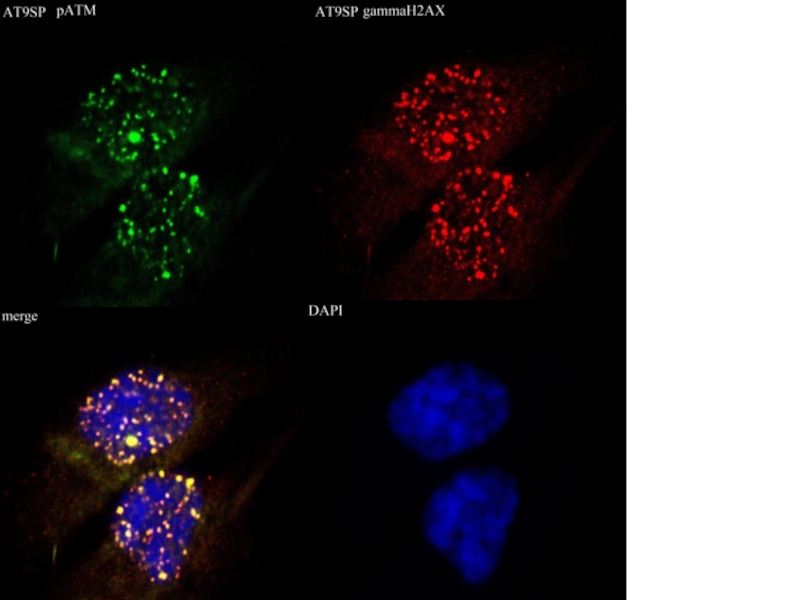
- 35. MOSAICS Мозаичная культура линий AT1SP и AT9SP: детекция фокусов Р-АТМ. Справа –DAPI.
- 36. SA-beta-GAL in AT-cells
- 37. Detection of γ-Н2АХ in nuclei of human
- 40. DNA damage caused by ionizing radiation is
- 41. 53ВР1 foci in human fibroblast nuclei VH-10
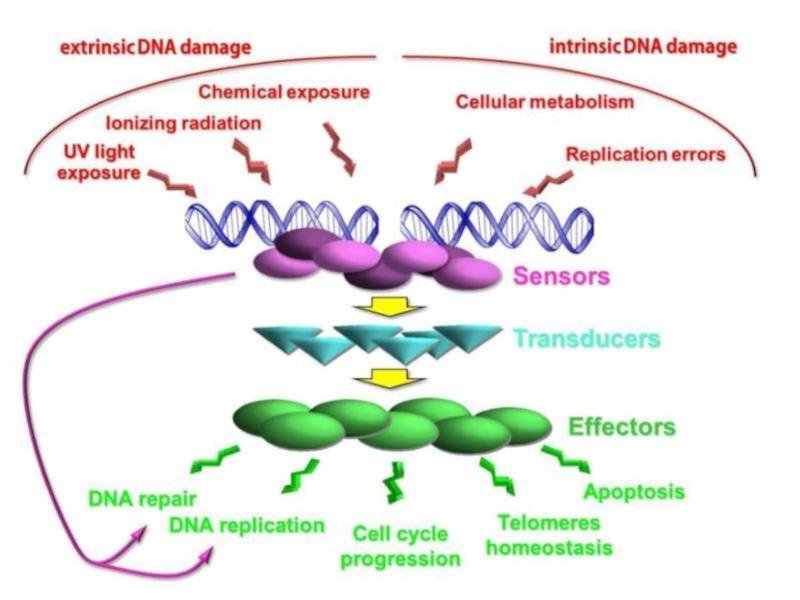
- 42. Detection of НР1-γ in nuclei of human
- 43. SIRT6 SIRT1
- 44. Detection of 3меК9Н3 in nuclei of human
- 45. 3meH3 fluorescence intensity in human fibroblasts In
- 46. Seckel syndrome (atr) O'Driscoll M, Ruiz-Perez VL,
- 47. Синдром Секкеля Микроцефалия, Умственная отсталость Карликовость Задержки
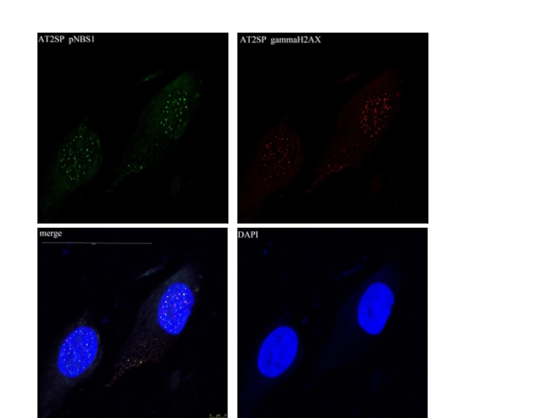
- 48. Совместная окраска антителами к рАТМ и активным
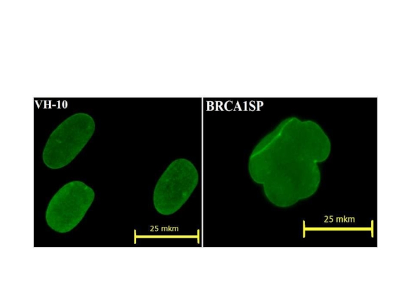
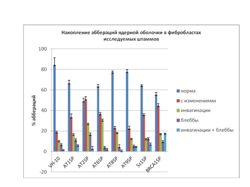
- 49. Ss1SP BRCA1SP
- 50. Ядерная оболочка, окраска антителами к ламину А\С
- 53. Mutations in TRAIP cause primordial dwarfism Harley
- 54. TRAIP localizes to sites of UV-induced DNA
- 55. PCNA-mutations
- 56. Excision repair systems (Spivak G., 2004)
- 58. Xeroderma pigmentosum (XP) XPA, XPB, XPD, XPC,
- 59. Lai et al., 2013. The influence of
- 60. Autosomal recessive CSA (WD-repeat protein, β-subunit
- 61. TTD
- 62. Анемия Фанкони FAA, FAB, FAC, FAD, FAE,
- 63. Hum Genomics. 2015 Nov 24;9(1):32. doi: 10.1186/s40246-015-0054-y. Update
- 64. Simplified model of the Fanconi anemia pathway
- 67. Age of onset of SCC in FA
- 68. Telomeres Telomeres, being the final fragments of
- 69. Age-dependent telomere length
- 70. Telomere length
- 71. Factor analysis of mean telomere length, genetic
- 72. Запретить
- 74. Комплекс генов анемии Фанкони
- 75. Simplified model of the Fanconi anemia pathway
- 76. Mechanism of ICL repair in the FA
- 77. FA/BRCA pathway and crosstalk between FA and
- 78. Overview of FA pathway genes identified in
- 79. Domain architecture and structure of FANCD2 and
- 80. Comparison of the SCF multi-subunit ubiquitin ligase
- 81. Models for FANCD2 and FANCI monoubiquitination The
- 82. Теломерные белки у человека
- 83. Data acquired in the course of study
- 84. Структурная организация белка Р53
- 85. SA-β-gal в первичных фибробластах мышей SHR
- 86. G1 чекпойнт
- 87. S-чекпойнт
- 88. G2-чекпойнт
- 89. Внутриядерная локализация белков во время ответа клетки на повреждение ДНК
- 90. Механизмы G2-ареста АТМ активирует СНК2 через фосфорелирование
- 91. Механизмы G2-ареста(2) После облучения резко падает уровень
- 92. Механизмы G2-ареста(3) PLK1 и PLK3 (Polo-like kinase).
- 93. Механизмы G2-ареста(4) Остановить вход в митоз при
- 94. Механизмы G2-ареста(5) Роль Р53 в поддержании G2-ареста
- 95. Механизмы G2-ареста(6) G2 чекпойнт-ответ разделяется на два
- 96. Репарация, спаренная с транскрипцией. TTD Повышенная фоточувствительность
- 97. Репарация межнитевых сшивок Схема процеса репарации сшивок
- 98. Репарация межнитевых сшивок
- 99. Репарация межнитевых сшивок

Слайд 2Primary non-transformed cell cultures tend to change with every passage. Population
doubling time increases, cell morphology alteres, larger rotund cells appear, which are regarded as older, in comparison with smaller oblong ones (Мikhelson, 1984; Lorenzini et al., 2005). Biochemical markers appear simultaneously with morphological alterations. They characterize aging cells in culture, defined as “aging markers”, which include changes in chromatine, nucleus and cytoplasmatic skeleton, high level of non-repaired DNA damages, etc. (Campisi, 2005); as well as in cell instability towards action of damaging agents, primarily hydrogen peroxide (Chen et al., 1998, Ryter et al., 2007).

")
")
Слайд 5The most frequently used marker of aging is lysosomal β-galactosidase (SA-β-gal)
associated with aging, its activity dramatically increasing in old cells (Dimri et al., 1995; Lee et al., 2006)
 associated with aging, its")
Слайд 6Primary fibroblasts of skin from donors of varios ages, and patients
with premature ageing with Hutchinson-Gilford syndrome (children’s progeria), and the Werner one (adult progeria), normally serve as model for the detection of ageing markers
Fibroblasts taken from old donors, and from the progeria patients, are currently regarded as containing more ageing markers, than those taken from young healthy donors (Scafidi, Misteli, 2006, Sedelnikova et al., 2008).
Fibroblasts of skin of other mammals (mice) may also be applied for the detection of ageing markers (taking into account the Hyflick limit)

Слайд 7Hutchinson-Gilford syndrome
Autosomal dominant, emerging “de novo”, getting, probably, from the father
site
15 (21)
LMNA 1q21.2
FACE-1/ZMPSTE24 1q34
Skin atrophy.
Bird’s face
Loss of subcutaneous fat
Hair loss.
Arteriosclerosis
Increased metabolic rate
Hypogonadism
Grow hormone insensitivity?
Slow growth.
Reduced replicative life span of cultured cells
Shot telomeres
DNA repair defects?
Silent mutation in (1-4)-β-Galactosyl-transferase gene (594C>T)
LMNA 1q21.2FACE-1/ZMPSTE24")

Слайд 9Nuclear lamina
Structural transformations of the nuclear lamina, occurring as a result
of the accumulation of aberrant product of the LMNA gene, progerin, were demonstrated in the course of the study of cells of patients with Hutchinson-Gilford syndrome, as well as of cell cultures, acquired from aged donors (Scaffidi, Misteli, 2006). Results acquired by these authors, taken in complex with the present-day data concerning participation of components of nuclear lamina, including lamin А in the wide range of cell processes. Allow to regard structural alterations of nuclear lamina as the basic process for induction of markers of ageing, associated with various levels of cell regulation .

Слайд 10 Scaffidi P., Misteli T. 2006. Lamin A-Dependent Nuclear Defects in Human
Aging.
Science 312 : 1059 – 1063
Farnezyl-protein-transpherase
Ras-converting enzyme or Zmpste24
S-adenosyl- metionin;
isiprenyl-carboxymethyl-transpherase
Zmpste24
lamine А
«progerin»
Rusinol,
Sinensky, 2006

")
Слайд 12 Chen et al., 2003. LMNA mutations in atypical Werner’s syndrome. Lancet
362 : 440–445.
Ramirez et al., 2006


Слайд 14Nuclear lamina (lamin A/C detection)
The nuclear defects (Scaffidi, Misteli, 2006; Smirnova
et al., 2008)
Healthy donor (11 у.) – 5 %
Healthy donor (87 y.) - 20%
Atypical Werner’s syndrome – 40%
Hutchinson-Gilford’s syndrome – 80%
Healthy donor (11 у.) – 5 %
Healthy donor (87 y.) - 20%
Atypical Werner’s syndrome – 40%
Hutchinson-Gilford’s syndrome – 80%
The nuclear defects (Scaffidi, Misteli, 2006; Smirnova et al., 2008)Healthy donor")
Слайд 15Accumulation of foci of γ-Н2АХ is observed in nuclei of aging
cells in culture, cells from old donors, and from Progeria patients (Sedelnikova et al., 2004,2008).
This phenomenon may be linked to both accumulation of either non-repaired DSBs or, modifications of chromatine, apprehended as DSBs by protein kinases АТМ and DNA-PK after DNA damage (Stiff et al., 2004), or else, ATR kinase during replication halt (Takahashi, Ohnishi, 2005); as well as by appearance of uncapping telomeres (Hao et al., 2004).
As a result, accumulation in cell population of cells with foci of γ-Н2АХ occurs, which forms an objective criterion of ageing on cell level.

Слайд 16Scaffidi P., Misteli T. 2006. Lamin A-dependent nuclear defects in human
aging. Science. 312:1059-1063

Слайд 17Cells change their epigenetic status with age. Heritable changes in gene
regulation with no changes in the DNA sequence itself are considered as epigenetic determinants. In recent years, the role of epigenetic mechanisms in the process of carcinogenesis (Jones, Baylin, 2002), as well as in cellular and organismal aging (Wilson, Jones, 1983; Issa, 2003; Fraga et al., 2005) was demonstrated.

Слайд 18Scaffidi P., Misteli T. 2006. Lamin A-dependent nuclear defects in human
aging. Science. 312:1059-1063

Слайд 19Werner syndrome
Autosomal recessive
53 (60)
Loss of WRN,
a RecQ family helicase.
Skin atrophy.
Hair graying/loss.
Arteriosclerosis
Osteoporosis
Muscle atrophy
Cataracts
Hyperlipidemia
Mild diabetes melitus, type 2
Hypogonadism
Cancer (sarcomas)
Slow growth.
Reduced replicative life span of cultured cells
Chromosome rearrangements
Sensitivity to 4-NQO and camptothecin (topoisomerase I poison)
Increased mutation rate, particulary DNA deletions
Rapid telomere shortening during cellular life span.
DNA repair defects
DNA replication defects (?)
 Loss of WRN, a RecQ family helicase.Skin atrophy. Hair")

")
Слайд 22Autosomal recessive
20 (40-50)
BLM-helicase (RecQ-homologue)
Increased metabolic rate
Slow growth.
Diabetes melitus
Hypogonadism
Neurodegeneration
Immunodeficiency
Cancer
Telangiectasias on
the face, forming the red “butterfly”
Low DNA synthesis (2 times) with the normal rate of DNA-polymerases α, β, γ
Chromosomes rearrangements
Icreased level of recombination
Low DNA synthesis (2 times) with the normal rate of DNA-polymerases α, β, γ
Chromosomes rearrangements
Icreased level of recombination
Bloom syndrome
BLM-helicase (RecQ-homologue)Increased metabolic rateSlow growth. Diabetes melitus HypogonadismNeurodegenerationImmunodeficiencyCancerTelangiectasias on the face, forming the")
")
")
 )")
Слайд 26In the study of cells from a patient with Hutchinson-Gilford syndrome,
besides the identification of all markers of aging described by Scaffidi P., Misteli T (2006), we observed a significant discrepancy between these cells and cells from old donors. The level of stable chromosomal aberrations in the investigated cells was not elevated and, by this the marker, normally showing the "real" biological age, our patient was consistent with her 9 years.

Слайд 27 The stable chromosome (1, 4, 8 or 12) aberration (SCA)
frequency
analysed by FISH in human blood lymphocytes
Age dynamics
The patients with Werner’s syndrome demonstrate increased SCA level, corresponding to premature aging of organisms.
The lymphocytes from patient with Hutchinson-Gilford syndrome have no time for SCA formation
 aberration (SCA) frequency analysed by FISH")
-50Skin atrophyDNA repair defectsCutix laxa")
Слайд 29The telomere fragments lenght
1.HeLa cells
2.Normal donor lymphocytes
3.AT2SP (ataxia-telangiectasia patient)
4.Cutis laxa
patient
Size in KB
Size in KB
4.Cutis laxa patient Size in KB")

Слайд 32Autosomal recessive
20 (40-50)
ATM, a protein kinase of the PI-3 kinase family
Neurodegeneration
Immunodeficiency
Cancer
(leukemias and lymphomas)
Occulocutaneous telangiectasias
Progeroid skin and hear changes
Hypogonadism
Defectif cell cycle chekpoint arrest
Reduced replicative life span
Chromosomal rearrangements
Sensitivity to ionizing radiation
Inoppropriate apoptosis
Shot telomeres
Delayed/absence P53 induction and accumulation after DNA damage
Defects in repair of DNA double-strand breaks
Defects in V(D)J recombination
Ataxia-telangiectasia (Luis-Bar syndrome)
ATM, a protein kinase of the PI-3 kinase familyNeurodegenerationImmunodeficiencyCancer (leukemias and lymphomas)Occulocutaneous telangiectasiasProgeroid")
Слайд 33Атаксия-телеангиэктазия АТМ
АТМ
Дефекты репаративного синтеза ДНК, нарушения клеточного цикла, высокая частота спонтанных
хромосомных аномалий, увеличенная чувствительность к ионизирующим излучениям и радиомиметикам, к УФ-свету и агентам сходного действия (таким, как 4-нит-рохинолиноксид)
Появляется у одного из 40 тыс. новорожденных, основные поражения отмечены в нервной и иммунной системах (мозжечковая атаксия, приводящая к нарушениям координации мышц, шатающейся походке и прогрессирующей умственной отсталости, кожным нарушениям, предрасположенности к раковым заболеваниям и др.)
Появляется у одного из 40 тыс. новорожденных, основные поражения отмечены в нервной и иммунной системах (мозжечковая атаксия, приводящая к нарушениям координации мышц, шатающейся походке и прогрессирующей умственной отсталости, кожным нарушениям, предрасположенности к раковым заболеваниям и др.)

 человека")


Слайд 37Detection of γ-Н2АХ in nuclei of human fibroblasts VH-10 (11 y),
donor 51 y. and АТ8SP (11 y.). For purposes of detection of the number of nuclei, the same field of vision is presented, after DAPI coloring.
1.08±1.07
4.06±1.28*
12.32±2.09**
, donor 51 y. and")
Слайд 40DNA damage caused by ionizing radiation is accompanied by ATM phosphorylation
of serine (S1219) in 53BP1 (Lee et al., 2008). This protein also plays an important role in the checkpoint response of the cell to damage (Iwabuchi et al., 2008, Eliezer et al., 2008). The appearance of foci 53ВР1, as well as foci of γ-Н2AХ can be regarded as one of the reliable markers of aging cells.
53ВР1 protein forms a traditional marker of repair processes (Wilson, Stern, 2008). Appearance and elimination of 53ВР1 foci after DNA damage tends to coincide with γ-Н2АХ dynamics, as they often colocalize in the zone of DSB
53ВР1 protein forms a traditional marker of repair processes (Wilson, Stern, 2008). Appearance and elimination of 53ВР1 foci after DNA damage tends to coincide with γ-Н2АХ dynamics, as they often colocalize in the zone of DSB
 in")
Слайд 4153ВР1 foci in human fibroblast nuclei VH-10 and АТ8SP
Diffuse coloring tends
to occur in coloring by 53ВР1 antibodies in intact cells, while clear-cut foci form after DNA damage. Such foci tend to form in AT patients’ cells in many intact cells, i.e. 16% and 18% respectedly, while there are only 6 % of such cells in control.

Слайд 42Detection of НР1-γ in nuclei of human fibroblasts VH-10 (11 y),
donor 51 y. и АТ8SP (11 y.). DAPI
170,71 ± 1,91
134,45 ± 4,95*
93,01 ± 3,55*
Fluorescence intensity
, donor 51 y. и")

Слайд 44Detection of 3меК9Н3 in nuclei of human fibroblasts VH-10 (11 y),
donor 52 y, AT6SP (23 y.) and АТ8SP (11 y.)
, donor 52 y, AT6SP")
Слайд 453meH3 fluorescence intensity in human fibroblasts
In the case of AT8SP, the
level of trimethyl derivatives of histone H3 (K9 and K27), in contrast to the cells from old donors and other progeria, is not falling, but rather rising, as it is shown in tumor cells.
In the case of AT6SP, the level of 3meK9H3 in contrast to the cells from AT8SP is falling as in the cells of old donors, but 3meK27H3 rather rising, as in AT8SP.
The quantity of 3meK9H3 may serve as a prognostic marker of gravity of the disease
In the case of AT6SP, the level of 3meK9H3 in contrast to the cells from AT8SP is falling as in the cells of old donors, but 3meK27H3 rather rising, as in AT8SP.
The quantity of 3meK9H3 may serve as a prognostic marker of gravity of the disease

Слайд 46Seckel syndrome (atr)
O'Driscoll M, Ruiz-Perez VL, Woods CG, Jeggo PA, Goodship
JA. 2003. A splicing mutation affecting expression of ataxia-telangiectasia and Rad3-related protein (ATR) results in Seckel syndrome. Nat Genet.,33:497-501
O'Driscoll M, Ruiz-Perez VL, Woods CG, Jeggo PA, Goodship JA. 2003. A splicing")
Слайд 47Синдром Секкеля
Микроцефалия, Умственная отсталость
Карликовость
Задержки развития Диспластический фенотип.
Microcephalic primordial dwarfism (MPD)
")
Слайд 48Совместная окраска антителами к рАТМ и активным киназным доменам АТМ/АTR здорового
донора VH-10 (вверху) и больной с синдромом Секкеля Sc1SP (внизу)
 и")


Слайд 53Mutations in TRAIP cause primordial dwarfism
Harley et al., 2016. TRAIP promotes DNA damage
response during genome replication and is mutated in primordial dwarfism.
Nat Genet.
48(1):36-43
The TRAF [tumor necrosis factor (TNF) receptor-associated factors] -interacting protein (TRAIP) functions as (RING)-type E3 ubiquitin ligase, but its physiological substrates are not yet known.
Nat Genet.
48(1):36-43
The TRAF [tumor necrosis factor (TNF) receptor-associated factors] -interacting protein (TRAIP) functions as (RING)-type E3 ubiquitin ligase, but its physiological substrates are not yet known.

Слайд 54TRAIP localizes to sites of UV-induced DNA damage
(a) TRAIP localizes to
DNA damage sites induced by UV laser microirradiation both in the absence and presence of pre-treatment with BrdU as a damage sensitizer. Representative images, before and after UV laser microirradiation. Scale bar, 5 μm. (b) GFP-TRAIP colocalizes with γH2AX and with RFP-PCNA at sites of UV laser-induced damage. Representative images of UV laser-irradiated GFP-TRAIP expressing cells immunostained for γH2AX (pre-sensitized with BrdU) or co-expressing RFP-PCNA (no BrdU pre-treatment) as indicated. Scale bar, 5 μm. (c, d) GFP-TRAIP is detected by a Proximity Ligation Assay (PLA) in close proximity to PCNA, an association enhanced after UV-induced damage. (c) Representative images of PLA signals/nucleus in doxycycline-inducible GFP-TRAIP HeLa cells before and after damage with 25 J/m2 UV-C. Scale bar, 5 μm. (d) Quantification of PLA signals/nucleus. Box plots, center line denote mean values, box 25/75 %, whiskers 5/95 %, data pooled from n=2 independent experiments, n>65 data points per condition per experiment; Mann Whitney rank sum test: *** p<0.001. (e) TRAIP accumulates at sites of localized UV damage, colocalising with RPA and γH2AX. Representative immunofluorescence images of MRC5 cells transfected with GFP-TRAIP or GFP alone after UV-C irradiation through 3 μm microfilters. Scale bar, 5 μm.
 TRAIP localizes to DNA damage sites induced")

")
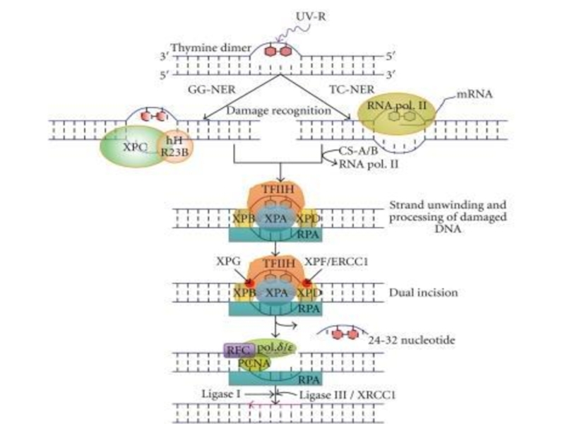
Слайд 58Xeroderma pigmentosum (XP)
XPA, XPB, XPD, XPC, XPE, XPF, XPG ,
XP-V не
выявлен дефект NER
Дефекты эксцизионной репарации нуклеотидов (нарушение вырезания, застройки брешей и др.). Разнообразные дефекты репарационных процессов.
При XP-вариант наблюдается изменение параметров репликации ДНК
Сверхчувствительность к УФ-свету,
ведущая к появлению красных пятен на коже, переходящих в незаживающие коросты и нередко в рак кожи; неврологические расстройства); поражения век, бровей и глаз. Распространение: 1 случай на 250000 человек в Европе и США; 1 :40000 человек в Японии
XPA, XPB, XPD, XPC, XPE, XPF, XPG ,XP-V не выявлен дефект NERДефекты эксцизионной")
Слайд 59Lai et al., 2013. The influence of DNA repair on neurological degeneration, cachexia,
skin cancer and internal neoplasms: autopsy report of four xeroderma pigmentosum patients (XP-A, XP-C and XP-D). Acta Neuropathol Commun. 1:4. doi: 10.1186/2051-5960-1-4.
XP patients studied. A and B: Case 1 XP-A patient (A) at age 17y with numerous freckle-like pigmented lesions on sun exposed skin and (B) at 37y with marked cachexia and thinning of subcutaneous tissues of face and chest. She had more that 100 surgical procedures on her face for removal of skin lesions. C: Case 2 XP-D patient at age 40y. She had been well protected from sun exposure since early childhood and had only few pigmented lesions and skin cancers. D: Case 3 XP-C patient, at age 29y with multiple freckle-like pigmented lesion on sun exposed skin and cheilitis. The patient underwent many surgical procedures for removal of skin cancers on her face. E and F: Case 4 XP-C patient, at age 28y (E) with multiple pigmented lesions, telangiectasia, cheilitis and corneal clouding. Multiple surgical procedures were performed on her face for removal of skin cancers and at age 48y (F) following exenteration of both orbits for treatment of ocular squamous cell carcinomas.

Слайд 60Autosomal recessive
CSA (WD-repeat protein, β-subunit GTP protein homologue)
CSB (ATPase of Swi2
family),
XPB and XPD (helicases in TFII-H),
XPG (yeast Rad2 homologue, endonuclease)
20 (40?)
Bird’s face
Loss of subcutaneous fat
Skin photosensitivity
Neurodegeneration
Hypogonadism
UV-sensitivity
Impaired transcription-coupled nucleotide excision repair
Decreased recovery of transcription after irradiation
General RNA-polymerase II transcription defects
Cockayne syndrome (CS)
CSB (ATPase of Swi2 family),XPB and XPD (helicases")

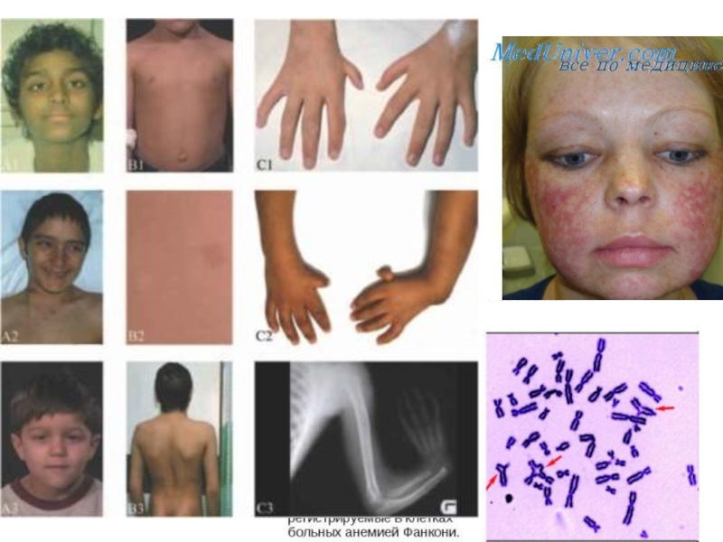
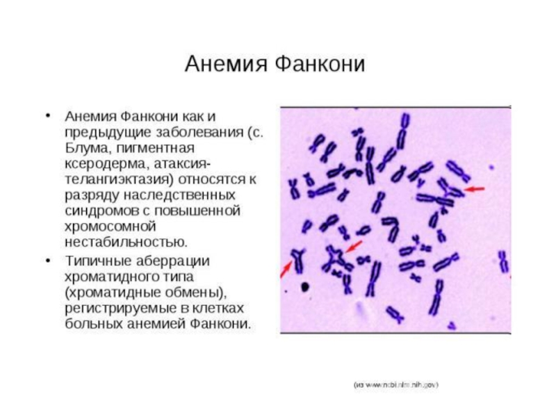
Слайд 62Анемия Фанкони
FAA, FAB, FAC, FAD, FAE, FAF, FAG -19 групп комплементации
Дефекты
репарации повреждений от химических мутагенов и канцерогенов (по не УФ-свста), обусловленные дефектами эндонуклеаз, дефектами распознавания кросс-сшивок ДНК;
пониженная способность к апоптозу после ионизирующего облучения; двукратное удлинение G2-фазы клеточного цикла
Сверхчувствительность к химическим мутагенам и канцерогенам, уменьшение количества всех клеточных элементов крови, различные аномалии врожденных способностей, деформация пальцев и другие виды скелетных нарушений, урогенитальные нарушения, микроцефалия, микрофтальмия, дефекты уха и потеря слуха, сердечные и гастроинтестинальные нарушения

Слайд 63Hum Genomics. 2015 Nov 24;9(1):32. doi: 10.1186/s40246-015-0054-y.
Update of the human and mouse Fanconi
anemia genes.
Dong HDong H, Nebert DWDong H, Nebert DW, Bruford EADong H, Nebert DW, Bruford EA, Thompson DCDong H, Nebert DW, Bruford EA, Thompson DC, Joenje HDong H, Nebert DW, Bruford EA, Thompson DC, Joenje H, Vasiliou V.
Fanconi anemia (FA) is a recessively inherited disease manifesting developmental abnormalities, bone marrow failure, and increased risk of malignancies. Whereas FA has been studied for nearly 90 years, only in the last 20 years have increasing numbers of genes been implicated in the pathogenesis associated with this genetic disease. To date, 19 genes have been identified that encode Fanconi anemia complementation group proteins, all of which are named or aliased, using the root symbol "FANC." Fanconi anemia subtype (FANC) proteins function in a common DNA repair pathway called "the FA pathway," which is essential for maintaining genomic integrity. The various FANC mutant proteins contribute to distinct steps associated with FA pathogenesis. Herein, we provide a review update of the 19 human FANC and their mouse orthologs, an evolutionary perspective on the FANC genes, and the functional significance of the FA DNA repair pathway in association with clinical disorders. This is an example of a set of genes--known to exist in vertebrates, invertebrates, plants, and yeast--that are grouped together on the basis of shared biochemical and physiological functions, rather than evolutionary phylogeny, and have been named on this basis by the HUGO Gene Nomenclature Committee (HGNC).
:32. doi: 10.1186/s40246-015-0054-y.Update of the human and mouse Fanconi anemia genes.Dong HDong H, Nebert DWDong")
Слайд 64Simplified model of the Fanconi anemia pathway
. Activation of FANCD2 and FANCI
by the FA core complex via monoubiquitination (orange circles) regulates downstream genes involved in recombination repair of DNA crosslinks.

Слайд 67Age of onset of SCC in FA patients with and without
HSCT
Forty-three of 83 female FA patients (51.8%) and 17 of 46 male FA patients (37.0%) developed SCC (average age at SCC diagnosis for 48 FA patients without HSCT 30.0 years, for 12 FA patients after HSCT 25.8 years).

Слайд 68Telomeres
Telomeres, being the final fragments of eukaryotic chromosomes, form one of
the most widely studied at present time in the framework of study of primary mechanisms of the organism ageing, potential factors, conditioning life span. Considerable interest directed at these specialized complexes, is conditioned by their unique functions in the securing of the cell genome (Blackburn, 2001). Apart from preventing the chromosome fusion, telomeres are responsible for their fixation at cell membrane (Podgornaya et al., 2000; Hediger et al., 2002; Rose et al., 2004), for the mitotic and meiotic segregation of chromosomes (Conrad et al.,1997; Kirk et al., 1997; Dynek, Smith, 2004), and for their meiotic coupling (Rockmill, Roeder, 1998), for the stabilization of broken chromosomes (Jager, Philippsen, 1989; Pennaneach et al., 2006), and for their defence against reparation systems (Shay, Wright, 2007; Mirsi et al., 2008), as well as being responsible for gene expression (Baur et al., 2001; Pedham et al., 2006).


Слайд 71Factor analysis of mean telomere length, genetic polymorphism, and age, by
senior age respondents
Regular correlation between telomere length with respondent age appears after having attained certain age, when adequate strategy of active longevity has been attained, and individual differences may be traced back.


Слайд 75Simplified model of the Fanconi anemia pathway
. Activation of FANCD2 and FANCI
by the FA core complex via monoubiquitination (orange circles) regulates downstream genes involved in recombination repair of DNA crosslinks.

Слайд 76Mechanism of ICL repair in the FA pathway on collision of
a replication fork with an ICL
Анемия Фанкони (A, B, C, D1, D2, E, F, G, I, K, L, M, N, O, P, and Q). Эти белки делятся на 3 группы: (1) белки корового комплекса; (2) FANCI и FANCD2 белки, составляющие комплекс ID2 и (3) эффекторные белки
(A) FA активируется во время фазы S при обнаружении ICLs или сходных повреждений ДНК. Коровый комплекс FA привлекается в зону повреждения MHF1-MHF2-FANCM комплексом.
(B) Комплекс ID2 моноубиквитинируется и связывается с зоной повреждения ДНК. Комплекс B-L-100 способствует этому убиквитинированию совместно с еще двумя субкомплексами (A-G-20 and C-E-F)
(C) Нуклеазы XPF/FANCQ-ERCC1 совместно с SLX4/FANCP, incise the Dпроводят ДНК-инцизию
(D) Моноубиквитинированный комплекс ID2 привлекает белки репарации, включая BRCA1, BRCA2/FANCD1, FANCJ, PALB2/FANCN, and RAD51C/FANCO.
(E) После успешной репарации комплекс, ID2 деубиквитинизируется USP1-UAF1 , что способствует его высвобождению из хроматина.

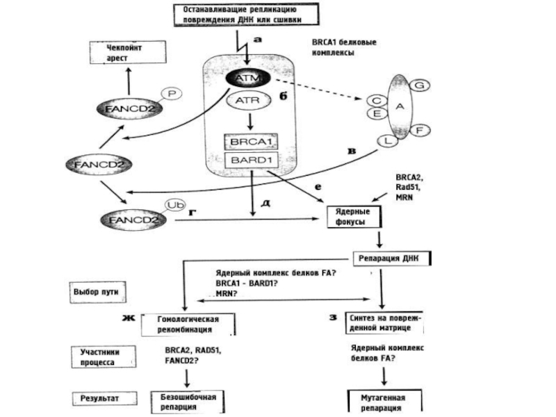
Слайд 77FA/BRCA pathway and crosstalk between FA and other DNA repair pathways
В
ответ на сигнал о повреждении ДНК (фосфорилирование ATR/ATM),через FA/BRCA патвэй [1], формируется коровый комплекс FA – состоящий из FANCA (A), FANCB (B), FANCC (C), FANCE (E), FANCF (F), FANCG (G), FANCM (M), и FANCL (L) белков плюс FAAP20, FAAP24, и FAAP100 (FAAP). Этот комплекс связывается UBE2T(T) через FANCL, моноубиквитинируя и активируя димер FANCD2/I. FANCD2/I (D2/I) переносится в зону повреждения и привлекает туда эе белки-эффекторы, включая BRCA1 (S), BRCA2 (D1), RAD51 (R), BRIP1 (J), PALB2 (N), RAD51C (O), SLX4 (P), and ERCC4 (Q), и ждругие репарационные факторы (FAN1). Через FANCM/BS патвэй [2], FA коровый комплекс связывается с комплексом BS путем взаимодействия FANCM-RMI1и TopoIIIα с BS, также привлекая его в район повреждения. Через FANCD2/ATM патвэй [3],, FANCD2 фосфорилируется ATM и колоколизуется с комплексом NMR , что вызывает S-арест. complex

Слайд 78Overview of FA pathway genes identified in eukaryotic lineages.
Representative species include
mammals (Homo sapiens, Mus musculus, and Gallus gallus), amphibian (African clawed toad, Xenopus laevis), fish (zebrafish, Danio rerio), sea squirt (Ciona intestinalis), insect (Drosophila melanogaster), worm (Caenorhabditis elegans), yeast (Saccharomyces cerevisiae), and plant (Arabidopsis thaliana). FANC genes are grouped into three classes. Group I includes nine genes that encode proteins that form the FA core complex; group II encodes FANCD2 and FANCI that form the D2/I complex; group III comprises eight genes that encode FA effector proteins that function downstream of D2/I complex. Lower eukaryotes tend to be missing orthologues of the FA core complex genes. A = FANCA, B = FANCB, C = FANCC, D2 = FANCD2, E = FANCE, F = FANCF, G = FANCG, I = FANCI, L = FANCL, M = FANCM, D1 = BRCA2/FANCD1, J = BRIP1/FANCJ, N = PALB2/FANCN, O = RAD51C/FANCO, P = SLX4/FANCP, Q = ERCC4/FANCQ/XPF, R = RAD51/FANCR, S = BRCA1/FANCS, T = UBE2T/FANCT. If we extend this gene family update to include prokaryotes, it might be noted that, whereas no orthologs of any of the 19 eukaryotic FANC genes exist in prokaryote genomes, RAD51 (as a nineteenth FANC member in living organisms) qualifies as a homologue of bacterial RecA

Слайд 79Domain architecture and structure of FANCD2 and FANCI
. (A) Schematic of
the FANCD2 protein indicating the amino-terminal NLS (nuclear localization signal) domain (green), CUE (coupling of ubiquitin conjugation to endoplasmic reticulum degradation) domain (maroon), PIP-box (PCNA-interacting protein motif) (orange), and the C-terminal EDGE motif (purple). Functionally-characterized phosphorylation sites (teal) and K561 monoubiquitination site (yellow) are indicated by small circles. (B) Schematic of the FANCI protein indicating the Leu (leucine zipper) domain (light blue), ARM (armadillo repeat) domain (pink), and C-terminal EDGE motif (purple) and NLS domain (green). The S/TQ motif (teal) and K523 site of monoubiquitination (yellow) are indicated by small circles. (C) Mouse Fanci-Fancd2 heterodimer crystal structure represented as both surface and ribbons with domains indicated. This structure was solved by the Pavletich group in 2011(PDB ID: 3S4W).
 Schematic of the FANCD2 protein indicating")
Слайд 80Comparison of the SCF multi-subunit ubiquitin ligase protein complex and the
FA core complex
. (A) The Skp1/Cullin/F-box protein (SCF) complex includes the Cullin protein, which acts as a scaffold to bridge the catalytic E3 ubiquitin ligase RBX1 to the adaptor protein Skp1, and the F-box protein. The F-box protein recognizes and recruits the target protein for ubiquitination by the E2 ubiquitin-conjugating enzyme, UBC. (B) We propose that the FANCA protein is structurally analogous to Cullin, and may link the E3 ubiquitin ligase FANCL with the putative adaptor protein FANCC. FANCC has been shown to interact with both FANCA and FANCE, indicating that it may function analogously to Skp1. FANCE may be analogous to the F-box protein. FANCE is known to interact directly with FANCD2 and may facilitate its monoubiquitination of FANCL and UBE2T.
")
Слайд 81Models for FANCD2 and FANCI monoubiquitination
The schematics depict several potential outcomes
upon monoubiquitination of FANCD2 and FANCI, which would preclude further ubiquitination. (A) The ID2 heterodimer inactivation model. Following monoubiquitination, ID2 heterodimerization occurs and is stabilized through a noncovalent interaction between monoubiquitin covalently linked to FANCI K523 and the FANCD2 CUE domain. There is also possibly a reciprocal interaction between monoubiquitinated FANCD2 K561 and an UBD in the carboxy-terminus of FANCI, shielding FANCD2 from further ubiquitination. (B) The FANCD2 self-inactivation model. Monoubiquitination could promote an intramolecular association between ubiquitin covalently attached to K561 and the amino-terminal CUE domain, resulting in a closed conformation. (C) The E3 ubiquitin ligase dissociation model. Once FANCL is autoubiquitinated, the ubiquitin moiety may interact noncovalently with the CUE domain on FANCD2 enabling monoubiquitination of FANCD2 on K561. This interaction is predicted to be weak and short-lived, leading to rapid dissociation of FANCL and FANCD2, precluding further ubiquitination.


Слайд 83Data acquired in the course of study of primary fibroblasts from
humans, are less contradictory, and provide a picture which is much more stable. It is necessary to assess more accurately the character of dispersion of all markers studied by decades, and to form a reliable scale.
It is just this observation which demonstrates that progeria is a deeply pathological aging and its analogy with the natural aging is limited.
It is just this observation which demonstrates that progeria is a deeply pathological aging and its analogy with the natural aging is limited.


Слайд 90Механизмы G2-ареста
АТМ активирует СНК2 через фосфорелирование триптофана в 68 положении, которая
в свою очередь фосфорелирует серин в 215 положении у CDC25C, что приводит к блокированию ее функций. Фосфорелированная форма CDC25C связывается с белком 14-3-3σ, что поддерживает еe каталитическую неактивность и способствует переходу в цитоплазму и секвестрированию.
Вторая ветвь G2-чекпойнта опосредуется через ATR/CHK1 активацию. При этом пути одновременно фосфорелируется-выключается белок CDC25А, а также фосфорелируется серин-549 белка Wee1 (пртеинкиназа), что облегчает его связывание с тем же белком 14-3-3σ и приводит к усилению ингибиторной активности киназ по отношению к CDC2(CDK1). Это придает второй ветви большую гибкость в контроле и консолидации G2-ареста.
Вторая ветвь G2-чекпойнта опосредуется через ATR/CHK1 активацию. При этом пути одновременно фосфорелируется-выключается белок CDC25А, а также фосфорелируется серин-549 белка Wee1 (пртеинкиназа), что облегчает его связывание с тем же белком 14-3-3σ и приводит к усилению ингибиторной активности киназ по отношению к CDC2(CDK1). Это придает второй ветви большую гибкость в контроле и консолидации G2-ареста.

Слайд 91Механизмы G2-ареста(2)
После облучения резко падает уровень мРНК циклина В, возможно из-за
ее повышенной нестабильности, причем этот эффект определяет протяженность G2-ареста.
Циклин В во время G1 и S фаз имеет цитоплазматическую локализацию и перемещается в ядро только к началу митоза. Белок 14-3-3σ приводит к секвестрированию циклина В в цитоплазме в ответ на повреждение ДНК.
Циклин В во время G1 и S фаз имеет цитоплазматическую локализацию и перемещается в ядро только к началу митоза. Белок 14-3-3σ приводит к секвестрированию циклина В в цитоплазме в ответ на повреждение ДНК.
После облучения резко падает уровень мРНК циклина В, возможно из-за ее повышенной нестабильности, причем")
Слайд 92Механизмы G2-ареста(3)
PLK1 и PLK3 (Polo-like kinase). Белки этого семейства принимают активное
участие в митозе, включая вход и выход из него. PLK1 является позитивным регулятором CDC25C-активности в необлученных клетках и, специфически фосфорелируя ее, способствует вхождению в митоз. PLK3, напротив, активируется АТМ в ответ на повреждение ДНК, взаимодействует с CDC25C, фосфорелируя ее по серину-216, что приводит к ингибированию ее активности.
PLK1 и PLK3 (Polo-like kinase). Белки этого семейства принимают активное участие в митозе, включая")
Слайд 93Механизмы G2-ареста(4)
Остановить вход в митоз при наличии повреждений в ДНК может
взаимодействие PCNA c Р21, CDC25C, и CDC2(Cdk1)-циклин В, но не одновременное, а последовательное. Связывание Р21 и CDC25C с комплексом PCNA-CDC2-циклин В является совершенно особым и не позволяет CDC25C дефосфорелировать CDК1 для активации митоза. Р21 может блокировать САК, которая активирует CDК1 путем фосфорелирования триптофана в 161 положении.
Остановить вход в митоз при наличии повреждений в ДНК может взаимодействие PCNA c Р21,")
Слайд 94Механизмы G2-ареста(5)
Роль Р53 в поддержании G2-ареста состоит в том, что он
активирует транскрипцию трех вовлеченных в него белков: GADD45, P21 и 14-3-3σ и подавляет транскрипцию CDC2(CDK1) и циклина В.
Есть данные о вовлеченности в G2-арест BRCA1, опосредованно через ATM/ATR или напрямую, через активацию CHK1, но механизм этого остается неясным.
G2-реакция клетки на облучение зависит от фазы цикла, в которую это произошло.
Есть данные о вовлеченности в G2-арест BRCA1, опосредованно через ATM/ATR или напрямую, через активацию CHK1, но механизм этого остается неясным.
G2-реакция клетки на облучение зависит от фазы цикла, в которую это произошло.
Роль Р53 в поддержании G2-ареста состоит в том, что он активирует транскрипцию трех вовлеченных")
Слайд 95Механизмы G2-ареста(6)
G2 чекпойнт-ответ разделяется на два различных пути. Один начинается сразу
же после облучения, захватывает клетки, облученные непосредственно в G2-фазе и является АТМ-зависимым, проходящим и независимым от дозы. Он приводит к резкому снижению митотического индекса.
Второй, который развивается позже, в клетках, облученных на более ранних стадиях клеточного цикла, является АТМ-независимым, зато зависимым от дозы и приводит к накоплению клеток в фазе G2.
Второй, который развивается позже, в клетках, облученных на более ранних стадиях клеточного цикла, является АТМ-независимым, зато зависимым от дозы и приводит к накоплению клеток в фазе G2.
G2 чекпойнт-ответ разделяется на два различных пути. Один начинается сразу же после облучения, захватывает")
Слайд 96Репарация, спаренная с транскрипцией. TTD
Повышенная фоточувствительность ДНК (примерно в 50% случаев
заболеваний), нарушение вырезания димеров тbмина или недостаточная скорость застройки брешей после вырезания нуклеотидов, возможны нарушения транскрипции
Нехватка серы в белках волос и их луковиц, ведущая к ломкости волос, "тигровость" волос (чередование светлых и темных полос по длине волоса, выявляемое под микроскопом); ихтиоз; часто умственная и физическая отсталость; дефекты полового развития; аномалии кожи и зубов; нередки раковые заболевания
Нехватка серы в белках волос и их луковиц, ведущая к ломкости волос, "тигровость" волос (чередование светлых и темных полос по длине волоса, выявляемое под микроскопом); ихтиоз; часто умственная и физическая отсталость; дефекты полового развития; аномалии кожи и зубов; нередки раковые заболевания
, нарушение вырезания димеров")
Слайд 97Репарация межнитевых сшивок
Схема процеса репарации сшивок у прокариот соответствует тем же
системам репарации - NER и HR