- Главная
- Разное
- Дизайн
- Бизнес и предпринимательство
- Аналитика
- Образование
- Развлечения
- Красота и здоровье
- Финансы
- Государство
- Путешествия
- Спорт
- Недвижимость
- Армия
- Графика
- Культурология
- Еда и кулинария
- Лингвистика
- Английский язык
- Астрономия
- Алгебра
- Биология
- География
- Детские презентации
- Информатика
- История
- Литература
- Маркетинг
- Математика
- Медицина
- Менеджмент
- Музыка
- МХК
- Немецкий язык
- ОБЖ
- Обществознание
- Окружающий мир
- Педагогика
- Русский язык
- Технология
- Физика
- Философия
- Химия
- Шаблоны, картинки для презентаций
- Экология
- Экономика
- Юриспруденция
Development of zygote презентация
Содержание
- 1. Development of zygote
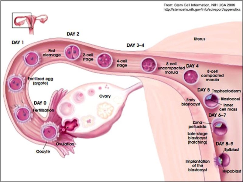
- 2. Zygote is the cell resulting from
- 4. Fertilization occurs in the ampullary part
- 5. Cleavage is a mitotic type of
- 7. Zygote immediately after its formation, in
- 8. This phase of development begins with
- 9. The 1st division of zygote gives rise
- 10. After three or four divisions, the
- 12. At this time, the morula consists
- 13. The inner cell mass will give
- 14. Morula enters uterine cavity about 4th day
- 15. The fluid-filled spaces soon fuse to
- 16. During the formation of blastocele the
- 17. The cells of the inner cell
- 19. The zygote lies free in the
- 20. At about 6th day after fertilization the
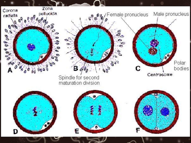
- 21. Zona pellucida Zona pellucida is created around
- 22. This Zona Pellucida is present in
- 23. Just before ovulation the primary oocyte
- 24. After ovulation the granulosa cells surrounding
- 25. During fertilization the sperms after passing
- 26. After fertilization Zona Pellucida contains ovum
- 27. Shortly after the morula enters the
- 29. After the blastocyst has floated in
- 30. IT IS IMPORTANT TO NOTE THAT

Слайд 2
Zygote is the cell resulting from the fertilization of a secondary
oocyte by a sperm.
The secondary oocyte finishes its second meiotic division immediately after the entry of the sperm and becomes ovum.
The secondary oocyte finishes its second meiotic division immediately after the entry of the sperm and becomes ovum.

Слайд 4
Fertilization occurs in the ampullary part of fallopian tube so zygote
is formed in the ampullary part of fallopian tube.
Immediately after its formation it starts:
1. Moving towards uterus
2. Multiplying mitotically
Immediately after its formation it starts:
1. Moving towards uterus
2. Multiplying mitotically

Слайд 5
Cleavage is a mitotic type of cell division where the daughter
cells are smaller than the parent cells.
The number of cells increases but the size of the daughter cells and the cell-mass does not increase.
The number of cells increases but the size of the daughter cells and the cell-mass does not increase.

Слайд 7
Zygote immediately after its formation, in the ampullary part, is guided
medially through the uterine tube toward the uterine cavity.
As the zygote, passes through the uterine tube, it undergoes mitotic cell divisions known as cleavage.
As the zygote, passes through the uterine tube, it undergoes mitotic cell divisions known as cleavage.

Слайд 8
This phase of development begins with the first mitotic division of
the zygote and ends with formation of blastocyst.
It extends for 6 to 7 days or a week.
It extends for 6 to 7 days or a week.

Слайд 9
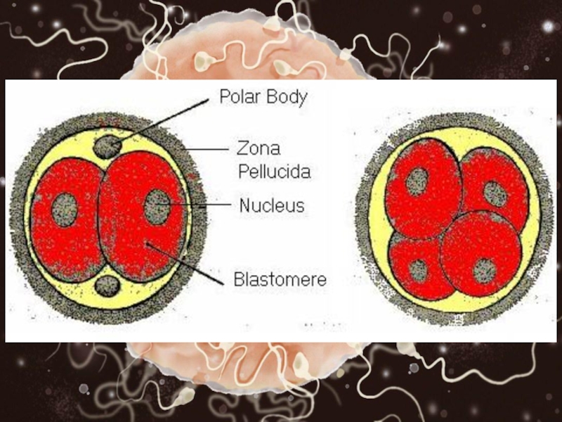
The 1st division of zygote gives rise to two daughter cells. They
are called blastomeres.
Each blastomere is half the size of parent cell. They develop about 30 hours after fertilization.
Subsequent divisions follow one another, forming progressively smaller blastomeres.
Each blastomere is half the size of parent cell. They develop about 30 hours after fertilization.
Subsequent divisions follow one another, forming progressively smaller blastomeres.

Слайд 10
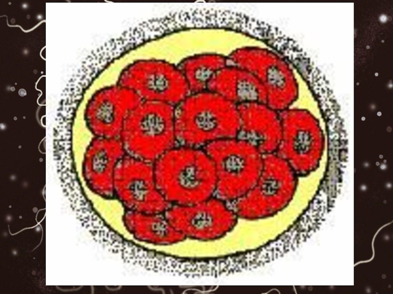
After three or four divisions, the zygote, looks like a mulberry
and is known as morula (L. Morus, mulberry).
It is a solid mass of 12 to 16 cells (blastomeres).
This stage is reached about three days after fertilization and the mass of cells is about to enter the uterus.
It is a solid mass of 12 to 16 cells (blastomeres).
This stage is reached about three days after fertilization and the mass of cells is about to enter the uterus.

Слайд 12
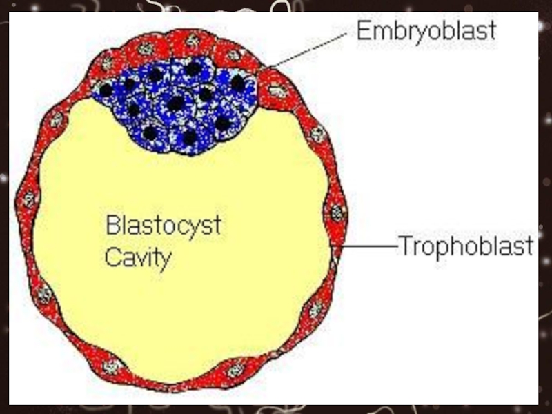
At this time, the morula consists of a group of centrally
located cells, the inner cell mass, and a surrounding layer, the outer cell mass.

Слайд 13
The inner cell mass will give rise to the tissues of
the embryo proper, while the outer cell mass forms the trophoblast which later contributes in the formation of placenta and other membranes.
Note also the degeneration process of zona pellucida.
Note also the degeneration process of zona pellucida.

Слайд 14
Morula enters uterine cavity about 4th day after fertilization. As the morula
enters the uterine cavity fluid begins to penetrate through the zona pellucida. Spaces appear between the inner cell mass and outer cell mass.

Слайд 15
The fluid-filled spaces soon fuse to form a single large space
or cavity, pushing the whole inner cell mass to one side.
The conceptus is now called blastocyst and the cavity is known as blastocyst cavity or blastocele.
The conceptus is now called blastocyst and the cavity is known as blastocyst cavity or blastocele.

Слайд 16
During the formation of blastocele the cells of outer cell mass
becomes flattened.
So the outer cell mass is now a single layer of flattened cells forming the wall of blastocyst and it is now given the name trophoblast (Gr. Trophe, nutrition), which will form placenta and associated membranes.
So the outer cell mass is now a single layer of flattened cells forming the wall of blastocyst and it is now given the name trophoblast (Gr. Trophe, nutrition), which will form placenta and associated membranes.

Слайд 17
The cells of the inner cell mass are attached to one
side of trophoblast and project into the blastocyst cavity.
They are now referred to as embryoblast, which will form the embryo.
They are now referred to as embryoblast, which will form the embryo.

Слайд 19
The zygote lies free in the uterine secretions for about two
days.
During this time the zona pellucida degenerates and disappears. The blastocyst hatches out and this is called hatching of blastocyst.
During this time the zona pellucida degenerates and disappears. The blastocyst hatches out and this is called hatching of blastocyst.

Слайд 20
At about 6th day after fertilization the embryonic pole of the blastocyst
becomes attached to the endometrium and start penetrating into it.

Слайд 21Zona pellucida
Zona pellucida is created around primary oocyte when primordial follicle
is transformed into primary follicle.
So Zona Pellucida is not present in primordial follicle while primary follicle is characterized by Zona Pellucida.
So Zona Pellucida is not present in primordial follicle while primary follicle is characterized by Zona Pellucida.

Слайд 22
This Zona Pellucida is present in all follicles (primary follicles, secondary
follicles, tertiary follicles and Gra’afian follicles) except primordial follicles.
Zona Pellucida surrounds primary oocyte and itself Zona Pellucida is surrounded by granulosa cells.
Zona Pellucida surrounds primary oocyte and itself Zona Pellucida is surrounded by granulosa cells.

Слайд 23
Just before ovulation the primary oocyte is converted into secondary oocyte
and it is always the secondary oocyte, which ovulates.
Now the Zona Pellucida contains secondary oocyte and first polar body.
Now the Zona Pellucida contains secondary oocyte and first polar body.

Слайд 24
After ovulation the granulosa cells surrounding the secondary oocyte and zona
pellucida are given the name corona radiata cells.

Слайд 25
During fertilization the sperms after passing through corona radiata, become attached
to Zona Pellucida. The enzymes released from the acrosome (esterases, acrosin, and neuraminidase) causes lysis of Zona Pellucida, thereby forming a path for the sperm through Zona Pellucida.

Слайд 26
After fertilization Zona Pellucida contains ovum with two polar bodies.
Zona pellucida
remains present around zygote and morula at different stages of development.

Слайд 27
Shortly after the morula enters the uterus (about 4 days after
fertilization), fluid starts appearing within the morula between the blastomeres.
This fluid passes from the uterine cavity through the zona pellucida to form spaces.
This fluid passes from the uterine cavity through the zona pellucida to form spaces.
, fluid starts appearing")

Слайд 29
After the blastocyst has floated in the uterine secretions for about
two days, the zona pellucida gradually degenerates, ruptures and disappears.
The blastocyst hatches out and this is called hatching of blastocyst.
The blastocyst hatches out and this is called hatching of blastocyst.

Слайд 30
IT IS IMPORTANT TO NOTE THAT THE LIFE OF ZONA PELLUCIDA
IS 15 DAYS IF THERE IS NO FERTILIZATION.
THE LIFE OF ZONA PELLUCIDA IS 20 DAYS IF WIFE BECOMES PREGNANT.
THE LIFE OF ZONA PELLUCIDA IS 20 DAYS IF WIFE BECOMES PREGNANT.