- Главная
- Разное
- Дизайн
- Бизнес и предпринимательство
- Аналитика
- Образование
- Развлечения
- Красота и здоровье
- Финансы
- Государство
- Путешествия
- Спорт
- Недвижимость
- Армия
- Графика
- Культурология
- Еда и кулинария
- Лингвистика
- Английский язык
- Астрономия
- Алгебра
- Биология
- География
- Детские презентации
- Информатика
- История
- Литература
- Маркетинг
- Математика
- Медицина
- Менеджмент
- Музыка
- МХК
- Немецкий язык
- ОБЖ
- Обществознание
- Окружающий мир
- Педагогика
- Русский язык
- Технология
- Физика
- Философия
- Химия
- Шаблоны, картинки для презентаций
- Экология
- Экономика
- Юриспруденция
Valvular Heart Diseases презентация
Содержание
- 1. Valvular Heart Diseases
- 3. Stages of Progression of Valvular Heart Disease
- 4. Innocent Murmurs Common in asymptomatic adults Characterized
- 5. Common Murmurs and Timing
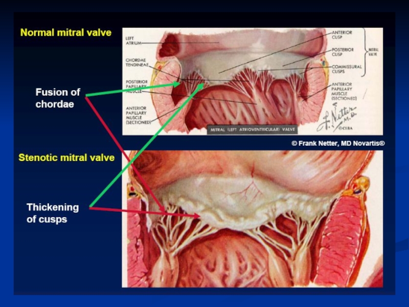
- 6. Mitral Valve Stenosis
- 7. Mitral Stenosis Etiology Rheumatic Heart Disease -99.8%
- 9. Pathophysiology
- 10. Pathophysiology Left atrial dilatation Allows larger volume
- 11. Symptoms Left sided failure Hemoptysis, URI Systemic embolism Palpitations Fatigue Right sided failure Hoarseness
- 12. Signs Loud S1 Opening snap following S2
- 13. Recognizing Mitral Stenosis Palpation: Small volume
- 14. Mitral stenosis murmur First heart sound (S1)
- 15. Lab Diagnosis EKG: A Fib, LAE, RVH
- 18. Echo - TTE
- 19. LAE LV AO Echo - TEE
- 20. Therapy Medical Diuretics: For pulmonary congestion, dyspnea
- 21. Mitral Valvuloplasty Percutaneous mitral balloon commissurotomy (PMBC)
- 25. Therapy Surgical Mitral commissurotomy: Effective long term improvement Mitral Valve Replacement Mechanical Bioprosthetic
- 26. MV Surgery Mitral valve surgery (repair, commissurotomy,
- 28. When to Perform Cardiac Catheterization in Valvular
- 29. Frequency of Echo Exam
- 30. Secondary Prevention of Rheumatic Fever Secondary prevention
- 31. Mitral Regurgitation
- 32. Etiology Valvular Myxomatous CT Disease Rheumatic Endocarditis
- 33. Pathophysiology
- 34. Symptoms Similar to MS Dyspnea, Orthopnea, PND
- 35. Signs Chronic MR Hyperdynamic, Displaced apex beat
- 36. Diagnosis EKG: LVH, LAE CXR: Cardiac enlargement Echo: Abnormal anatomy,
- 39. Echocardiography
- 40. Echo assessment of severity Color Doppler –
- 41. Therapy MEDICAL Diuretics: reduce vol. Overload Vasodilators:
- 45. MV Repair 1. Mitral valve repair is
- 46. Mitral Valve Prolapse
- 47. What is Mitral Valve Prolapse? Abnormal Mitral
- 48. Pathophysiology Forms Functional Common LV is small,
- 49. Symptoms Most patients: None Chest pain Palpitations Easy fatigability Arrhythmias TIA MR
- 50. Signs Mid-systolic Click Systolic murmur with co-existent MR Other connective tissue disorders
- 51. Diagnosis EKG: Non specific ST-T changes CXR: Usually normal
- 52. Therapy Functional MVP Reassurance Periodic clinical follow-up


Слайд 4Innocent Murmurs
Common in asymptomatic adults
Characterized by
Grade I – II @ LSB
Systolic
ejection pattern
Normal intensity & splitting of second sound (S2)
No other abnormal sounds or murmurs
No evidence of LVH, and no ↑ with Valsalva
S1 S2

Слайд 5
Common Murmurs and
Timing
Systolic Murmurs
Aortic stenosis
Mitral insufficiency
Mitral valve prolapse
Tricuspid insufficiency
Diastolic
Murmurs
Aortic insufficiency
Mitral stenosis
Aortic insufficiency
Mitral stenosis
S1 S2 S1


Слайд 7Mitral Stenosis
Etiology
Rheumatic Heart Disease -99.8% of cases
Normal Valve area: >4 cm2
Critical
MS: <1 cm2


Слайд 10Pathophysiology
Left atrial dilatation
Allows larger volume at low pressure
Prone to A. Fib
Thrombi
may form and embolize
Pulmonary artery vasoconstriction
PVR increases
Pressure overload to RV
RV dilates
PI, TR
Leads to RVH and RV failure
Pulmonary artery vasoconstriction
PVR increases
Pressure overload to RV
RV dilates
PI, TR
Leads to RVH and RV failure

Слайд 11Symptoms
Left sided failure
Hemoptysis, URI
Systemic embolism
Palpitations
Fatigue
Right sided failure
Hoarseness

Слайд 12Signs
Loud S1
Opening snap following S2
Narrow pulse pressure
Diastolic murmur
Atrial Fibrillation
Pulmonary congestion; Right
sided failure
Sternal lift, Loud S2, Elevated Jugular pressure, edema, hepatomegaly
Sternal lift, Loud S2, Elevated Jugular pressure, edema, hepatomegaly

Слайд 13Recognizing Mitral
Stenosis
Palpation:
Small volume pulse
Tapping apex-palpable S1
+/- palpable opening snap (OS)
RV
lift
Palpable S2
ECG:
LAE, AFIB, RVH, RAD
Palpable S2
ECG:
LAE, AFIB, RVH, RAD
Auscultation:
Loud S1- as loud as S2 in aortic area
A2 to OS interval inversely proportional to severity
Diastolic rumble: length proportional to severity
In severe MS with low flow- S1, OS & rumble may be inaudible
RV liftPalpable S2ECG: LAE, AFIB,")
Слайд 14Mitral stenosis murmur
First heart sound (S1) is accentuated and snapping
Opening snap
(OS) after aortic valve closure
Low pitch diastolic rumble at the apex
Pre-systolic accentuation (esp. if in sinus rhythm)
Low pitch diastolic rumble at the apex
Pre-systolic accentuation (esp. if in sinus rhythm)
S1 S2 OS S1
 is accentuated and snappingOpening snap (OS) after aortic valve")
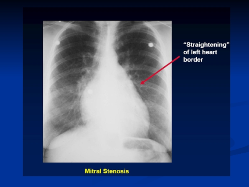
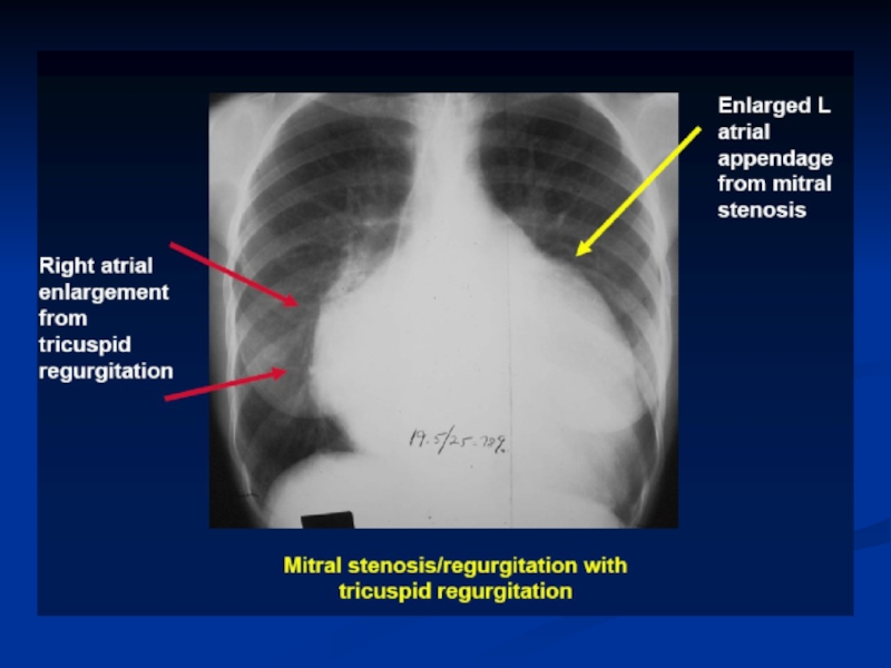
Слайд 15Lab Diagnosis
EKG: A Fib, LAE, RVH
CXR: Large LA, Pulm venous congestion,
RV dilatation, interstitial/alveolar edema
Echo: Valve orifice, calcification, pliability, size of the chambers, other valvular disease, quantification of stenosis and pulm. HTN
Cardiac Catheterization: Pressures and area
Echo: Valve orifice, calcification, pliability, size of the chambers, other valvular disease, quantification of stenosis and pulm. HTN
Cardiac Catheterization: Pressures and area


Слайд 20Therapy
Medical
Diuretics: For pulmonary congestion, dyspnea and orthopnea
Rate control in A Fib:
Beta blockers, Ca channel blockers, amiodarone, propafenone, digitalis?
Anticoagulation: In A Fib
Balloon Valvuloplasty
Effective long term improvement
Anticoagulation: In A Fib
Balloon Valvuloplasty
Effective long term improvement

Слайд 21Mitral Valvuloplasty
Percutaneous mitral balloon commissurotomy (PMBC) is recommended for symptomatic patients
with severe MS (mitral valve area <1.5 cm2, stage D) and favorable valve morphology in the absence of left atrial thrombus or moderate-to-severe MR
Percutaneous mitral balloon commissurotomy may be considered for symptomatic patients with mitral valve area greater than 1.5 cm2 if there is evidence of hemodynamically significant MS based on pulmonary artery wedge pressure greater than 25 mm Hg or mean mitral valve gradient greater than 15 mm Hg during
exercise.
 is recommended for symptomatic patients with severe MS (mitral")


Слайд 25Therapy
Surgical
Mitral commissurotomy:
Effective long term improvement
Mitral Valve Replacement
Mechanical
Bioprosthetic

Слайд 26MV Surgery
Mitral valve surgery (repair, commissurotomy, or valve
replacement) is indicated in
severely symptomatic patients
(NYHA class III to IV) with severe MS (mitral valve area £1.5
cm2, stage D) who are not high risk for surgery and who are not
candidates for or who have failed previous percutaneous mitral
balloon commissurotomy
Concomitant mitral valve surgery may be considered for patients
with moderate MS (mitral valve area 1.6 cm2 to 2.0 cm2)
undergoing cardiac surgery for other indications.
 is indicated in severely symptomatic patients(NYHA class")

Слайд 28When to Perform Cardiac Catheterization in Valvular Patient?
No “routine” cardiac
catheterization
Cardiac catheterization for hemodynamic assessment is recommended in symptomatic patients when noninvasive tests are inconclusive or when there is a discrepancy between the findings on noninvasive testing and physical examination regarding severity of the valve lesion.
Cardiac catheterization for hemodynamic assessment is recommended in symptomatic patients when noninvasive tests are inconclusive or when there is a discrepancy between the findings on noninvasive testing and physical examination regarding severity of the valve lesion.


Слайд 30Secondary Prevention of Rheumatic Fever
Secondary prevention of rheumatic fever is indicated
in patients
with rheumatic heart disease, specifically mitral stenosis (MS)
with rheumatic heart disease, specifically mitral stenosis (MS)


Слайд 32Etiology
Valvular
Myxomatous CT Disease
Rheumatic
Endocarditis
Chordae
Annulus
Calcification
Papillary Muscles
CAD (Ischemia, Infarction)
Infiltrative disorders
LV Dilatation & Functional Prolapse
Infiltrative disordersLV Dilatation & Functional Prolapse")

Слайд 34Symptoms
Similar to MS
Dyspnea, Orthopnea, PND
Fatigue
Pulmonary HTN, Right sided failure
Systemic embolization in
A Fib

Слайд 35Signs
Chronic MR
Hyperdynamic, Displaced apex beat
Apical holosystolic murmur
Pounding pulse
Variable Pulm. HTN
Acute
MR
Marked pulmonary congestion
Short systolic murmur
Small pulse
Marked pulm. HTN; Loud single S2
Giant V wave in LA pressure tracing
Marked pulmonary congestion
Short systolic murmur
Small pulse
Marked pulm. HTN; Loud single S2
Giant V wave in LA pressure tracing

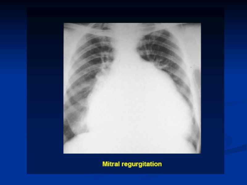
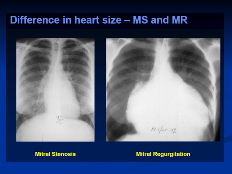
Слайд 36Diagnosis
EKG: LVH, LAE
CXR: Cardiac enlargement
Echo: Abnormal anatomy, chamber size, EF, Qualitative assessment of MR
and Pulmonary HTN, suitability for repair
Cardiac Catheterization: Measure pulmonary arterial & Wedge pressures, EF, Severity of MR
Cardiac Catheterization: Measure pulmonary arterial & Wedge pressures, EF, Severity of MR


Слайд 40Echo assessment of severity
Color Doppler – may be misleading
Calculations
Effective regurgitant orifice
Regurgitant
Volume, Regurgitant fraction
Pulmonary venous flow reversal
Pulmonary venous flow reversal

Слайд 41Therapy
MEDICAL
Diuretics: reduce vol. Overload
Vasodilators: Increase forward output and decrease LV size
Digitalis:
Control HR, Inotrope in Chronic MR
Anticoagulants: A Fib
Anticoagulants: A Fib
SURGICAL: Indicated for severe symptoms and LV failure
Valve repair: Preserves LV function
Valve Replacement:
Bioprosthetic
Mechanical


Слайд 45MV Repair
1. Mitral valve repair is performed at a lower operative
mortality rate than MVR. Although no RCTs exist, virtually every clinical report, including data from the
STS database, indicates that operative risk (30–day mortality) for repair is about half that of MVR.
2. LV function is better preserved following repair preserving the integrity of the mitral valve apparatus versus following MVR.
3. Repair avoids the risks inherent to prosthetic heart valves, that is, thromboembolism or anticoagulant induced hemorrhage for mechanical valves or structural deterioration for bioprosthetic valves.


Слайд 47What is Mitral Valve Prolapse?
Abnormal Mitral Valve mechanism which results in
billowing of one or both mitral leaflets into the Left atrium towards the end of systole
3-5% of population
2:1 Female preponderance
3-5% of population
2:1 Female preponderance

Слайд 48Pathophysiology
Forms
Functional
Common
LV is small, Hyperdynamic
Valve is normal
Organic (Myxomatous Degeneration)
Uncommon
LV: Nl to Large
Thickened
& Bulging valve leaflets
UncommonLV: Nl to LargeThickened & Bulging valve leaflets")


Слайд 51Diagnosis
EKG: Non specific ST-T changes
CXR: Usually normal
Echo: Mitral valve anatomy, leaflet thickness, degree of
prolapse, assessment of MR, LV function.

Слайд 52Therapy
Functional MVP
Reassurance
Periodic clinical follow-up
Organic MVP
Treat MR
Anticoagulation, if h/o TIA
B-blockers for palpitations
Endocarditis
prophylaxis: not anymore
ICD for Vtach
MVR for severe MR
ICD for Vtach
MVR for severe MR