- Главная
- Разное
- Дизайн
- Бизнес и предпринимательство
- Аналитика
- Образование
- Развлечения
- Красота и здоровье
- Финансы
- Государство
- Путешествия
- Спорт
- Недвижимость
- Армия
- Графика
- Культурология
- Еда и кулинария
- Лингвистика
- Английский язык
- Астрономия
- Алгебра
- Биология
- География
- Детские презентации
- Информатика
- История
- Литература
- Маркетинг
- Математика
- Медицина
- Менеджмент
- Музыка
- МХК
- Немецкий язык
- ОБЖ
- Обществознание
- Окружающий мир
- Педагогика
- Русский язык
- Технология
- Физика
- Философия
- Химия
- Шаблоны, картинки для презентаций
- Экология
- Экономика
- Юриспруденция
Tumor pathogenesis. (Lecture 7) презентация
Содержание
- 1. Tumor pathogenesis. (Lecture 7)
- 2. Growth factors Epidermal growth factor Endothelial
- 3. Genes controlling growth The genes which
- 4. Mutagenic factors chemical - pro-carcinogens (substances that
- 5. Mutagenic factors physical (ionizing radiation, ultraviolet rays);
- 6. Epigenetic carcinogenesis Transformation of the normal
- 7. The main causes of cancer Smoking,
- 8. Role of host factors and environment Microsomal
- 9. Definitions Neoplasia A pathologic process in which
- 10. General pathogenesis of tumor mutation CLONE mutation
- 11. General Pathogenesis of tumors Stage 1.
- 12. General Pathogenesis of tumors Stage 3. TUMOR
- 13. General pathogenesis of tumors Initiation Promotion Progression
- 14. Types of neoplasms Benign – less autonomy,
- 15. Common characteristics of neoplasms Neoplasia is
- 16. Common characteristics of neoplasms Absence of cell
- 17. Common characteristics of neoplasms Inadequate Differentiation Neoplastic
- 18. Characteristics Unique to Malignant Neoplasms Loss of
- 19. Characteristics Unique to Malignant Neoplasms Morphological anaplasia.
- 20. Characteristics Unique to Malignant Neoplasms Biochemical anaplasia.
- 21. Characteristics Unique to Malignant Neoplasms Functional anaplasia
- 22. Characteristics Unique to Malignant Neoplasms Loss of
- 23. Characteristics Unique to Malignant Neoplasms Invasion Malignant
- 24. Characteristics Unique to Malignant Neoplasms Invasive malignancies
- 25. Characteristics Unique to Malignant Neoplasms Metastasis The
- 26. Invasion and metastasis of malignant tumor cells
- 27. Invasion and metastasis of malignant tumor cells
- 28. Invasion and metastasis of malignant tumor cells
- 29. Metastasis of tumor cells Malignant tumor cells
- 30. Other Differences between Benign and Malignant Neoplasms
- 31. Other Differences between Benign and Malignant Neoplasms
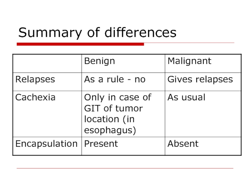
- 32. Summary of differences
- 33. Summary of differences
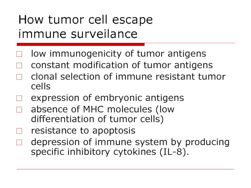
- 34. How tumor cell escape immune surveilance
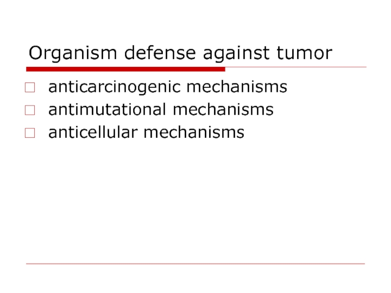
- 35. Organism defense against tumor anticarcinogenic mechanisms antimutational mechanisms anticellular mechanisms
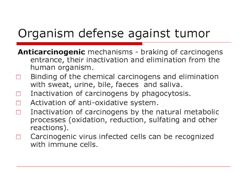
- 36. Organism defense against tumor Anticarcinogenic mechanisms -
- 37. Organism defense against tumor Antimutational mechanisms provide
- 38. Organism defense against tumor TNF is released
- 39. Interrelations between the host organism and the
- 40. Interrelations between the host organism and the
- 41. Interrelations between the host organism and the
- 42. Cancer grading and staging The
- 43. Cancer grading and staging Staging of a
- 44. Treatment of Neoplasms Surgery.” Situations in which
- 45. Treatment of neoplasms Immunotherapy. Stimulation of immune
Слайд 2Growth factors
Epidermal growth factor
Endothelial growth factor
Fibroblast growth factor
Platelet-derived growth
factor
Nerve growth factor
Insulin-like growth factors
Hepatocyte growth factor
Transforming growth factor
Interleukins 1-7
Nerve growth factor
Insulin-like growth factors
Hepatocyte growth factor
Transforming growth factor
Interleukins 1-7
Слайд 3Genes controlling growth
The genes which are controlling growth factors synthesis
are named proto-oncogenes.
If gene controlling growth factor is mutant or hyper activated it is named oncogen.
Tumor suppressor gene or anti-oncogenes - their protein product inhibits mitosis
If gene controlling growth factor is mutant or hyper activated it is named oncogen.
Tumor suppressor gene or anti-oncogenes - their protein product inhibits mitosis
Слайд 4Mutagenic factors
chemical - pro-carcinogens (substances that can convert to carcinogens) and
carcinogens.
Aflatoxin (from fungus Aspergillus flavus) growing on stored grains, nuts and peanut butter
Cooking protein-rich food at high temperatures can lead to the formation of many potent carcinogens.
Industrial and tobacco smoke contain benzopyrene, tobacco-specific nitrosamines such as nitrosonornicotine
Formaldehyde —embalming and making plastics.
Слайд 5Mutagenic factors
physical (ionizing radiation, ultraviolet rays);
biological - viruses. The viral
particles can carry a gene that encodes for an overactive oncogene called viral-oncogene and the infected cell is transformed into tumor cell.
When tumor development occurs due to above mentioned cause carcinogenesis is named mutational.
When tumor development occurs due to above mentioned cause carcinogenesis is named mutational.
Слайд 6Epigenetic carcinogenesis
Transformation of the normal cel to tumor one without
mutations by stimulating mitosis may be caused by:
certain hormones
chronic tissue injury
agents that cause inflammation
certain viruses.
certain hormones
chronic tissue injury
agents that cause inflammation
certain viruses.
Слайд 7The main causes of cancer
Smoking,
Dietary imbalances (excess fat and calories;
inadequate intake of fruits, vegetables, fiber, and calcium)
Chronic infections leading to chronic inflammation. Chronic inflammation is a major cause of cancer in the world because it releases powerful oxidants which both stimulate cell division and are mutagens.
Chronic infections leading to chronic inflammation. Chronic inflammation is a major cause of cancer in the world because it releases powerful oxidants which both stimulate cell division and are mutagens.
Слайд 8Role of host factors and environment
Microsomal enzymes in the liver degrade
a large part of a pro-carcinogens to non-carcinogenic products.
Naturally occurring compounds, such as indole, flavones, and related compounds that occur in vegetables have a protective action in animals exposed to carcinogenic polycyclic hydrocarbons.
Endogenous (and exogenous) sex hormones are important factors apparently in the promotion stage of human carcinomas of breast, endometrium, and prostate.
Слайд 9Definitions
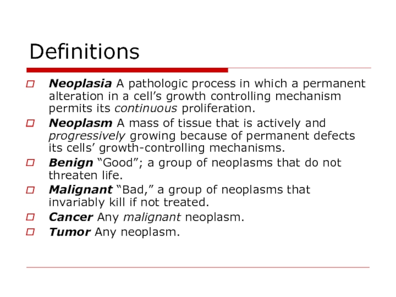
Neoplasia A pathologic process in which a permanent alteration in a
cell’s growth controlling mechanism permits its continuous proliferation.
Neoplasm A mass of tissue that is actively and progressively growing because of permanent defects its cells’ growth-controlling mechanisms.
Benign “Good”; a group of neoplasms that do not threaten life.
Malignant “Bad,” a group of neoplasms that invariably kill if not treated.
Cancer Any malignant neoplasm.
Tumor Any neoplasm.
Слайд 11General Pathogenesis of tumors
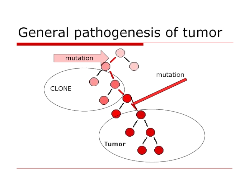
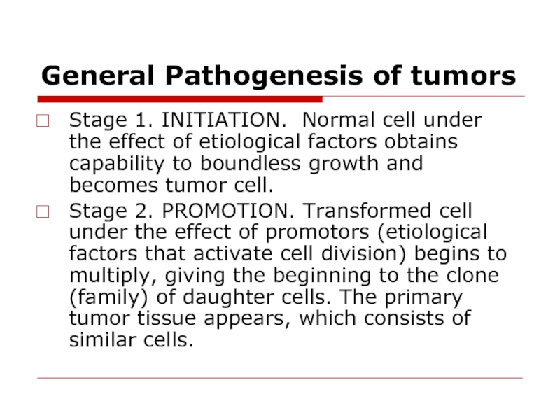
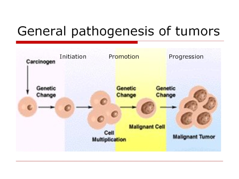
Stage 1. INITIATION. Normal cell under the
effect of etiological factors obtains capability to boundless growth and becomes tumor cell.
Stage 2. PROMOTION. Transformed cell under the effect of promotors (etiological factors that activate cell division) begins to multiply, giving the beginning to the clone (family) of daughter cells. The primary tumor tissue appears, which consists of similar cells.
Stage 2. PROMOTION. Transformed cell under the effect of promotors (etiological factors that activate cell division) begins to multiply, giving the beginning to the clone (family) of daughter cells. The primary tumor tissue appears, which consists of similar cells.
Слайд 12General Pathogenesis of tumors
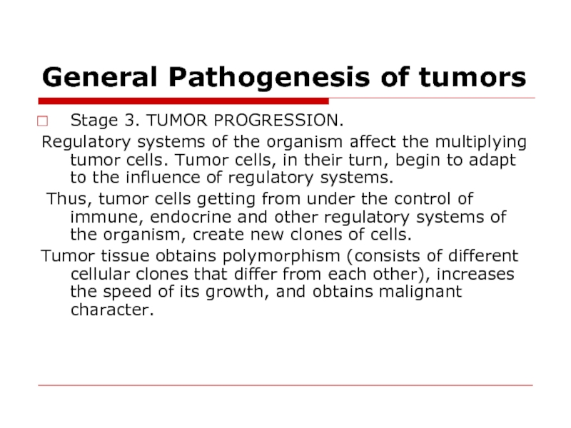
Stage 3. TUMOR PROGRESSION.
Regulatory systems of the
organism affect the multiplying tumor cells. Tumor cells, in their turn, begin to adapt to the influence of regulatory systems.
Thus, tumor cells getting from under the control of immune, endocrine and other regulatory systems of the organism, create new clones of cells.
Tumor tissue obtains polymorphism (consists of different cellular clones that differ from each other), increases the speed of its growth, and obtains malignant character.
Слайд 14Types of neoplasms
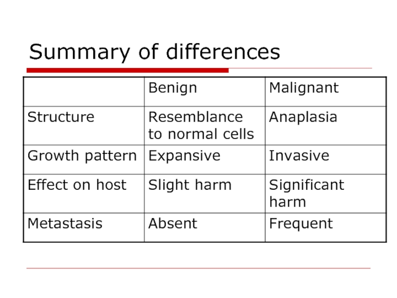
Benign – less autonomy, usually not invasive, does not
metastasize, and generally produces no great harm if treated adequately.
Malignant – manifests a greater degree of autonomy, is capable of invasion and metastatic spread, may be resistant to treatment, and may cause death.
Malignant – manifests a greater degree of autonomy, is capable of invasion and metastatic spread, may be resistant to treatment, and may cause death.
Слайд 15Common characteristics of neoplasms
Neoplasia is an irreversible new growth
Growth
controlling mechanisms are impaired in neoplastic cells.
The difference between hyperplasia and neoplasia:
Hyperplasia is characterized by cellular proliferation, once its cause is removed, growth stops. Therefore, in hyperplasia growth is “active” but not “progressive.”
Neoplastic cells continue to divide even when their causes are removed.
The difference between hyperplasia and neoplasia:
Hyperplasia is characterized by cellular proliferation, once its cause is removed, growth stops. Therefore, in hyperplasia growth is “active” but not “progressive.”
Neoplastic cells continue to divide even when their causes are removed.
Слайд 16Common characteristics of neoplasms
Absence of cell division limit.
Normal cells have
division limit
Tumor cells are free to divide unlimited amount of times, they are theoretically immortal.
The doubling rate of benign cells is relatively slow.
The growth rate is much faster in malignant neoplasms.
Tumor cells are free to divide unlimited amount of times, they are theoretically immortal.
The doubling rate of benign cells is relatively slow.
The growth rate is much faster in malignant neoplasms.
Слайд 17Common characteristics of neoplasms
Inadequate Differentiation
Neoplastic cells do not become as specialized
as do normal cells.
Malignant neoplasms are composed of less-differentiated cells than benign ones.
Benign neoplasms are composed of cells that more closely resemble normal, mature, adult, differentiated cells.
Malignant neoplasms are composed of cells that more closely resemble primitive, immature, juvenile, undifferentiated, stem cells.
Слайд 18Characteristics Unique to Malignant Neoplasms
Loss of differentiation is known as “anaplasia.”
Anaplasia
is seen only in malignant neoplasms
Anaplasia may be severe or mild. The more anaplasia that is observed, the more malignant a neoplasm is.
There are several types of anaplasia: morphological, biochemical, physical, chemical, functional.
Anaplasia may be severe or mild. The more anaplasia that is observed, the more malignant a neoplasm is.
There are several types of anaplasia: morphological, biochemical, physical, chemical, functional.
Слайд 19Characteristics Unique to Malignant Neoplasms
Morphological anaplasia.
Pleomorphism (pleo- = many; -morphism
= shape).
Abnormally large nuclei (increased nuclear-cytoplasmic ratio).
Abnormally dark nuclei—hyperchromatism. (hyper- = increased; -chromatism = color).
Abnormally large and multiplied nucleoli.
The quantity and quality of other cell organelles may be changed too. (mitochondrions and ribosomes).
Abnormally large nuclei (increased nuclear-cytoplasmic ratio).
Abnormally dark nuclei—hyperchromatism. (hyper- = increased; -chromatism = color).
Abnormally large and multiplied nucleoli.
The quantity and quality of other cell organelles may be changed too. (mitochondrions and ribosomes).
Слайд 20Characteristics Unique to Malignant Neoplasms
Biochemical anaplasia.
Carbohydrate atypia –shift to anaerobic glycolysis.
negative Paster’s effect - great amount of sub-oxidized products occurs after decomposition of carbohydrates, particularly, lactic acid.
Protein atypia - appearance of unusual proteins during certain kinds of tumors. The presence of these proteins, also called embryony proteins or Abbel’s proteins, is a diagnostic test for hepatoma identification.
Protein atypia - appearance of unusual proteins during certain kinds of tumors. The presence of these proteins, also called embryony proteins or Abbel’s proteins, is a diagnostic test for hepatoma identification.
Слайд 21Characteristics Unique to Malignant Neoplasms
Functional anaplasia
Normal cells stop their division
when they contact surrounding cells, and that is called contact inhibition.
Tumor cells do not stop their division even when they contact surrounding cells. Thus they destroy healthy cells and grow into the surrounding tissue.
Autonomy (independence from the organism)
Tumor cells get out of control of regulating systems of the organism (nervous, endocrine, immune).
Tumor cells do not stop their division even when they contact surrounding cells. Thus they destroy healthy cells and grow into the surrounding tissue.
Autonomy (independence from the organism)
Tumor cells get out of control of regulating systems of the organism (nervous, endocrine, immune).
Слайд 22Characteristics Unique to Malignant Neoplasms
Loss of polaruty and specialized functions
Normal differentiated
cells are polar and possess specialized functions.
Anaplastic cells lose their polarity and specialized functions.
The more severe is the anaplasia, the more polarity and specialized functions are impaired. The less severe the anaplasia, the more cells show polarity and specialized functions.
Anaplastic cells lose their polarity and specialized functions.
The more severe is the anaplasia, the more polarity and specialized functions are impaired. The less severe the anaplasia, the more cells show polarity and specialized functions.
Слайд 23Characteristics Unique to Malignant Neoplasms
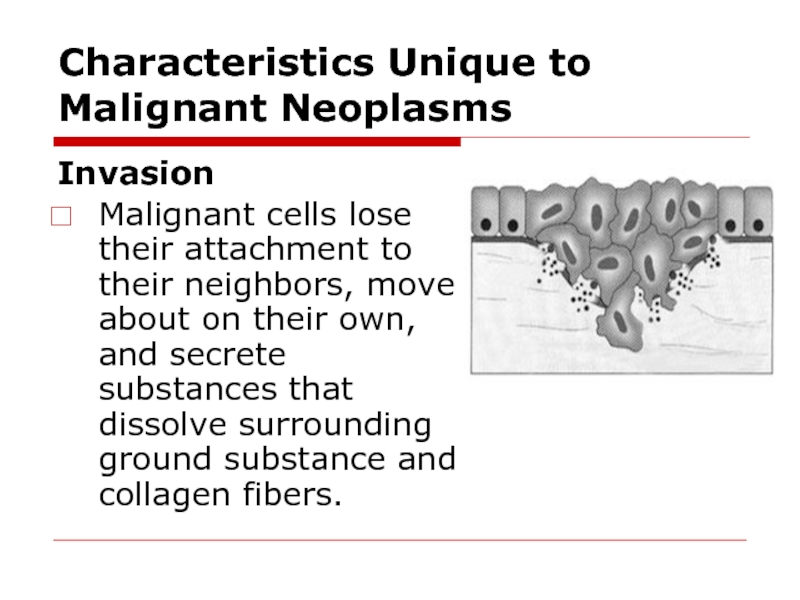
Invasion
Malignant cells lose their attachment to their
neighbors, move about on their own, and secrete substances that dissolve surrounding ground substance and collagen fibers.
Слайд 24Characteristics Unique to Malignant Neoplasms
Invasive malignancies are difficult to eradicate.
There is
no line of demarcation around malignant neoplasms
As malignant cells invade they infiltrate important structures—nerves, ducts, bone marrow.
Malignant cells can invade lymphatic and blood vessels providing ready access to body regions far from their point of origin.
As malignant cells invade they infiltrate important structures—nerves, ducts, bone marrow.
Malignant cells can invade lymphatic and blood vessels providing ready access to body regions far from their point of origin.
Слайд 25Characteristics Unique to Malignant Neoplasms
Metastasis
The tendency of malignant neoplasms to spread
far is called metastasis (meta- = change, -stasis = location).
The term “metastasis” is only used in reference to malignant neoplasms.
Invasion is a prerequisite for tumor cell metastasis
The presence of metastasis is a major factor in determining the prognosis of a malignant neoplasm.
Metastasis involves three steps or processes: tumor cell invasion, embolization, and extravasation
Слайд 26Invasion and metastasis of malignant tumor cells
Tumor cell invasion.
The sequence
of events:
detachment of tumor cells
from the primary tumor;
attachment to basement membrane matrix;
degradation of basement membrane matrix;
locomotion and infiltration of tumor cells;
degradation of extracellular matrix;
degradation of vascular basement membrane matrix.
detachment of tumor cells
from the primary tumor;
attachment to basement membrane matrix;
degradation of basement membrane matrix;
locomotion and infiltration of tumor cells;
degradation of extracellular matrix;
degradation of vascular basement membrane matrix.
Слайд 27Invasion and metastasis of malignant tumor cells
Tumor Cell Embolization.
Malignant tumor
cells may invade lymphatic or blood vessels and enter the circulation.
Only an extremely small number of embolized cells are able to establish metastatic lesions.
The survival and growth of metastatic cells depends on the selection of cancer cells possessing specific properties needed for metastatic growth
Only an extremely small number of embolized cells are able to establish metastatic lesions.
The survival and growth of metastatic cells depends on the selection of cancer cells possessing specific properties needed for metastatic growth
Слайд 28Invasion and metastasis of malignant tumor cells
Tumor Cell Extravasation. The sequence
of mechanisms includes:
adhesion to endothelial cells
endothelial cell retraction
migration
degradation of matrix
locomotion.
adhesion to endothelial cells
endothelial cell retraction
migration
degradation of matrix
locomotion.
Слайд 29Metastasis of tumor cells
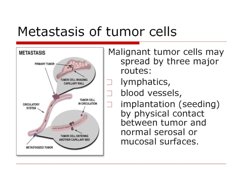
Malignant tumor cells may spread by three major
routes:
lymphatics,
blood vessels,
implantation (seeding) by physical contact between tumor and normal serosal or mucosal surfaces.
lymphatics,
blood vessels,
implantation (seeding) by physical contact between tumor and normal serosal or mucosal surfaces.
Слайд 30Other Differences between Benign and Malignant Neoplasms
Benign Neoplasms growth pattern is
known as expansive growth. Sometimes a fibrous connective tissue band surrounding the neoplasm—a capsule. When they arise from some surface, benign neoplasms almost always grow outward from the surface—a pattern known an exophytic growth.
Benign neoplasms never invade surrounding tissues; they never metastasize; are composed of well-differentiated cells that resemble the tissue from which they originated. Cells of benign neoplasms stay attached to each other. Because of their cohesiveness, they do not enter blood or lymphatic vessels to spread elsewhere.
Benign neoplasms never invade surrounding tissues; they never metastasize; are composed of well-differentiated cells that resemble the tissue from which they originated. Cells of benign neoplasms stay attached to each other. Because of their cohesiveness, they do not enter blood or lymphatic vessels to spread elsewhere.
Слайд 31Other Differences between Benign and Malignant Neoplasms
Malignant neoplasms grow rapidly and
have the capacity to invade surrounding tissues. They are not well demarcated from the surrounding tissues—there is no capsule. If they arise from some surface, malignant neoplasms grow inward invading the underlying tissues, a pattern called endophytic growth.
Metastatic malignant neoplasms cause severe body wasting—cachexia. Cachexia is loss of weight, muscle atrophy, fatigue, weakness and anorexia. Cachexia is often seen in end-stage cancer, and in that context is called "cancer cachexia".
Metastatic malignant neoplasms cause severe body wasting—cachexia. Cachexia is loss of weight, muscle atrophy, fatigue, weakness and anorexia. Cachexia is often seen in end-stage cancer, and in that context is called "cancer cachexia".
Слайд 34How tumor cell escape
immune surveilance
low immunogenicity of tumor antigens
constant modification
of tumor antigens
clonal selection of immune resistant tumor cells
expression of embryonic antigens
absence of MHC molecules (low differentiation of tumor cells)
resistance to apoptosis
depression of immune system by producing specific inhibitory cytokines (IL-8).
clonal selection of immune resistant tumor cells
expression of embryonic antigens
absence of MHC molecules (low differentiation of tumor cells)
resistance to apoptosis
depression of immune system by producing specific inhibitory cytokines (IL-8).
Слайд 35Organism defense against tumor
anticarcinogenic mechanisms
antimutational mechanisms
anticellular mechanisms
Слайд 36Organism defense against tumor
Anticarcinogenic mechanisms - braking of carcinogens entrance, their
inactivation and elimination from the human organism.
Binding of the chemical carcinogens and elimination with sweat, urine, bile, faeces and saliva.
Inactivation of carcinogens by phagocytosis.
Activation of anti-oxidative system.
Inactivation of carcinogens by the natural metabolic processes (oxidation, reduction, sulfating and other reactions).
Carcinogenic virus infected cells can be recognized with immune cells.
Слайд 37Organism defense against tumor
Antimutational mechanisms provide revealing, elimination or inhibition of
oncogenes activity with the help of tumor suppressor gene (anti-oncogenes) or DNA reparative systems.
Anticellular mechanisms provide recognition and destruction of tumor cells or inhibition of their division process.
Non-immune cellular mechanisms - phagocytes and NK cells.
Non-immune humoral mechanisms - tumor necrosis factor alpha (TNF).
Immune mechanisms are represented by cellular immunity (cytotoxic T-cells – T-killers) and humoral immunity (specific antibodies formation).
Слайд 38Organism defense against tumor
TNF is released primarily by WBC and endothelium.
Stimulating of the corticotropin releasing hormone (CRH) release in the hypothalamus
Suppressing appetite (hence its name "cachexin").
Fever.
Activates liver function (C-reactive protein).
Oncolytic effect of TNF is provided due to its ability:
To attract neutrophils
To stimulate phagocytosis and activate “respiratory burst” in phagocytes
To activate thrombogenesis in the vessels of tumor
Слайд 39Interrelations between the host organism and the tumor
Peculiarities of tumor
Location
is of critical importance with both benign and malignant tumors (e.g. adenoma compress hypophysis; carcinoma within the common bile duct may induce fatal biliary tract obstruction).
The production of hormones. (pancreatic adenoma often produces hyperinsulinism).
Ulceration through a surface with consequent bleeding or secondary infection
Intussusception— leading to ulceration of the mucosa or intestinal obstruction or infarction.
The production of hormones. (pancreatic adenoma often produces hyperinsulinism).
Ulceration through a surface with consequent bleeding or secondary infection
Intussusception— leading to ulceration of the mucosa or intestinal obstruction or infarction.
Слайд 40Interrelations between the host organism and the tumor
Cancer Cachexia is a
progressive loss of body weight accompanied by profound weakness, anorexia, and anemia.
Small, localized cancers therefore are generally silent and produce no cachexia, but there are exceptions.
The origins of cancer cachexia:
Anorexia - due to abnormalities in taste and in the central control of appetite.
High basal metabolic rate.
TNF suppresses appetite and inhibits the action of lipoprotein lipase, thereby inhibiting the release of free fatty acids from lipoproteins.
Слайд 41Interrelations between the host organism and the tumor
The paraneoplastic syndromes: hypercalcemia,
Cushing's syndrome, and nonbacterial thrombotic endocarditis.
Cushing’s syndrome as a paraneoplastic phenomenon is usually related to ectopic production by the cancer of ACTH or ACTH-like polypeptides.
The mediation of hypercalcemia may be caused by the cancer secretion of a parathyroid hormone-like peptide.
Paraneoplastic syndromes may take many other forms, such as hypercoagulability leading to venous thrombosis and nonbacterial thrombotic endocarditis.
Слайд 42Cancer grading and staging
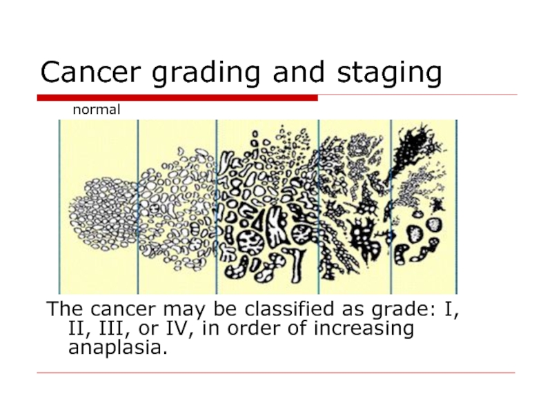
The cancer may be classified as
grade: I, II, III, or IV, in order of increasing anaplasia.
normal
Слайд 43Cancer grading and staging
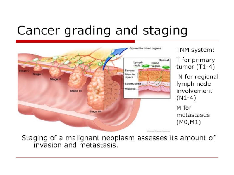
Staging of a malignant neoplasm assesses its amount
of invasion and metastasis.
TNM system:
T for primary tumor (T1-4)
N for regional lymph node involvement (N1-4)
M for metastases (M0,M1)
Слайд 44Treatment of Neoplasms
Surgery.” Situations in which little surrounding tissue needs to
be sacrificed is called “simple surgical excision”.
Surgery performed for lesions that have invaded is often described as “radical” because much surrounding tissue is sacrificed.
Radiation kills cells which are dividin. Radiation therapy and surgery are often used together.
Chemotherapy. Certain chemicals affect cell which are in mitosis process.
Surgery performed for lesions that have invaded is often described as “radical” because much surrounding tissue is sacrificed.
Radiation kills cells which are dividin. Radiation therapy and surgery are often used together.
Chemotherapy. Certain chemicals affect cell which are in mitosis process.
Слайд 45Treatment of neoplasms
Immunotherapy. Stimulation of immune system to attack cancer cells
(BCG).
Hormone Therapy. Some neoplasms are hormone-dependent.
Cryotherapy is used for surface lesions where the freezing agent can be controlled accurately.
Electosurgery resembles scalpel surgery with two exceptions. First, an electric current makes a wire loop hot enough to cut tissue. Second, the tissue around the edge of the surgical specimen is incinerated.
