E.
VM-409
Checked: Zhakipbekova. A.
- Главная
- Разное
- Дизайн
- Бизнес и предпринимательство
- Аналитика
- Образование
- Развлечения
- Красота и здоровье
- Финансы
- Государство
- Путешествия
- Спорт
- Недвижимость
- Армия
- Графика
- Культурология
- Еда и кулинария
- Лингвистика
- Английский язык
- Астрономия
- Алгебра
- Биология
- География
- Детские презентации
- Информатика
- История
- Литература
- Маркетинг
- Математика
- Медицина
- Менеджмент
- Музыка
- МХК
- Немецкий язык
- ОБЖ
- Обществознание
- Окружающий мир
- Педагогика
- Русский язык
- Технология
- Физика
- Философия
- Химия
- Шаблоны, картинки для презентаций
- Экология
- Экономика
- Юриспруденция
Treatment of atresia ani презентация
Содержание
- 1. Treatment of atresia ani
- 2. Atresia ani is a genetic disease of
- 4. A number of forms have been characterized
- 6. Rectovaginal fistula formation are sometimes observed in
- 8. Atresia ani and coli are congenital defects
- 9. Intestinal atresias are the most common abdominal
- 18. If your horse has any type of
- 19. Atresia ani in horses is an uncommon
- 20. Symptoms of Atresia Ani in Horses
- 22. Causes of Atresia Ani in Horses
- 26. Type I - The veterinarian will use
- 27. Recovery of Atresia Ani in Horses
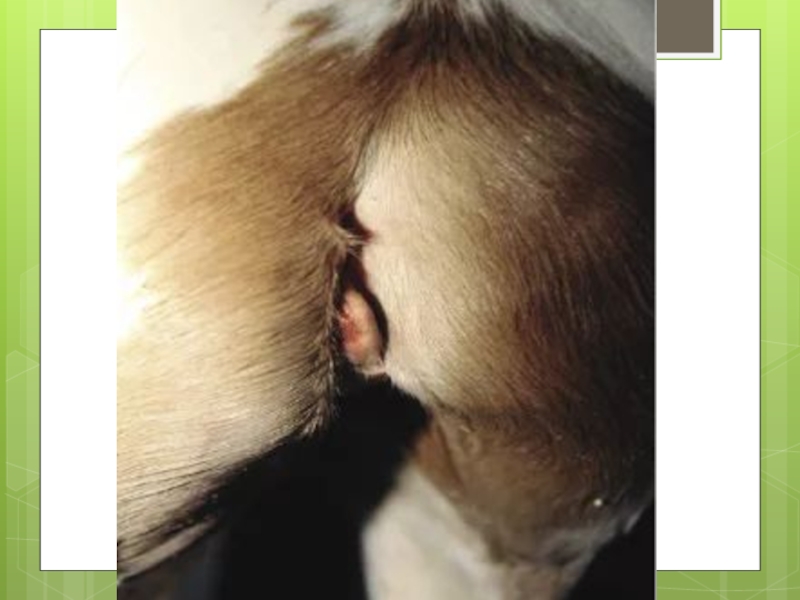
Слайд 2Atresia ani is a genetic disease of dogs characterized by malformation
or non-formation of the anus.
It is the most common anorectal anomaly in dogs and an increased incidence is found in females and in several breeds, including Miniature Poodles, Toy Poodles and Boston Terriers
It is the most common anorectal anomaly in dogs and an increased incidence is found in females and in several breeds, including Miniature Poodles, Toy Poodles and Boston Terriers
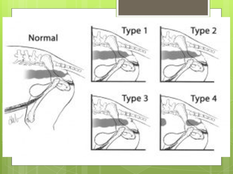
Слайд 4 A number of forms have been characterized including:
Type I - congenital
anal stenosis
Type II - imperforate anus alone
Type III - imperforate anus combined with more cranial termination of the rectum as a blind pouch
Type IV - discontinuity of the proximal rectum with normal anal and terminal rectal development
Type II - imperforate anus alone
Type III - imperforate anus combined with more cranial termination of the rectum as a blind pouch
Type IV - discontinuity of the proximal rectum with normal anal and terminal rectal development
Слайд 6Rectovaginal fistula formation are sometimes observed in these affected patients.
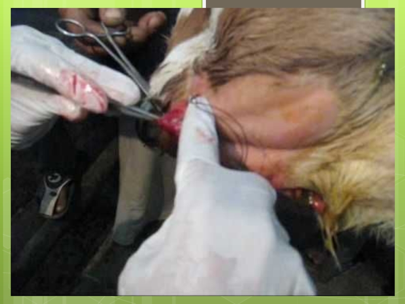
Surgical repair
is the treatment of choice, but postoperative complications can occur, including fecal incontinence, persistent megacolon, anal stricture, recurrent cystitis and colonic atony secondary to prolonged preoperative distension.
Surgical repair of type I or II atresia ani using balloon dilation usually results in long-term survival and fecal continence in most cases.
Canine patients with type III AA have a poorer outcome and require more extensive surgical repair
Surgical repair of type I or II atresia ani using balloon dilation usually results in long-term survival and fecal continence in most cases.
Canine patients with type III AA have a poorer outcome and require more extensive surgical repair
Слайд 8Atresia ani and coli are congenital defects that result in an
inability to pass feces since birth. Atresia is a term that describes a body orifice that is abnormally small or completely closed. In the case of atresia ani, an animal either has a very small opening or no opening at all at the anus due to a failure of the anal membrane to break down. Atresia coli is similar in that the colon or large bowel has a section missing and thus the animal cannot defecate. Intestinal atresias are very serious conditions.
Слайд 9Intestinal atresias are the most common abdominal disease in calves less
than 8 days old. Calves born with a form of intestinal atresia can appear normal at birth, but they develop signs 24-48 hours later and most often die within hours of birth if the problem is not surgically corrected. It appears that there is some breed predisposition among cattle to the development of atresia coli. Atresia ani is an inherited abnormality.
Слайд 11
Etiology:
Both atresia ani and atresia coli are congenital defects meaning that they are defects that occurred during fetal development that animals are born with. There is some evidence that suggests that Holstein-Friesian calves are at greater risk for atresia coli than all other breeds. Another possible source of atresia coli is damage to the amniotic vesicle during early pregnancy palpation leading to poor blood supply to the fetal intestine. Atresia ani is an inheritable defect in cattle.
Both atresia ani and atresia coli are congenital defects meaning that they are defects that occurred during fetal development that animals are born with. There is some evidence that suggests that Holstein-Friesian calves are at greater risk for atresia coli than all other breeds. Another possible source of atresia coli is damage to the amniotic vesicle during early pregnancy palpation leading to poor blood supply to the fetal intestine. Atresia ani is an inheritable defect in cattle.
Слайд 13
Symptoms:
Symptoms of atresia coli usually don’t appear until 24-48 hours after birth. Calves with atresia ani can show severe signs within hours of birth. The affected calf will be unable to pass any feces so it takes some time for feces to build up and cause signs. Typical signs of intestinal atresia include decreased appetite, progressive bilateral abdominal distention, the absence of feces, episodes of straining to defecate, and occasionally, blood-tinges mucous can be found around the rectum.
Symptoms of atresia coli usually don’t appear until 24-48 hours after birth. Calves with atresia ani can show severe signs within hours of birth. The affected calf will be unable to pass any feces so it takes some time for feces to build up and cause signs. Typical signs of intestinal atresia include decreased appetite, progressive bilateral abdominal distention, the absence of feces, episodes of straining to defecate, and occasionally, blood-tinges mucous can be found around the rectum.
Слайд 15
Diagnosis:
Abdominal radiographs of calves with atresia coli will show enlarged loops of bowel and no feces present in the rectum. Sometimes, giving a barium enema prior to radiographing the abdomen can help determine the level of the defect in cases of atresia coli. Animals with atresia ani are usually easier to diagnose since they will show signs early on and sometimes the imperforate membrane can be palpated. On radiographs, animals with atresia ani will show a distended, feces-filled colon with an abrupt end at the level of the rectum.
Abdominal radiographs of calves with atresia coli will show enlarged loops of bowel and no feces present in the rectum. Sometimes, giving a barium enema prior to radiographing the abdomen can help determine the level of the defect in cases of atresia coli. Animals with atresia ani are usually easier to diagnose since they will show signs early on and sometimes the imperforate membrane can be palpated. On radiographs, animals with atresia ani will show a distended, feces-filled colon with an abrupt end at the level of the rectum.
Слайд 16
Prevention:
Preventing intestinal atresia in cattle cannot be fully accomplished because the causes of this abnormality are not fully understood. However, there are measures to take to reduce the incidence in a herd. Given the Holsteins have an increased incidence of atresia coli and it is evidence to suggest it may be an autosomal recessive trait in the breed, it is advisable to cull any affected calves and not attempt surgical correction of atresia coli. Additionally, the possibility that damage to the blood supply to the developing fetal intestine during the first 6 weeks of pregnancy can cause atresia coli, warrants gentle palpation techniques during this sensitive period of gestation.
Preventing intestinal atresia in cattle cannot be fully accomplished because the causes of this abnormality are not fully understood. However, there are measures to take to reduce the incidence in a herd. Given the Holsteins have an increased incidence of atresia coli and it is evidence to suggest it may be an autosomal recessive trait in the breed, it is advisable to cull any affected calves and not attempt surgical correction of atresia coli. Additionally, the possibility that damage to the blood supply to the developing fetal intestine during the first 6 weeks of pregnancy can cause atresia coli, warrants gentle palpation techniques during this sensitive period of gestation.
Слайд 17
Treatment:
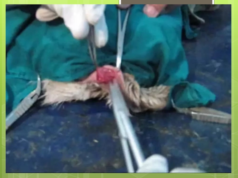
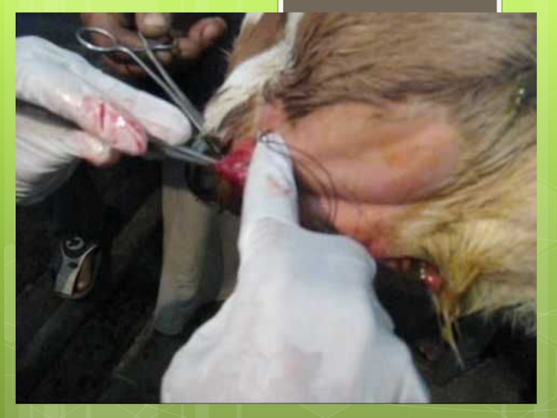
Treating atresia ani and atresia coli involves surgically opening the blocked orifice. The defect in atresia coli is usually at the level of the spiral loop of the ascending colon so correction involves removing the defective section of bowel and reattaching the normal sections. Unfortunately, surgical correction of atresia coli has a poor overall success rate, with a short-term survival rate of less than 50% and a long-term survival rate of less than 35%. It is also somewhat controversial to fix the defect in Holstein calves because of the possibility that the animal will reach breeding age and perpetuate the defect in the cattle population. Atresia coli in Holsteins is a heritable defect so most Holstein calves born with this abnormality are euthanized. Surgical correction of atresia ani similarly involves surgically opening the blind-ended sac ending at the rectum and suturing around the newly created orifice.
Treating atresia ani and atresia coli involves surgically opening the blocked orifice. The defect in atresia coli is usually at the level of the spiral loop of the ascending colon so correction involves removing the defective section of bowel and reattaching the normal sections. Unfortunately, surgical correction of atresia coli has a poor overall success rate, with a short-term survival rate of less than 50% and a long-term survival rate of less than 35%. It is also somewhat controversial to fix the defect in Holstein calves because of the possibility that the animal will reach breeding age and perpetuate the defect in the cattle population. Atresia coli in Holsteins is a heritable defect so most Holstein calves born with this abnormality are euthanized. Surgical correction of atresia ani similarly involves surgically opening the blind-ended sac ending at the rectum and suturing around the newly created orifice.
Слайд 18If your horse has any type of atresia ani, he will
be in obvious pain within 12 hours due to the fact that it cannot defecate. Body waste will build up in the intestine until it ruptures if it is not treated right away. Any of these types are serious and must be treated by a veterinary professional who specializes in horses. Surgery is needed to create the anal opening and connect the rectum to where it belongs, but there are many complications. Some of these complications are colonic atony, recurring cystitis (urinary tract infection), anal stricture (narrowing of the anal canal), persisting megacolon (severe constipation and inflammation of the colon), and fecal incontinence (inability to control fecal matter).
Слайд 19Atresia ani in horses is an uncommon congenital defect in which
the anus and rectum are not formed properly. The word atresia means absence of a natural opening and ani means anus, which is where it got the name atresia ani. This condition is a birth defect causing serious colic and retention of feces because there is no anal opening for the horse to defecate from. Veterinary experts describe this as the hindgut failing to connect with the perineum correctly to create the rectum and anus. This is a serious and dangerous condition that requires immediate surgery for your horse to survive. There are four types of atresia ani, which are congenital narrowing of the anal canal without a formed opening for the anus, imperforate anus less than 1.5 centimeters from a blind rectal pouch, imperforated anus more than 1.5 centimeters from a blind rectal pouch, and a blind rectal pouch with a normal rectum.
Слайд 20Symptoms of Atresia Ani in Horses
The symptoms of atresia ani may
differ due to the four different types. However, the general signs that your horse is suffering from atresia ani are:
Swelling of the abdomen that continues to get bigger
Abdominal pain that can become excruciating as it progresses
Absence of bowel movements
Lack of response to enema
In females, the feces may come from the vulva (vaginal opening)
Swelling of the abdomen that continues to get bigger
Abdominal pain that can become excruciating as it progresses
Absence of bowel movements
Lack of response to enema
In females, the feces may come from the vulva (vaginal opening)
Слайд 21
Types
Type I (membrane atresia) - A membrane or diaphragm blocks the intestinal opening
Type II (cord atresia) - The ends are joined by a cord of tissue.
Type III (blind end atresia) - An intestinal section is entirely in blind end atresia which leaves two blind ends and a V-shaped intestinal defect.
Type IV (multiple atresia) - More than one small bowel atresias of any of the above types
Слайд 22Causes of Atresia Ani in Horses
All the types of atresia ani
are genetic birth defects
The anus and rectum are not formed properly
Colic and retention of feces will occur
Waste will build up in the intestine until it ruptures
This condition causes a need for corrective surgery
The anus and rectum are not formed properly
Colic and retention of feces will occur
Waste will build up in the intestine until it ruptures
This condition causes a need for corrective surgery
Слайд 23
Treatment of Atresia
Ani in Horses
Unfortunately, in some cases, there is nothing that can be done to save the horse and euthanasia will be suggested to stop the suffering. You are more than welcome to get a second opinion or to see a specialist that studies atresia ani, but the longer you wait, the longer your horse is in excruciating pain. Some of the treatment plans include: Fluids The first step will be to give your horse an intravenous line with ringer lactate solution and electrolytes to rehydrate your pet. This also makes it easier for the physician to administer medications when needed. Flunixin meglumine may be given for pain.
Слайд 24
Fluids
The first step will be to give your horse an intravenous line with ringer lactate solution and electrolytes to rehydrate your pet. This also makes it easier for the physician to administer medications when needed. Flunixin meglumine may be given for pain.
The first step will be to give your horse an intravenous line with ringer lactate solution and electrolytes to rehydrate your pet. This also makes it easier for the physician to administer medications when needed. Flunixin meglumine may be given for pain.
Слайд 25
Surgery
A midline laparoscopic operation is performed to repair the defect. The details vary depending on the type of atresia ani.
A midline laparoscopic operation is performed to repair the defect. The details vary depending on the type of atresia ani.
Слайд 26Type I - The veterinarian will use balloon dilation to expand
the anal canal so the fecal matter can pass more easily.
Type II and III - The veterinarian will have to do at least two operations for these more serious types of defect. The first (colostomy) is done by creating a small opening in the skin and muscles of the abdominal wall. A colostomy bag will be attached for the fecal matter to drain into until the horse is big enough for the second surgery, which is about six months. In the second (and hopefully final) surgery, the veterinarian will move the colon to a different spot and the rectal pouch will be put into place where a new anal opening will be made.
Type IV - With this serious type of atresia ani, the abdomen will need to be completely opened up to isolate and attach the distal colon and rectum.
Слайд 27Recovery of Atresia Ani in Horses
Horses with type I atresia ani
have a good chance of a full recovery and long life. With types II and III, the prognosis really is contingent upon which part of the colon is affected, but the success rate is low so the prognosis is poor. If your horse has type IV, the chances are grave, but possible.
