- Главная
- Разное
- Дизайн
- Бизнес и предпринимательство
- Аналитика
- Образование
- Развлечения
- Красота и здоровье
- Финансы
- Государство
- Путешествия
- Спорт
- Недвижимость
- Армия
- Графика
- Культурология
- Еда и кулинария
- Лингвистика
- Английский язык
- Астрономия
- Алгебра
- Биология
- География
- Детские презентации
- Информатика
- История
- Литература
- Маркетинг
- Математика
- Медицина
- Менеджмент
- Музыка
- МХК
- Немецкий язык
- ОБЖ
- Обществознание
- Окружающий мир
- Педагогика
- Русский язык
- Технология
- Физика
- Философия
- Химия
- Шаблоны, картинки для презентаций
- Экология
- Экономика
- Юриспруденция
The value of protocol biopsies in renal allografts презентация
Содержание
- 1. The value of protocol biopsies in renal allografts
- 2. INTRODUCTION Protocol biopsy of an allografted
- 3. INTRODUCTION A significant number of cases with
- 4. WHY Banff – CLASSIFICATION (METHODS) We
- 5. Diagnostic categories in Banff classification 1997
- 6. Diagnostic categories in Banff classification 1997 Kidney
- 7. NUMERICAL CODES Glomerulitis (G) 0, 1, 2, 3
- 8. Differential diagnosis of other entities 1.
- 9. Diagnostic categories according to Banff-classification criteria Normal
- 10. Diagnostic categories according to Banff-classification criteria Acute
- 11. Diagnostic categories according to Banff-classification criteria Acute vascular rejection (grade 2a/b): Intimal arteritis
- 12. Diagnostic categories according to Banff-classification criteria Severe acute rejection- grade 3: transmural arteritis
- 13. Diagnostic categories according to Banff-classification criteria Chronic
- 14. Diagnostic categories according to Banff-classification criteria Chronic allograft nephropathy: intimal fibrosis, transplant glomerulopathy.
- 15. Diagnostic categories according to Banff-classification criteria De novo glomerulonephritis
- 16. Diagnostic categories according to Banff-classification criteria Recurrent disease
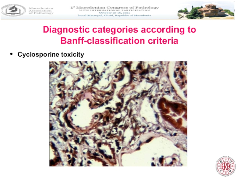
- 17. Diagnostic categories according to Banff-classification criteria
- 18. Diagnostic categories according to Banff-classification criteria Cyclosporine toxicity
- 19. Banff classification 07 – updates and future
- 20. Banff classification 07 – updates and future
- 21. Banff classification 07 – updates and future
- 22. Banff classification 07 – updates and future
- 23. INTRODUCTION In Macedonia, most cases usually underwent
- 24. MATERIALS AND METHODS A total of
- 25. MATERIALS AND METHODS The immunosuppressive regimen
- 26. RESULTS The mean age
- 27. RESULTS Signs of acute rejection
- 28. RESULTS It is of interest
- 29. RESULTS Immunohistochemical study for cell proliferation by
- 30. RESULTS Evaluation of the apoptosis showed significant
- 31. DISCUSSION We demonstrated histopathological findings in
- 32. DISCUSSION Study of protocol biopsies from stable
- 33. DISCUSSION Findings of recurrent disease and cyclosporine
- 34. CONCLUSION AND RECOMMENDATIONS There are three possible
- 35. CONCLUSION AND RECOMMENDATIONS There are three possible
- 36. CONCLUSION AND RECOMMENDATIONS There are three possible
- 37. CONCLUSION AND RECOMMENDATIONS SCR results in
- 38. THANK YOU FOR YOUR ATTENTION
Слайд 2INTRODUCTION
Protocol biopsy of an allografted kidney has been introduced in many
Biopsy may also detect clinically unsuspected lesions, such as drug induced nephropathy, recurrent original disease, ischemic tubular injury.
The information provided by different centers suggests that acute lesions tend to reach their maximum during the initial months after transplantation, and the incidence of chronic lesions is low during the first month, progressively increasing thereafter.
Seron D et al: Kidney Int 1997;51: 310-316;
Rush DN et al: Transplantation 1995; 59: 511-514
Слайд 3INTRODUCTION
A significant number of cases with acute rejection after kidney transplantation
Early diagnosis of CAN as major cause of late renal allograft loss is important to determine treatment strategies.
Protocol biopsies can also provide useful information early in the evaluation process, often before clinical signs of CAN appear and influence clinical management.
Besides that, it allows research on the pathobiology of kidney transplants.
Слайд 4WHY Banff – CLASSIFICATION (METHODS)
We need standardized interpretation of the allograft
Methods included are:
a) analysis of the data in the studies that use Banff classification;
b) using published data acquired in so called collaborative clinical studies for transplantation;
c) international conferences.
Слайд 5Diagnostic categories in Banff classification 1997
Kidney International, Vol.55(1999), pp713-723
1. Normal
2. Hyperacute antibody mediated rejection (immediate and accelerated)
3.Borderline changes (very mild acute rejection): mild to moderate focal mononuclear inflammatory substrate with foci of mild tubulitis (1-4 cells)
4. Acute rejection:
Grade 1A- mild acute rejection (>25% of parenchyma affected) + moderate tubulitis (>4 cells / tubular cross section);
Grade 1B –significant interstitial infiltration and foci of severe tubulitis (>10 mononuclear cells / tubular cross section)
Grade 2A Mild to moderate arteritis (v1)
Grade 2B – severe intimal arteritis (v2)
Grade 3 – transmural arteritis and/or arterial fibrinoid change and necrosis of medial smooth muscle cells (v3) + lymphocytic inflammation
Слайд 6Diagnostic categories in Banff classification 1997
Kidney International, Vol.55(1999), pp713-723
5. Chronic /
Grade 1 – mild interstitial fibrosis and tubular atrophy with or without specific changes suggesting rejection.
Grade 2 - Moderate interstitial fibrosis and tubular atrophy
Grade 3 – Severe interstitial fibrosis and tubular atrophy and tubular loss
6. Other –changes not considered to be due to rejection
Слайд 7NUMERICAL CODES
Glomerulitis (G) 0, 1, 2, 3
Interstitial mononulear infiltration
Tubulitis (T) 0, 1, 2, 3
Vasculitis (V) 0, 1, 2, 3
Hyaline arteriolar thickening (AH) 0, 1, 2, 3
Chronic transplant glomerulopathy (CG) 0, 1, 2, 3
Interstitial fibrosis with mononuclear inflammation (CI) 0, 1, 2, 3
Tubular atrophy and loss (CT) 0, 1, 2, 3
Fibrous intimal thickening and
fragmentation of the intima(CV) 0, 1, 2, 3
Acute and chronic codes are used together
Adequacy of the specimen: not satisfied; marginal; adequate.
Minimal number of sections: 7 slides with 3 HE, 3 PAS and
1 Trichrome.
Слайд 8Differential diagnosis of other entities
1. Post-transplant lymphoproliferative disorder
2. Nonspecific changes: interstitial
3. Acute tubular necrosis
4. Acute interstitial nephritis
5. Changes associated with the application of cyclosporine
6. Subcapsular injury
7. Pre-transplant acute endothelial lesion
8. Papillary necrosis
9. De novo glomerulonephritis
10. Recurrent disease
11. Pre-existing disease
12. Other (viral infection, thromboses, obstruction, lymphocele, urine leak)
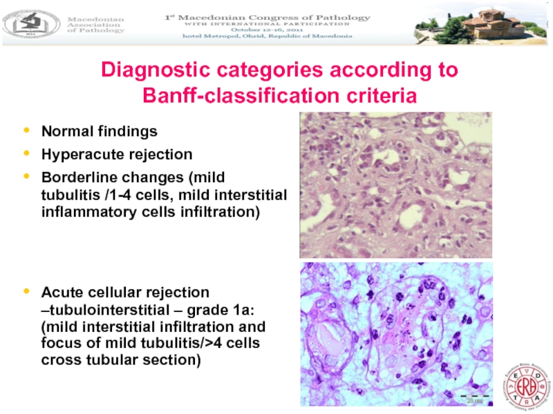
Слайд 9Diagnostic categories according to Banff-classification criteria
Normal findings
Hyperacute rejection
Borderline changes (mild tubulitis
Acute cellular rejection –tubulointerstitial – grade 1a: (mild interstitial infiltration and focus of mild tubulitis/>4 cells cross tubular section)
Слайд 10Diagnostic categories according to Banff-classification criteria
Acute cellular rejection –
grade 1b:
Слайд 11Diagnostic categories according to Banff-classification criteria
Acute vascular rejection (grade 2a/b): Intimal
Слайд 12Diagnostic categories according to Banff-classification criteria
Severe acute rejection- grade 3:
Слайд 13Diagnostic categories according to Banff-classification criteria
Chronic allograft nephropathy: tubular atrophy and
Слайд 14Diagnostic categories according to Banff-classification criteria
Chronic allograft nephropathy: intimal fibrosis, transplant
Слайд 17Diagnostic categories according to Banff-classification criteria
Transmission
Transitional cell carcinoma from donor
Arteriosclerosis and calcinosis of the media
Слайд 19Banff classification 07 – updates and future directions American Journal of
1. Normal
2. Antibody mediated changes (may coincide with categories 3, 4, 5 and 6) C4d deposition without morphologic evidence of active rejection.
- Acute antibody mediated reaction (C4d+) –
acute active lesions (Type and grade);
- Chronic active antibody mediated rejection (C4d+)
chronic active lesions .
3. Borderline changes suspicious for acute T-cell mediated rejection (may coincide with categories 2 and 5 and 6). There are foci of tubulitis (t1, t2 or t3) with mild interstitial infiltration (i0 or i1) or i2 and i3 with mild (t1) tubulitis.
Слайд 20Banff classification 07 – updates and future directions
4. T cell mediated
Acute T cell mediated rejection (Type/Grade):
Grade 1A – significant interstitial infiltration (>25% of parenchyma affected, i2, i3) + moderate tubulitis(t2);
Grade 1B –significant interstitial infiltration (>25% parenchyma affected, i2, i3) and foci of severe tubulitis (t3);
Grade 2A Mild to moderate arteritis (v1)
Grade 2B – severe intimal aretritis (v2);
Grade 3 – transmural arteritis and/or arterial fibrinoid change and necrosis of medial smooth muscle cells (v3) + lymphocytic inflammation
Chronic active T cell mediated rejection – chronic allograft arteriopathy (intimal fibrosis with mononuclear cell infiltration in fibrosis, formation of neo-intima).
Слайд 21Banff classification 07 – updates and future directions
5. Interstitial fibrosis and
Grade 1 – mild interstitial fibrosis and tubular atrophy without/with specific changes suggesting rejection.
Grade II - Moderate interstitial fibrosis and tubular atrophy
Grade III – Severe interstitial fibrosis and tubular atrophy with tubular loss
Other –changes not considered to be due to rejection
Слайд 22Banff classification 07 – updates and future directions American Journal of Transplantation
1. Inclusion of: peritubular capillaritis grading (0,1,2,3);
2. C4d scoring (negative, mild, focal, diffuse);
3. Interpretation of C4d deposition without morphological evidence of active rejection;
4. Application of the Banff criteria to zero time and protocol biopsies;
5. Introduction of a new scoring for total interstitial inflammation
(ti score);
6. Establishment of collaborative working groups addressing issues like isolated `v` lesion and introduction of omics technologies;
7. Future combination of graft biopsy and molecular parameters.
Слайд 23INTRODUCTION
In Macedonia, most cases usually underwent biopsy at the time of
In this study we analyzed the main histopathological changes in the protocol biopsies from grafts with stable renal function, taken at one and six months using Banff Classification 1997, for a three year period.
We also determined proliferative and apoptotic indexes in renal protocol biopsies to obtain insights into their role in CAN changes.
Слайд 24MATERIALS AND METHODS
A total of 28 paired biopsy specimens from allografted
All specimens were fixed in 10% formalin, embedded in paraffin
Paraffin sections were stained with H&E, PAS, Masson trichrome and Silver methenamine-PAS. Biopsies were considered adequate when they contained more than 7 glomeruli and at least one artery.
Additional immunohistochemical staining for Ki67 by LSAB immunoperoxidase was done, as well as ApoPtag in situ hybridization for the detection of apoptosis.
Слайд 25MATERIALS AND METHODS
The immunosuppressive regimen consisted of methylprednisolone and Daclizumab as
Clinical parameters analyzed included age, sex, source of donor, serum urea/creatinine level, urine volume and presence of proteinuria. Histological diagnosis was made according to the criteria of the Banff working classification.
In present study we included cases with serum creatinine <200 µmol/L and proteinuria <1g/24 hours at the time of the first biopsy, which was defined as “stable” graft function.
Слайд 26RESULTS
The mean age of the recipients was 35.2+/-8.3 years
Male to female
The mean living donor age was 58.5+/-13.4 years.
Histopathological diagnosis included six specimens (21,4%) with normal findings in the 1 and 6 months biopsies.
Borderline changes were found in 10/28 (35,7%) and 10/28 (35,7%) at 1 and 6 months biopsies, respectively.
Слайд 27RESULTS
Signs of acute rejection were found in 13/28 (46,4%) and 12/28
There was significant increase of CAN in the second allograft biopsy after six months: - In the first biopsy we found mild degree of CAN in 14 specimens (50%) and moderate in 2 specimens (7,1%).
- On second biopsy CAN was detected in 23 cases (82%) (11 with mild CAN and 12 with moderate CAN).
Слайд 28RESULTS
It is of interest that in three cases there were signs
We correlated these findings with the serum creatinine levels (sCr): We found significantly increased levels of sCr at 6 months after transplantation, while the calculated creatinine clearance (cCrcl) and proteinuria were significantly lower compared to the one month values for the respective group.
Слайд 29RESULTS
Immunohistochemical study for cell proliferation by Ki 67 showed greater proliferative
Proliferation was almost absent in the biopsies taken at 1 month after transplantation.
Слайд 30RESULTS
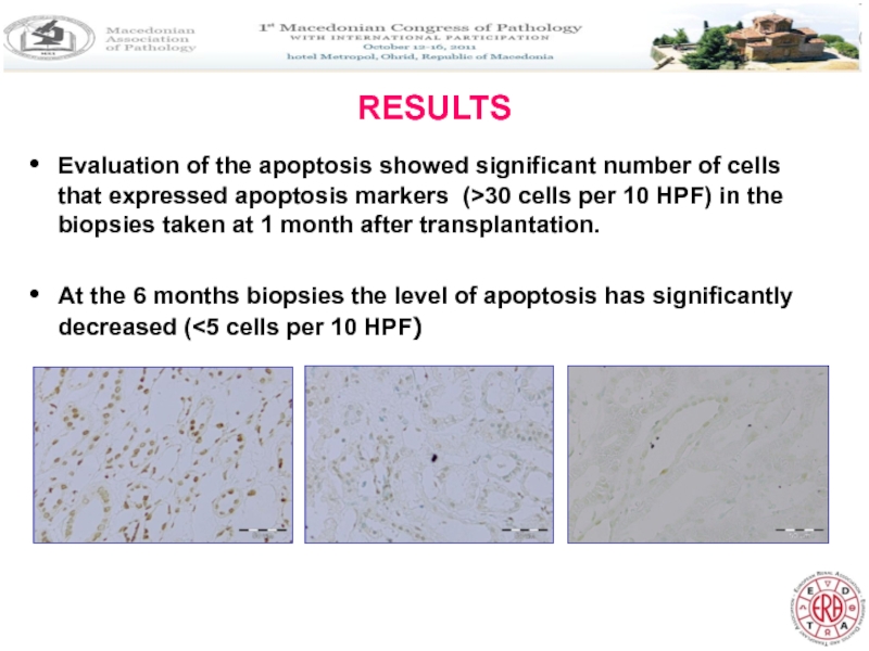
Evaluation of the apoptosis showed significant number of cells that expressed
At the 6 months biopsies the level of apoptosis has significantly decreased (<5 cells per 10 HPF)
Слайд 31DISCUSSION
We demonstrated histopathological findings in grafted kidneys, which clinically showed adquate
Our results showed presence of high percentages of BR and AR in allograft biopsies, which means they do not necessarily cause clinically recognizable graft dysfunction. Higher percentages of BR and SR in this study, compared to previous reports, might be due to the different sampling time for the biopsies, as well as to the lower number of patients included in the study.
Слайд 32DISCUSSION
Study of protocol biopsies from stable grafts had revealed an unexpectedly
In favor of this concept is the significant increase of CAN in the second biopsy taken at 6 months after transplantation, in our study.
Слайд 33DISCUSSION
Findings of recurrent disease and cyclosporine nephrotoxicity are important because of
High level of apoptosis expression in early protocol biopsies shows that apoptosis might be one of the pathways in the development of CAN, together with epithelial-mesenchymal transformation.
Слайд 34CONCLUSION AND RECOMMENDATIONS
There are three possible stategies:
1. No biopsy: This is
Слайд 35CONCLUSION AND RECOMMENDATIONS
There are three possible strategies:
2. Biopsies in high-risk individuals:
Слайд 36CONCLUSION AND RECOMMENDATIONS
There are three possible strategies:
3. Universal biopsy policy:
Protocol biopsies
- Advantages include simplicity of implementation; detection of unsuspected SCR in low risk individuals; early detection of other diagnoses (BK nephropathy, transmission).
- Disadvantages include costs, consumption of clinical and pathology resources, total number of adverse events incurred – which would increase with higher throughput programs, despite a low per procedure risk.
Слайд 37CONCLUSION AND RECOMMENDATIONS
SCR results in chronic tubulointerstitial damage, impaired renal dysfunction
Hence, one could make a clinical screening strategy – either as protocol biopsy in all recipients, or in high-risk individudals only, as an alternative to blanket use of heavy immunosuppression.
This decision is both a clinical and an economic decision, influenced by the prevalence of SCR and potential gains of treatment, against costs and resource utilization.
Nankivell BJ et al: American Journal of transplantation 2006; 6: 2006-2012
