- Главная
- Разное
- Дизайн
- Бизнес и предпринимательство
- Аналитика
- Образование
- Развлечения
- Красота и здоровье
- Финансы
- Государство
- Путешествия
- Спорт
- Недвижимость
- Армия
- Графика
- Культурология
- Еда и кулинария
- Лингвистика
- Английский язык
- Астрономия
- Алгебра
- Биология
- География
- Детские презентации
- Информатика
- История
- Литература
- Маркетинг
- Математика
- Медицина
- Менеджмент
- Музыка
- МХК
- Немецкий язык
- ОБЖ
- Обществознание
- Окружающий мир
- Педагогика
- Русский язык
- Технология
- Физика
- Философия
- Химия
- Шаблоны, картинки для презентаций
- Экология
- Экономика
- Юриспруденция
The Heart презентация
Содержание
- 1. The Heart
- 2. Imaging of the heart
- 4. PA view of normal chest. RA, right
- 5. Lateral view of normal chest. RV, right
- 7. Echocardiography 1.
- 8. 3. Patient is positioned in a 45
- 11. Apical four-chamber transthoracic echocardiogram in a
- 13. Doppler examination
- 14. Apical continuous-wave Doppler trace in a
- 15. Cardiac catheterization This procedure
- 16. Right heart catheterization This can
- 17. The site of the catheter tip can
- 18. Left heart catheterization The
- 19. Isotope
- 20. demonstrates a partially reversible perfusion defect in
- 22. Axial composite image
- 24. Arteriography
- 26. (B) Coronary arteriogram, same projection and patient
- 27. Normal aortogram of transverse arch in patient
- 28. Intravenous digital subtraction
- 30. Perfusion scanning gives the estimation of the
- 32. MRI image
- 34. Cardiac
- 35. Posterioanterior Projection
- 36. the left border has
- 38. Lateral Projection
- 41. Cardiac Size — normal is 1/2 or
- 42. Technical
- 43. Cardiothoracic ratio (CT) It is
- 45. The causes for increased
- 46. Expiratory phase on a PA radiograph. Note
- 47. AP (A) and PA (B) radiographs of
- 48. Common causes of cardiomegaly Valvular heart
- 49. Causes of small
- 50. Enlargement of the heart
- 51. The common causes of
- 52. The left ventricle enlarges to the left
- 54. Lateral view shows the left
- 55. The common causes of right
- 56. Right ventricle when enlarges, it does so
- 57. Direct signs of right ventricular
- 58. Indirect signs
- 60. The common causes of left atrial
- 61. On the anterior view the right atrium
- 62. left atrial enlargement
- 63. The causes of the right atrium
- 64. Right atrium enlargement
- 65. Essential hypertension
- 66. Left heart enlargement is common in prolonged
- 67. The pulmonary artery and the conus are
- 68. Chronic nephritis
- 69. Pericardial effusion
- 70. Radiological features
- 71. CT: may also identify the aetiology, e.g.
- 74. Cardiac
- 75. Radiological features
- 76. interstitial pulmonary oedema: initially, prominence of the
- 77. Valvular diseases of heart
- 78. In posterioanterior view
- 79. In right
- 80. Elevation of left main bronchus due to
- 81. Rheumatic mitral
- 82. Mitral regurgitation
- 83. In mild regurgitation heart size may remain
- 84. Aortic valve stenosis
- 85. Aortic regurgitation
- 86. * The ventricle enlarges mainly downwards and
- 87. Coarctation of aorta
- 89. Pulmonary stenosis
- 90. Pulmonary regurgitation
- 91. Isolated pulmonary regurgitation is a benign lesion
- 92. Venous hypertension When
- 93. When the capillary pressure exceeds the normal
- 94. Deep septal lines are caused by edema
- 95. In hilar edema, fluid collects in the
- 96. When the pulmonary venous pressure reaches 30
- 97. Pulmonary hemosiderosis is due to focal deposition
- 98. Fallot’s
- 99. Plain radiograph features: the
- 101. Ventricular septal defect
- 102. Chest radiograph:
- 103. Atrial septal
- 104. Chest
- 105. Atrial septal defect
- 106. Cardiac

Слайд 2 Imaging of the heart will be considered
1. Simple x-ray
2. Screening
3. Cardiac catheterization
4. Angiocardiography
5. Coronary arteriography
6. Ultrasound
7. Isotope scan
8. MRI

Слайд 3 Simple x-ray
A simple
1. size of the heart
2. enlargement of individual chambers
3. condition of the lung fields

Слайд 4PA view of normal chest. RA, right atrium; RDPA, right descending pulmonary

Слайд 5Lateral view of normal chest. RV, right ventricle; RSS, retrosternal clear space;

Слайд 6 Screening
Calcification is most commonly seen:
in the mitral or aortic valves
but may also be seen in atheromatous coronary arteries, in the mitral annulus
or in a left atrium containing mural thrombus

Слайд 7 Echocardiography
1. Echocardiography is a highly versatile
2. Echocardiography is performed from the transthoracic route using a sector probe.

Слайд 83. Patient is positioned in a 45 degree semierect position rotated
4. Two-dimensional imaging gives direct information about the anatomy and physiology of the heart
5. M-mode is a one-dimensional evaluation useful for precise measurement and timing of cardiac events.


Слайд 11 Apical four-chamber transthoracic echocardiogram in a patient with


Слайд 13 Doppler examination
Doppler evaluation allows
1. of different flow velocities within the cardiac chambers and in the outflow tracts
2. calculation of the cardiac output, ejection fraction

Слайд 14 Apical continuous-wave Doppler trace in a patient with dynamic left

Слайд 15 Cardiac catheterization
This procedure requires the introduction of a

Слайд 16 Right heart catheterization
This can be performed percutaneously or after

Слайд 17The site of the catheter tip can be confirmed by taking

Слайд 18 Left heart catheterization
The usual technique of left heart

Слайд 19 Isotope scanning
Technetium-99m pyrophosphate accumulates in

Слайд 20demonstrates a partially reversible perfusion defect in th interventricular

Слайд 21 CT scan
1. the atherosclerotic disease of the coronary vessels
2. myocardial calcifications and aneurysmal
3. dilatations and dissection of aorta
4. CT is the investigation of choice for the evaluation: of cardiac tumors like myxoma, for pericardial diseases like effusion and pericardial tumors and dissection of aorta


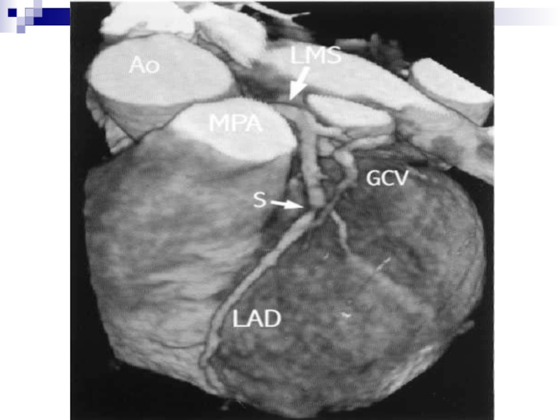
Слайд 24 Arteriography
Vascular access is usually obtained


Слайд 26(B) Coronary arteriogram, same projection and patient as in (A), obtained 1
 Coronary arteriogram, same projection and patient as in (A), obtained 1 day later. The")
Слайд 27Normal aortogram of transverse arch in patient suspected of having traumatic
Aortogram in")
Слайд 28 Intravenous digital subtraction
This technique is utilized to visualize the arterial system by injection of a bolus of contrast into the superior vena cava. After passage through the heart and lungs, the dilute contrast may be imaged in the arterial circulation by computer subtraction. Resolution is not as detailed as conventional arteriography, but can be an effective investigation in many clinical situations.

Слайд 29
MRI is fast gaining popularity as the investigation of choice in most cardiac pathologies. Assessment of the flow velocities in different cardiac chambers and outflow tracts helps in estimating the ejection fraction, cardiac output.

Слайд 30Perfusion scanning gives the estimation of the surviving and infracted myocardium
Cardiac tumors and pericardial diseases are also better evaluated with MRI.
MRI is the investigation of choice in the evaluation of congenital heart diseases, can help in quantifying shunt.


Слайд 34 Cardiac pulsation
Normally, pulsation on the
On the right side the lower border formed by right auricle shows a faint contraction of not more than 1 mm. Pulsation is greater in children than in adults and increases after exercise.

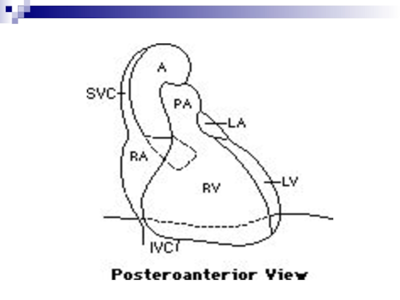
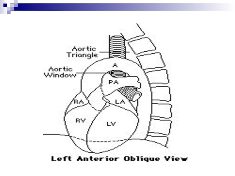
Слайд 35 Posterioanterior Projection
the upper right border is
1. the SVC
2. the lower cardiac border is formed by the RA

Слайд 36 the left border has three well-defined segments:
1.
2. the main pulmonary artery lies immediately below the aortic knob
3. LV and the apex (the LA appendage lies between the pulmonary artery segment and the LV and is usually not seen as a separate bulge)


Слайд 42 Technical Factors
• The heart appears
• Film during expiration — simulates pulmonary edema and the heart appears larger.
• One should check side markers for dextrocardia.
• One should check the clavicles for angulation.
• Over penetrated films may miss heart failure.

Слайд 43 Cardiothoracic ratio (CT)
It is a simple method of estimating
Estimation of CT ratio should always be done in erect PA view.
Normal:
for adults 50%
for neonates 60%
Cardiomegaly is diagnosed on frontal chest PA radiographs when the CT ratio exceeds 50%.
It is a simple method of estimating cardiac enlargement.Estimation of CT ratio")

Слайд 45 The causes for increased CT ratio due to
poor inspiration
supine position
prone position
AP radiographs, or with a short focus film distance

Слайд 46Expiratory phase on a PA radiograph. Note the low lung volumes, apparent

Слайд 47AP (A) and PA (B) radiographs of the chest in same
 and PA (B) radiographs of the chest in same patient on same day.")
Слайд 48 Common causes of cardiomegaly
Valvular heart diseases like mitral stenosis, mitral
Pericardial diseases like pericardial effusion
Myocardial diseases like ventricular aneurisms
Congenital cardiac diseases like atria septal defect, ventricle septal defect


Слайд 50 Enlargement of the heart
It may be general, involving

Слайд 51 The common causes of the left
hypertension
aortic regurgitation
aortic stenosis
coronary arteriosclerosis
acute/chronic nephritis
cardiac aneurism
coarctation of aorta

Слайд 52The left ventricle enlarges to the left and posteriorly and only


Слайд 54 Lateral view shows the left ventricle extending behind the

Слайд 55 The common causes of right ventricle
mitral stenosis
congestive failure
chronic pulmonary diseases
tricuspid regurgitation
Fallot’s tetralogy

Слайд 56Right ventricle when enlarges, it does so by a broadening of

Слайд 57 Direct signs of right ventricular
upward and outward displacement of the ventricular border
elevation of the apex
an upper longer arc above the apex and a lower shorter arc turning medially below the apex

Слайд 58 Indirect signs are:
prominent right atrial border
dilated
signs of pulmonary hypertension


Слайд 60The common causes of left atrial
ischemic heart disease
mitral stenosis
mitral regurgitation
aortic obstruction and regurgitation
systemic hypertension
left heart tumor

Слайд 61On the anterior view the right atrium forms less than the


Слайд 63 The causes of the right atrium
Shunts into right atrium (left ventricular – right atrial shunt, ruptured aortic sinus into right atrium)
Pulmonary obstruction and regurgitation
Pulmonary arterial hypertension
tricuspid obstruction and regurgitation
Right – sided cardiomyopathy
right atrial tumors


Слайд 65 Essential hypertension
It is
In most cases there is unfolding and pseudoenlargement of aorta.
The ascending part appears wider and longer.
The aortic knuckle becomes higher.

Слайд 66Left heart enlargement is common in prolonged hypertension.
The apex lies
When failure does occur the heart enlarges to the left and right in the transverse diameter greater than the long diameter.

Слайд 67The pulmonary artery and the conus are somewhat dilated.
The enlargement

Слайд 68 Chronic nephritis
The heart is enlarged in
Feature in chronic nephritis than in essential hypertension. Pulmonary edema occurs.

Слайд 69 Pericardial effusion
A pericardial effusion is a

Слайд 70 Radiological features
Chest film: illustrates a symmetrically
Echocardiography: the investigation of choice. Effusions are visible as echo-free areas surrounding the heart.

Слайд 71CT: may also identify the aetiology, e.g. mediastinal malignancy.
MRI: accurate for

Слайд 72 Causes
Infective
viral
bacterial
tuberculosis
Uraemia
Posmyocardial infarction
Myxoedema
Malignancy
bronchial and mediastinal tumors with pericardial invasion
Collagen vascular diseases
systemic lupus erythematosus
rheumatoid arthritis


Слайд 74 Cardiac failure
Cardiac failure is said

Слайд 75 Radiological features
On a
cardiac enlargement
upper-lobe vascular prominence: from raised pulmonary venous pressure
pleural effusions: seen as blunting at the costophrenic angels, but as the effusions become larger, there is a homogeneous basal opacity with a concave upper border

Слайд 76interstitial pulmonary oedema: initially, prominence of the upper-lobe and narrowing of
alveolar pulmonary oedema: with further increases in venous pressure, fluid transgresses into the alveolar spaces (alveolar shadowing) with haziness and blurring in the perihilar regions; in severe cases, pulmonary oedema develops throughout both lung fields. The outer thirds of the lungs may be spared, the bilateral central oedema being described as “bat’s wing”

Слайд 77 Valvular diseases of heart
Mitral stenosis presenting in infancy or early childhood is due to congenital lesion. It takes years to develop mitral stenosis after rheumatic fever. Mitral stenosis produces a pressure load on the left atrium and ultimately on the right ventricle.

Слайд 78 In posterioanterior view
An enlarged left auricle is
Double heart shadow in many cases can be seen to the right of the spine. Left border of the heart becomes straight and is known as mitralization.
Small aortic knuckle is caused partly by a true hypoplasia of aorta and partly by right ventricular rotation.

Слайд 79 In right oblique view
The enlargement
On barium swallow a bolus passes normally down to a point just below the left main bronchus when it seems to halt abruptly. Barium bolus then fills slowly the lower third of the oesophagus which is curved sharply backwards. This sign is more obvious in expiration than in inspiration.

Слайд 80Elevation of left main bronchus due to enlarged left atrium may
Horizontally Kerley “B” lines are more often noted. These lines are usually persistent. Other more fluid signs such as mottling, hilar edema and pleural effusion may develop which disappear on treatment.

Слайд 81
Rheumatic mitral stenosis. This frontal film shows
left atrial appendage (arrow).

Слайд 82 Mitral regurgitation
Mitral incompetence may result from

Слайд 83In mild regurgitation heart size may remain normal.
In late cases,

Слайд 84 Aortic valve stenosis
Heart is never more than slightly enlarged unless there is regurgitation.
Left border is often more rounded or longer than normal with a low apex, a shape characteristic of left ventricular enlargement.
Poststenotic dilatation of aorta is seen as a localized bulge to the right above the right atrium.
Calcification of the valves is almost invariable in males over the age of 40 years.

Слайд 85 Aortic regurgitation
Congenital regurgitation is usually due

Слайд 86* The ventricle enlarges mainly downwards and many cause no increase
* A prominent appendix is particularly suggestive of rheumatic valve disease.
* Dilatation of ascending aorta is more diffuse.
* Calcification of the valve is less common and less extensive with pure regurgitation than in stenosis.
* A few plaques are occasionally seen but obvious calcification always means a mitral lesion.

Слайд 87 Coarctation of aorta
It is a congenital narrowing

Слайд 88 X-ray shows:
*
* descending aorta may lie far off to the left off to the left of the spine
* rib notching is an important finding
* plethora with or without edema suggest a shunt in addition to coarctation
* in adults aortic knuckle becomes prominent

Слайд 89 Pulmonary stenosis
Pulmonary valve stenosis is
The heart is usually normal in size with severe stenosis but may be slightly enlarged in childhood as a result of marked hypertrophy of the right ventricle with elevated apex.
Gross enlargement is seen only with congestive cardiac failure.
Right atrium appears prominent.
Poststenotic dilatation of pulmonary trunk and or the left branch occurs in 90% cases.
Pulmonary oligaemia is noted.


Слайд 91Isolated pulmonary regurgitation is a benign lesion unless associated with pulmonary
Elderly patients on chronicity may develop congestive failure. When the pulmonary trunk is large with normal size heart, idiopathic dilatation is due to pulmonary regurgitation.

Слайд 92 Venous hypertension
When there is an increase in
Earliest change is dilatation of upper zone vessels. More often both veins and arteries are widened, all vessels above the hilum are little wider than those at lower levels. Vessels may measure more than 3mm in diameter.

Слайд 93When the capillary pressure exceeds the normal plasma osmotic pressure to
Kerley “B” lines are dense, short, straight horizontal lines most commonly seen in the bases. They result from thickening of the interlobar septa. Unlike vessels these do not branch. After treatment these lines disappear but may occasionally persist due to fibrous replacement of edema fluid and deposition of hemosiderin. Thus they become thinner and sharp.

Слайд 94Deep septal lines are caused by edema of deep tissue probably
Edema if the perivascular loose connective tissue blurs the edges of the segmental vessels.

Слайд 95In hilar edema, fluid collects in the loose connective tissue. The
The lung field may show a generalized loss of translucency with or without fine generalized mottling.
Pleural effusion is commonly found. Small effusion may be noted without septal lines and is the only sign of edema. Larger effusions are usually seen a higher venous pressure and are common in left ventricle failure than in mitral valve disease.

Слайд 96When the pulmonary venous pressure reaches 30 mmHg, edema fluid may

Слайд 97Pulmonary hemosiderosis is due to focal deposition of hemosiderin. The lung
Pulmonary ossific nodules are also formed following organization of intraalveolar edema. The nodules are dense and irregularly round or oval and rarely a small central medullary space may be visible. These vary from 1 to 10 mm most commonly seen in lower zones. These increase slowly in number.

Слайд 98 Fallot’s tetralogy
Consists of:
ventricular septal defect
right
pulmonary stenosis
right ventricular hypertrophy

Слайд 99 Plain radiograph features:
the heart is usually is not
the pulmonary vasculature shows pulmonary oligemia
the classic “cour en sabot” silhouette is due to combination of a deeply concave pulmonary bay and elevation from the diaphragm of slightly angular cardiac apex due to right ventricular hypertrophy
the ascending aorta is typically enlarged and prominent on plain radiograph


Слайд 101 Ventricular septal defect
is abnormal opening
ventricles.
Types:
membranous
muscular

Слайд 102 Chest radiograph:
left atrium is enlarged
associated hypertrophy
increased pulmonary vascular markings (plethora)

Слайд 103 Atrial septal defect
Types:
osteum secondum
osteum primum

Слайд 104 Chest radiograph:
enlargement of right atrium
pulmonary vascular prominence in lung field (plethora)


Слайд 106 Cardiac tumors
metastasis from bronchogenic carcinoma,
left atrial myxoma is the most common primary tumor of the

Слайд 107 Myxoma:
most common
in echocardiography, a polypoidal and mobile mass with heterogeneous echotexture is seen
on Ct scan, a heterogeneous mass lesion noted in the left atrium showing inhomogeneous enhancement