THE GENERAL DIAGNOSTICS OF TUBERCULOSIS. SPECIAL METHODS OF EXPOSURE AND DIAGNOSTICS OF TUBERCULOSIS (MICROBIOLOGICAL DIAGNOSTICS, TUBERCULIN SKIN TEST, ROENTGENOLOGIC DIAGNOSTICS)
- Главная
- Разное
- Дизайн
- Бизнес и предпринимательство
- Аналитика
- Образование
- Развлечения
- Красота и здоровье
- Финансы
- Государство
- Путешествия
- Спорт
- Недвижимость
- Армия
- Графика
- Культурология
- Еда и кулинария
- Лингвистика
- Английский язык
- Астрономия
- Алгебра
- Биология
- География
- Детские презентации
- Информатика
- История
- Литература
- Маркетинг
- Математика
- Медицина
- Менеджмент
- Музыка
- МХК
- Немецкий язык
- ОБЖ
- Обществознание
- Окружающий мир
- Педагогика
- Русский язык
- Технология
- Физика
- Философия
- Химия
- Шаблоны, картинки для презентаций
- Экология
- Экономика
- Юриспруденция
The general diagnostics of tuberculosis. (Lecture 2) презентация
Содержание
- 1. The general diagnostics of tuberculosis. (Lecture 2)
- 2. ANAMNESIS Here is a set of questions
- 3. COMPLAINS If a patient has any of
- 4. COMPLAINS Tuberculosis patients may complain of general
- 5. Cough
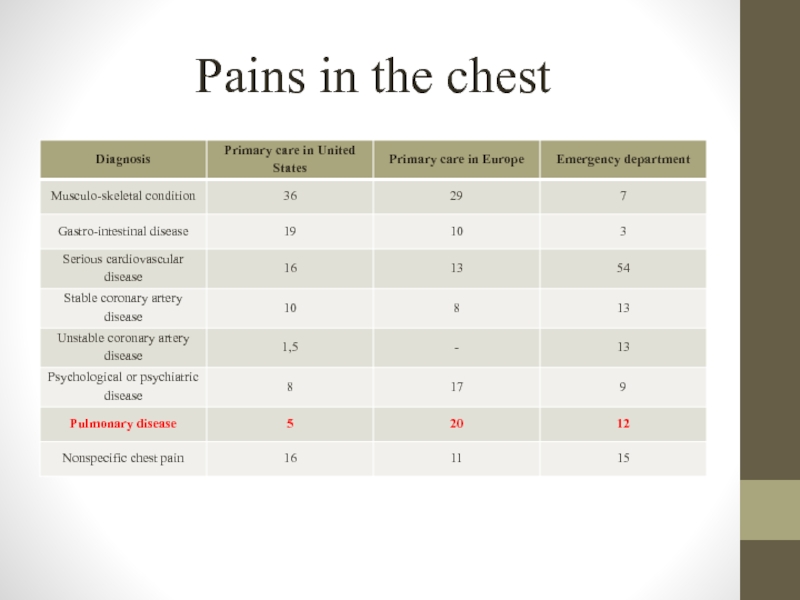
- 6. Pains in the chest
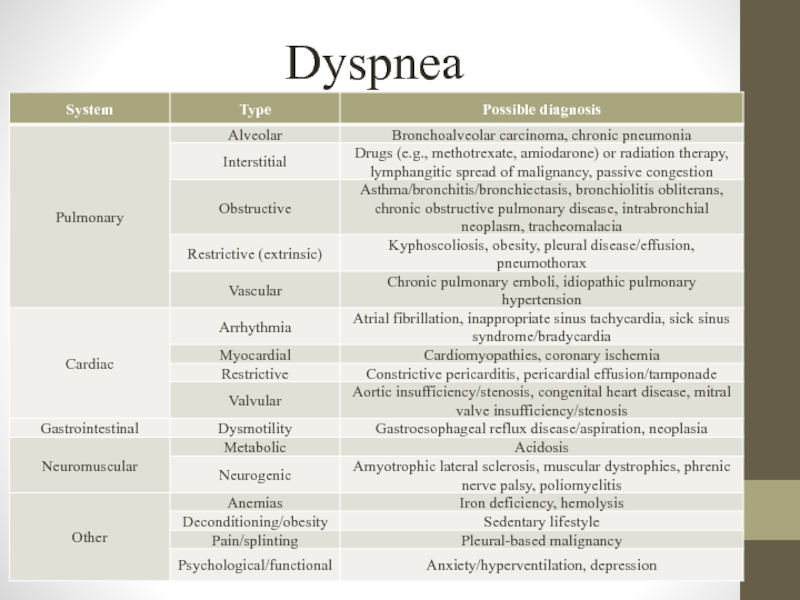
- 7. Dyspnea
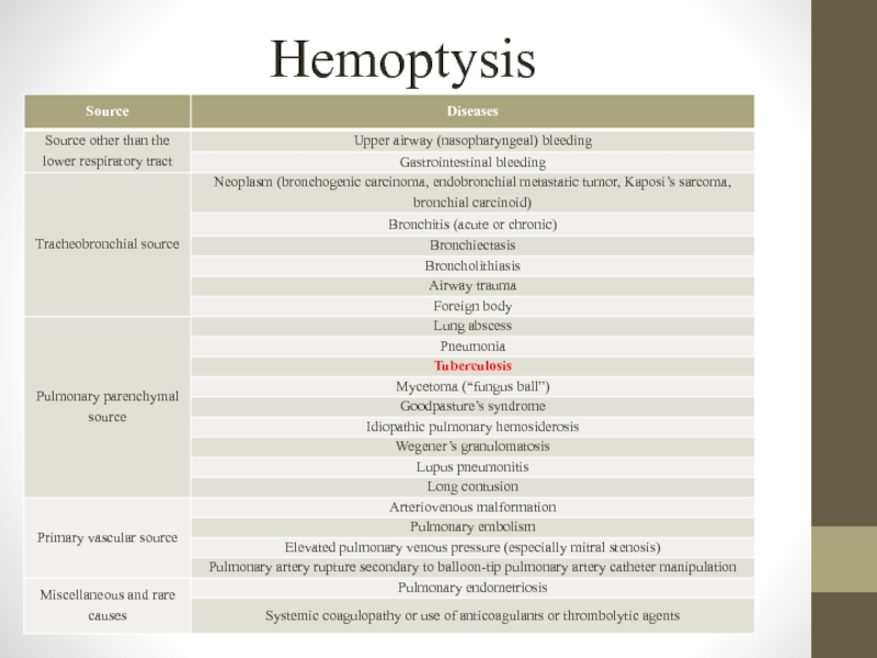
- 8. Hemoptysis
- 9. Physical investigation
- 10. Physical investigation
- 11. TUBERCULOSIS SCREENING METHODS Tuberculosis screening methods should
- 12. Tuberculin skin test Tuberculin includes purified protein
- 13. Delayed type hypersensitivity If patient has infected
- 14. Delayed type hypersensitivity In persons infected with
- 15. Delayed type hypersensitivity In reaction antigen -
- 16. Delayed type hypersensitivity There is a different
- 17. Mass tuberculinodiagnostics targets: identification of newly MBT-infected
- 18. TUBERCULIN PREPARATIONS To tuberculin preparations are related:
- 19. Mantoux test For routine tuberculinodiagnostics as
- 20. Mantoux test In carrying out immunization
- 21. Mantoux test For the Mantoux test
- 22. Mantoux test results estimation Negative
- 23. Mantoux test results estimation Doubtful
- 24. Mantoux test results estimation Positive
- 25. Mantoux test results estimation Hyperergic – indurations
- 26. Mantoux test results estimation
- 27. Mantoux skin test “virage” the first
- 28. Differences between postvaccinal and infectious allergies
- 29. Differences between postvaccinal and infectious allergies Postvaccinal
- 30. Contraindications to Mantoux skin test - Skin
- 31. False-Positive Reactions It may be positive TST
- 32. False-Negative Reactions It may be negative TST
- 33. Boosted Reactions and Serial Tuberculin Testing In
- 34. Diaskintest Diaskintest – it`s a new screening
- 35. Diaskintest Advantages More specific and sensitive test,
- 36. QuantiFERON-TB Gold and QuantiFERON-TB Gold PLUS General
- 37. QuantiFERON-TB Gold and QuantiFERON-TB Gold PLUS Baseline
- 38. QuantiFERON-TB Gold and QuantiFERON-TB Gold PLUS
- 39. QuantiFERON-TB Gold and QuantiFERON-TB Gold PLUS Advantages:
- 40. T-SPOT test General principles T-SPOT (Oxford Immunotec,
- 41. T-SPOT test
- 42. T-SPOT test Baseline epidemiological data As for
- 43. T-SPOT test - The test result is
- 44. Screening fluorography In Ukraine screening fluorography examination
- 45. Screening fluorography “Obligatory contingents” include: Students of
- 46. Screening fluorography “high risk” group by medical
- 47. Screening fluorography “high risk” group by social
- 48. Sputum samples collection In order to collect
- 49. Sputum samples collection The ideal situation is
- 50. The quantity and quality of collected sputum
- 51. Ziehl-Neelsen staining method This method is based
- 52. Ziehl-Neelsen staining method
- 53. Storage of specimens In order to increase
- 54. Storage of specimens Aseptic material should be
- 55. Transportation of specimens For safe transportation of
- 56. Homogenisation and decontamination of specimens The frequency
- 57. CULTURE MEDIA There are 4 main groups
- 58. Egg-based media The advantages of egg-based media:
- 59. Agar-based media These media are prepared in
- 60. Liquid media Liquid media offer a considerable
- 61. Culture examination In evaluating culture results of
- 62. Culture examination Estimating results register the following
- 63. Characterization of M. tuberculosis colonies Cultures should
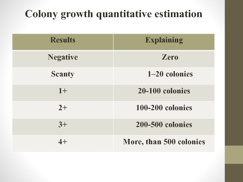
- 64. Colony growth quantitative estimation
- 65. BACTEC MGIT 960 system BACTEC MGIT 960
- 66. BACTEC MGIT 960 system Advantages of the
- 67. BACTEC MGIT 960 system An important component
- 68. BACTEC MGIT 960 system Firstly you should
- 69. Identification of Mycobacterium tuberculosis Growth in
- 70. Identification of Mycobacterium tuberculosis Detection of
- 71. Biochemical tests of identification Niacin test Niacin
- 72. Biochemical tests of identification Determining ability to
- 73. Biochemical tests of identification Thermostability catalase Catalase
- 74. Drug susceptibility testing for Mycobacterium tuberculosis complex
- 75. Drug susceptibility testing for Mycobacterium tuberculosis complex
- 76. Indirect methods of determining the sensitivity of
- 77. The proportion method on Löwenstein-Jensen medium
- 78. Indirect absolute concentrations drug susceptibility testing The
- 79. Drug susceptibility testing in liquid media (MGIT
- 80. Investigation of the sensitivity of mycobacteria to
- 81. Absolute concentration method DST results counted
- 82. Nitrate reductase assay The nitrate reductase
- 83. GeneXpertMTB/RIF test Test system GeneXpertMTB/RIF is recommended
- 84. GeneXpertMTB/RIF test The principle of the PCR
- 85. GeneXpertMTB/RIF test Results in the detection of
- 86. GenoType test system The GenoType test is
- 87. GenoType test system Indications to GenoType test:
- 88. GenoType test system Results interpretation. M.
- 89. GenoType test system Results interpretation. M.
- 90. THANK YOU FOR YOUR ATTENTION!
Слайд 1Zaporizhzhia State Medical University Department of phthisiology and pulmonology R.N. Yasinskyi (PhD, assistant of
Слайд 2ANAMNESIS
Here is a set of questions that are to be addressed
Whether the given patient was prior infected by tuberculosis?
Whether his/her relatives were infected by tuberculosis?
Whether the patient had contact with tuberculous patients or animals (household, professional, industrial contact)?
Whether the patient is registered in a tuberculosis dispensary due to: tuberculin testing or hypersensitive reaction to the test, contact with tuberculous patients, and no clear diagnosis of tuberculosis.
When the patient had the X-ray examination?
Whether the patient was invited after the X-ray examination for additional research?
Whether he was in a prison or lived with someone who was in a prison.
Whether the patient is homeless, a refugee, migrant or being in unfavorable social conditions?
Слайд 3COMPLAINS
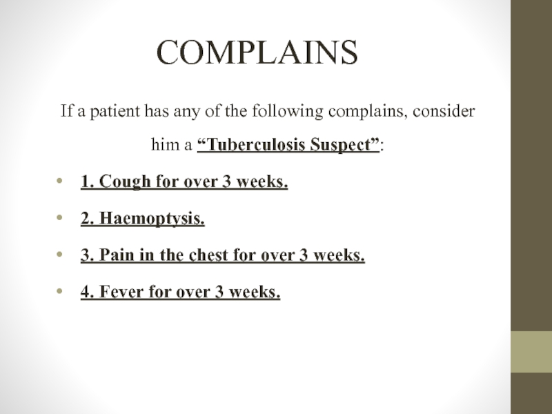
If a patient has any of the following complains, consider him
1. Cough for over 3 weeks.
2. Haemoptysis.
3. Pain in the chest for over 3 weeks.
4. Fever for over 3 weeks.
Слайд 4COMPLAINS
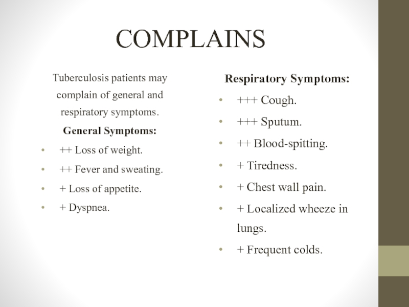
Tuberculosis patients may complain of general and respiratory symptoms.
General Symptoms:
++ Loss
++ Fever and sweating.
+ Loss of appetite.
+ Dyspnea.
Respiratory Symptoms:
+++ Cough.
+++ Sputum.
++ Blood-spitting.
+ Tiredness.
+ Chest wall pain.
+ Localized wheeze in lungs.
+ Frequent colds.
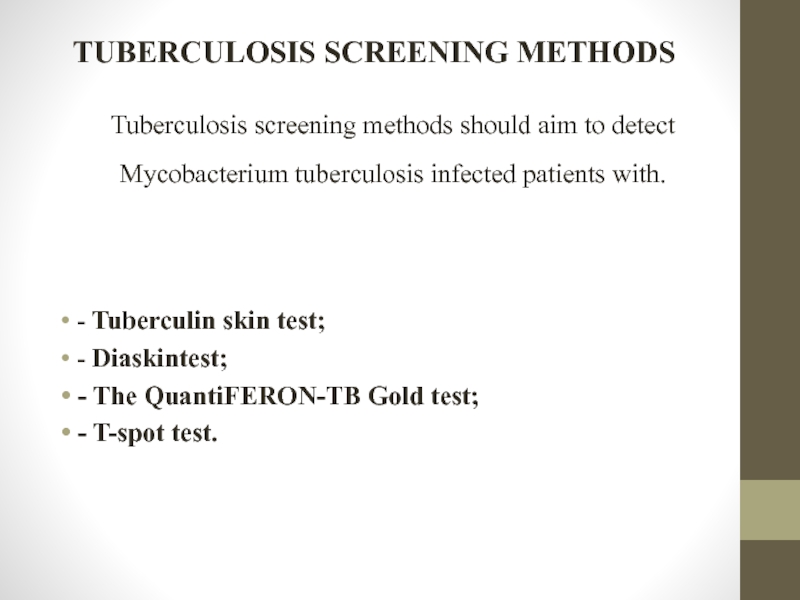
Слайд 11TUBERCULOSIS SCREENING METHODS
Tuberculosis screening methods should aim to detect Mycobacterium tuberculosis
- Tuberculin skin test;
- Diaskintest;
- The QuantiFERON-TB Gold test;
- T-spot test.
Слайд 12Tuberculin skin test
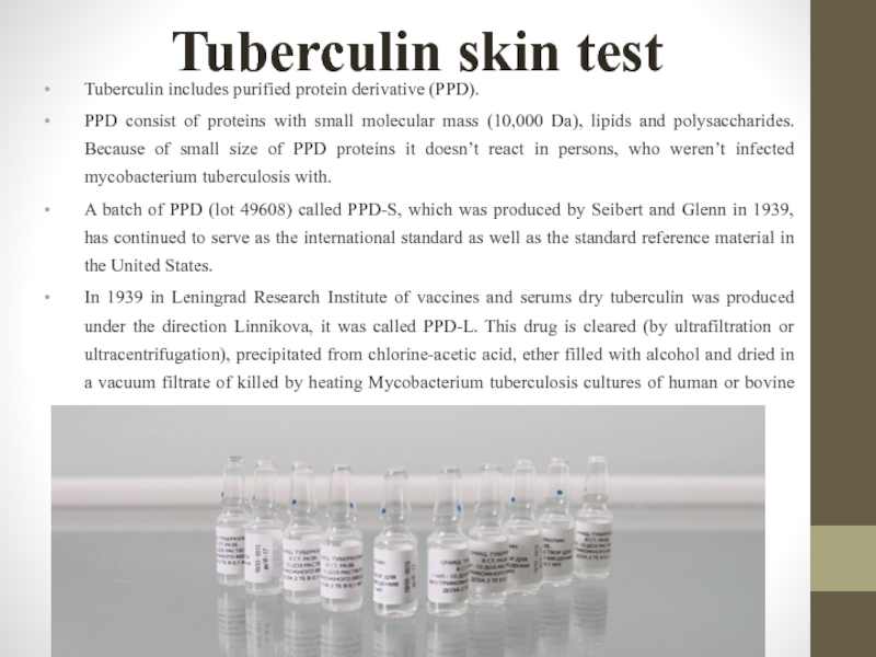
Tuberculin includes purified protein derivative (PPD).
PPD consist of proteins
A batch of PPD (lot 49608) called PPD-S, which was produced by Seibert and Glenn in 1939, has continued to serve as the international standard as well as the standard reference material in the United States.
In 1939 in Leningrad Research Institute of vaccines and serums dry tuberculin was produced under the direction Linnikova, it was called PPD-L. This drug is cleared (by ultrafiltration or ultracentrifugation), precipitated from chlorine-acetic acid, ether filled with alcohol and dried in a vacuum filtrate of killed by heating Mycobacterium tuberculosis cultures of human or bovine type.
Слайд 13Delayed type hypersensitivity
If patient has infected with mycobacterium tuberculosis – allergy
Слайд 14Delayed type hypersensitivity
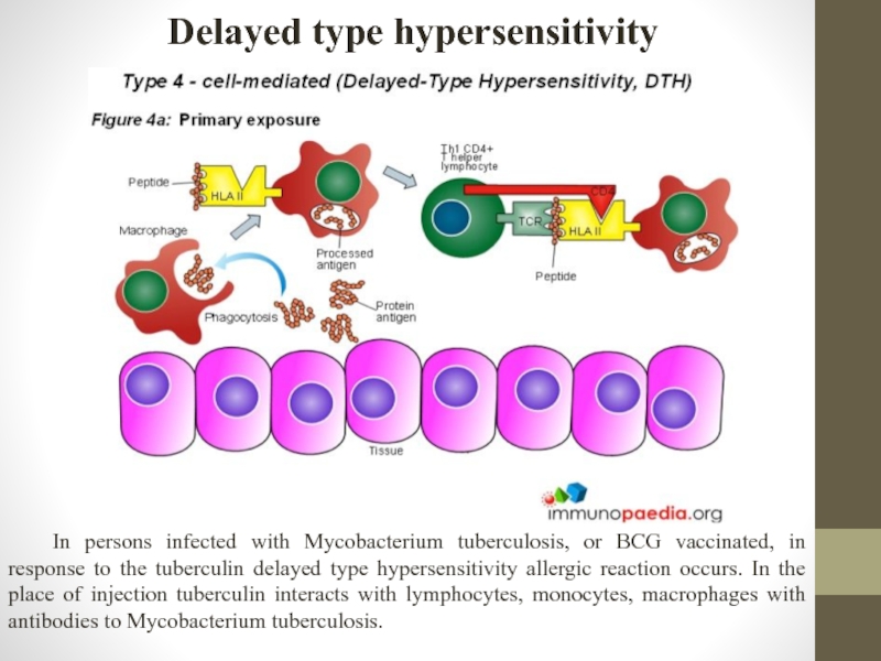
In persons infected with Mycobacterium tuberculosis, or BCG vaccinated,
Слайд 15Delayed type hypersensitivity
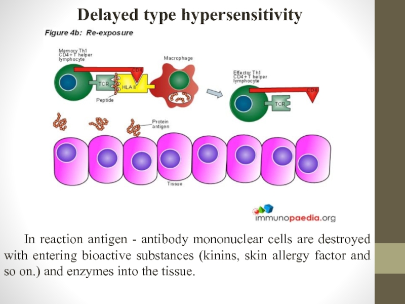
In reaction antigen - antibody mononuclear cells are destroyed
Слайд 16Delayed type hypersensitivity
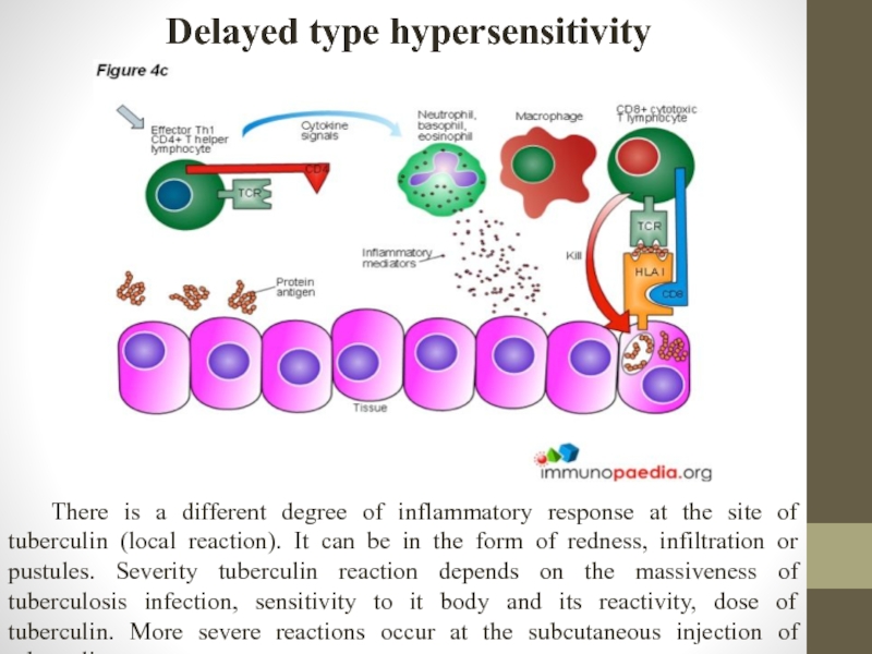
There is a different degree of inflammatory response at
Слайд 17Mass tuberculinodiagnostics targets:
identification of newly MBT-infected persons ("Virage" of tuberculin tests);
identify
selection contingent of children to be revaccination against tuberculosis and vaccination if children that have not been vaccinated in the hospital aged 2 months or more;
early diagnosis of tuberculosis in children and adolescents;
identifying epidemiological indicators of tuberculosis (MBT infection of the population, the annual MBT infection risk).
Слайд 18TUBERCULIN PREPARATIONS
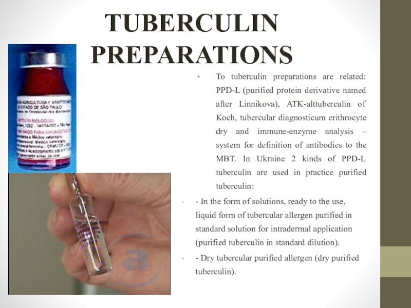
To tuberculin preparations are related: PPD-L (purified protein derivative named
- In the form of solutions, ready to the use, liquid form of tubercular allergen purified in standard solution for intradermal application (purified tuberculin in standard dilution).
- Dry tubercular purified allergen (dry purified tuberculin).
Слайд 19Mantoux test
For routine tuberculinodiagnostics as the only tuberculin reaction used
Tuberculin tests carried out annually regardless of the previous result. The use of tuberculinodiagnostics for the early detection of tuberculosis should allow the possibility for the comparison of sensitivity to tuberculin in dynamics, number and timing of BCG vaccinations, the presence and size of post-vaccinated scars, contact with TB patients, the appearance of clinical signs of disease.
Слайд 20Mantoux test
In carrying out immunization schedule approved by the Health
In order to early detection of TB Mantoux test with 2 TU carried out in all vaccinated children from 4 year to 14 year of age and adolescents regularly annually once a year, regardless of the previous result.
Слайд 21Mantoux test
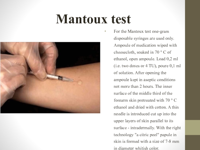
For the Mantoux test one-gram disposable syringes are used
Слайд 22Mantoux test results estimation
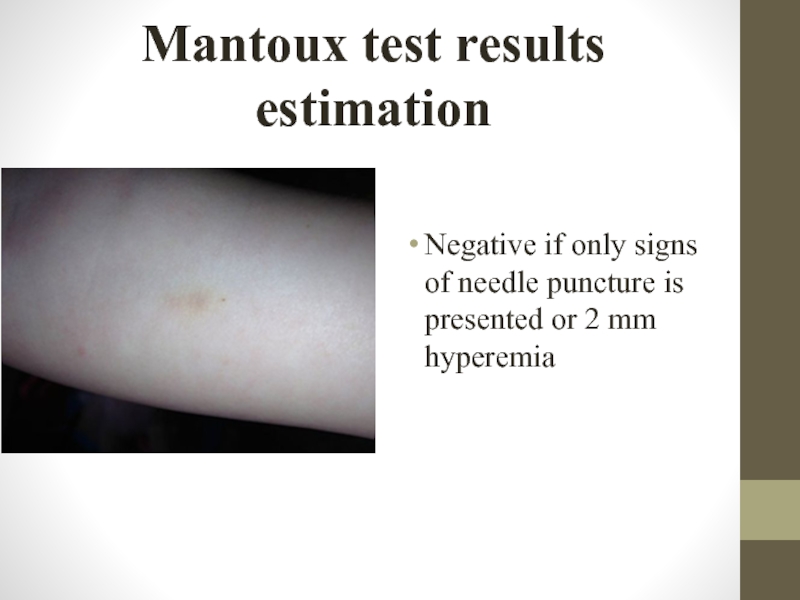
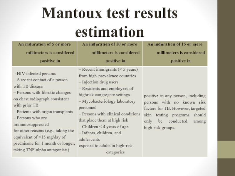
Negative if only signs of needle puncture is
Слайд 24Mantoux test results estimation
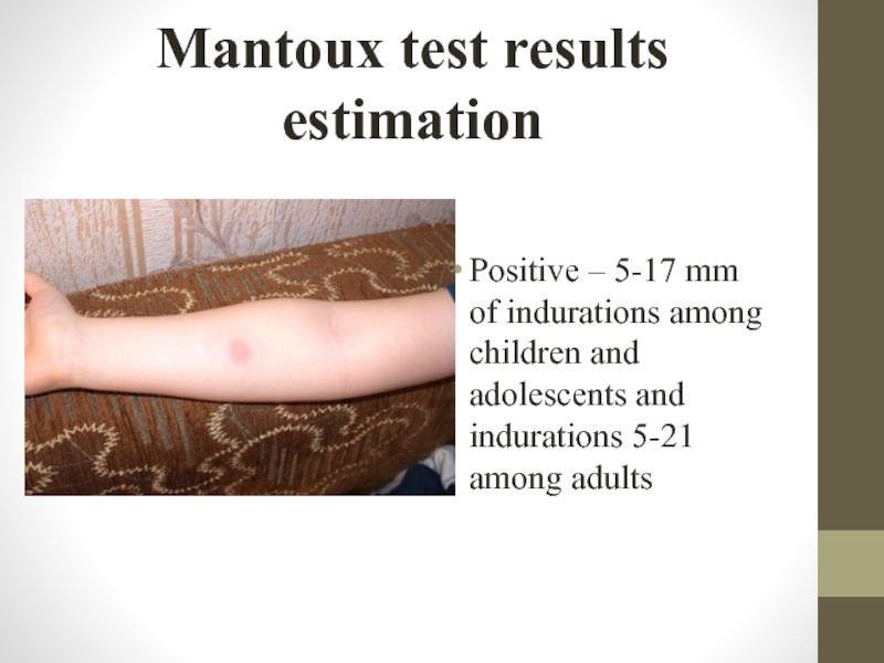
Positive – 5-17 mm of indurations among children
Слайд 25Mantoux test results estimation
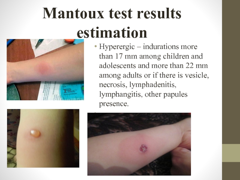
Hyperergic – indurations more than 17 mm among
Слайд 27Mantoux skin test “virage”
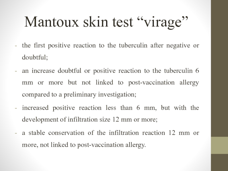
the first positive reaction to the tuberculin
an increase doubtful or positive reaction to the tuberculin 6 mm or more but not linked to post-vaccination allergy compared to a preliminary investigation;
increased positive reaction less than 6 mm, but with the development of infiltration size 12 mm or more;
a stable conservation of the infiltration reaction 12 mm or more, not linked to post-vaccination allergy.
Слайд 28Differences between postvaccinal and infectious allergies
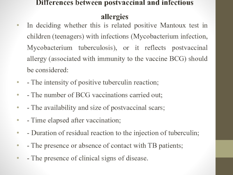
In deciding whether this is related
- The intensity of positive tuberculin reaction;
- The number of BCG vaccinations carried out;
- The availability and size of postvaccinal scars;
- Time elapsed after vaccination;
- Duration of residual reaction to the injection of tuberculin;
- The presence or absence of contact with TB patients;
- The presence of clinical signs of disease.
Слайд 29Differences between postvaccinal and infectious allergies
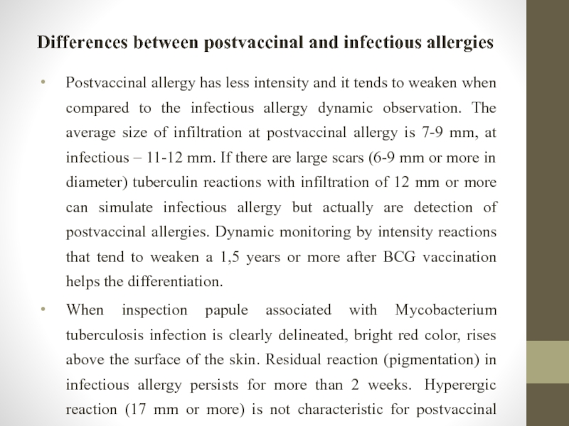
Postvaccinal allergy has less intensity and
When inspection papule associated with Mycobacterium tuberculosis infection is clearly delineated, bright red color, rises above the surface of the skin. Residual reaction (pigmentation) in infectious allergy persists for more than 2 weeks. Hyperergic reaction (17 mm or more) is not characteristic for postvaccinal allergy.
Слайд 30Contraindications to Mantoux skin test
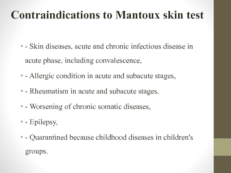
- Skin diseases, acute and chronic infectious
- Allergic condition in acute and subacute stages,
- Rheumatism in acute and subacute stages,
- Worsening of chronic somatic diseases,
- Epilepsy,
- Quarantined because childhood diseases in children's groups.
Слайд 31False-Positive Reactions
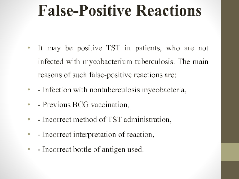
It may be positive TST in patients, who are not
- Infection with nontuberculosis mycobacteria,
- Previous BCG vaccination,
- Incorrect method of TST administration,
- Incorrect interpretation of reaction,
- Incorrect bottle of antigen used.
Слайд 32False-Negative Reactions
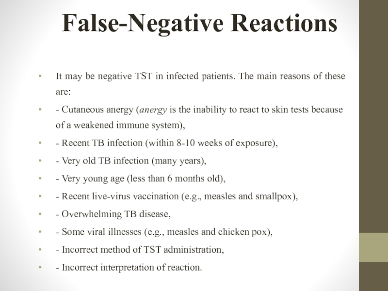
It may be negative TST in infected patients. The main
- Cutaneous anergy (anergy is the inability to react to skin tests because of a weakened immune system),
- Recent TB infection (within 8-10 weeks of exposure),
- Very old TB infection (many years),
- Very young age (less than 6 months old),
- Recent live-virus vaccination (e.g., measles and smallpox),
- Overwhelming TB disease,
- Some viral illnesses (e.g., measles and chicken pox),
- Incorrect method of TST administration,
- Incorrect interpretation of reaction.
Слайд 33Boosted Reactions and Serial Tuberculin Testing
In most individuals, PPD skin test
Boosted reactions also are particularly common in individuals exposed to other mycobacteria or who have been vaccinated with BCG. If repeated tuberculin testing is anticipated, as in health care workers, for example, a two-step method is recommended. In this method, persons who have a negative initial PPD skin test undergo a second tuberculin test 1–3 weeks after the first. The results from the second test should be considered to be the “correct” result, i.e., those individuals with a positive reaction on the second test should be considered to be previously infected, and those with a negative reaction on the second test should be considered uninfected. In these uninfected persons, a positive result on any future PPD skin test should be interpreted as a skin test conversion. Repeated skin testing with tuberculin will not induce a positive skin test reaction in individuals who have no cellular immunity to the antigens in PPD.
Слайд 34Diaskintest
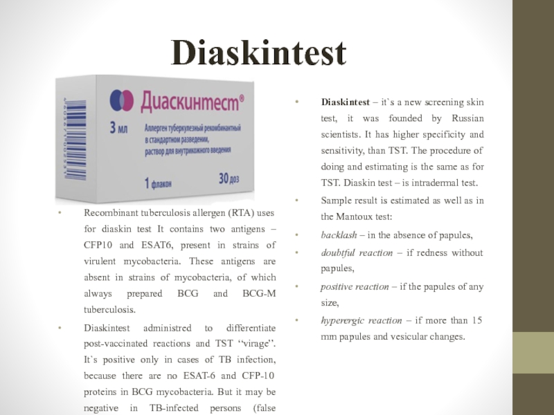
Diaskintest – it`s a new screening skin test, it was founded
Sample result is estimated as well as in the Mantoux test:
backlash – in the absence of papules,
doubtful reaction – if redness without papules,
positive reaction – if the papules of any size,
hyperergic reaction – if more than 15 mm papules and vesicular changes.
Recombinant tuberculosis allergen (RTA) uses for diaskin test It contains two antigens – CFP10 and ESAT6, present in strains of virulent mycobacteria. These antigens are absent in strains of mycobacteria, of which always prepared BCG and BCG-M tuberculosis.
Diaskintest administred to differentiate post-vaccinated reactions and TST “virage”. It`s positive only in cases of TB infection, because there are no ESAT-6 and CFP-10 proteins in BCG mycobacteria. But it may be negative in TB-infected persons (false negative reactions).
Слайд 35Diaskintest
Advantages
More specific and sensitive test,
there are no false positive reactions.
Disadvantages
It
There are the same contraindications to diascintest as to Mantoux test.
Слайд 36QuantiFERON-TB Gold and QuantiFERON-TB Gold PLUS
General principles
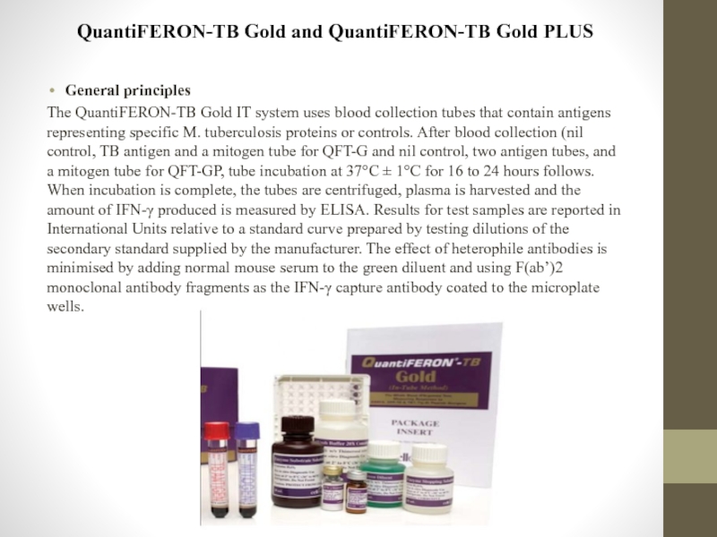
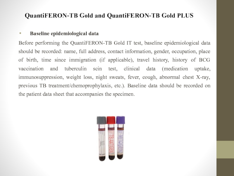
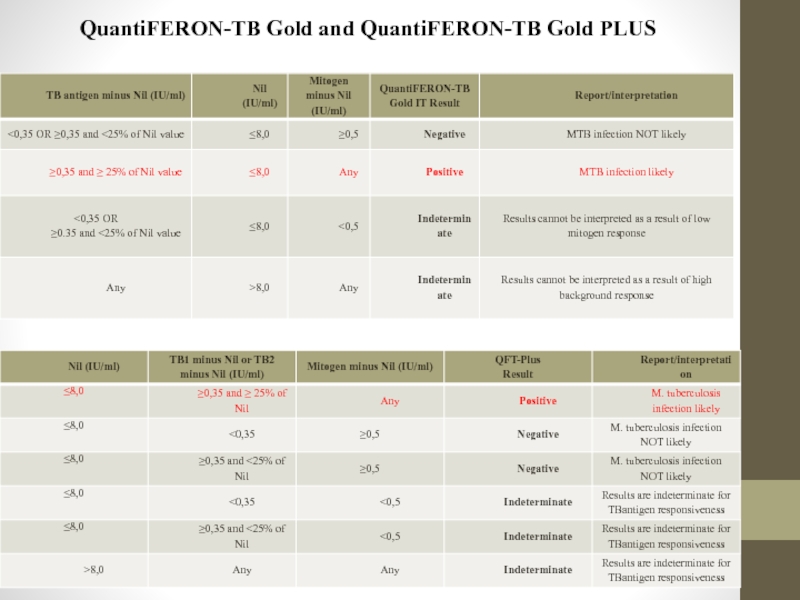
The QuantiFERON-TB Gold IT system
Слайд 37QuantiFERON-TB Gold and QuantiFERON-TB Gold PLUS
Baseline epidemiological data
Before performing the QuantiFERON-TB
Слайд 39QuantiFERON-TB Gold and QuantiFERON-TB Gold PLUS
Advantages:
greater sensitivity;
higher specificity;
there are
there are no contraindications.
Disadvantages.
This test, as TST can`t differentiate latent TB infection and active TB, distinguish reactivation from reinfection.
These tests needed for expensive equipment and are not cheep for patients.
Слайд 40T-SPOT test
General principles
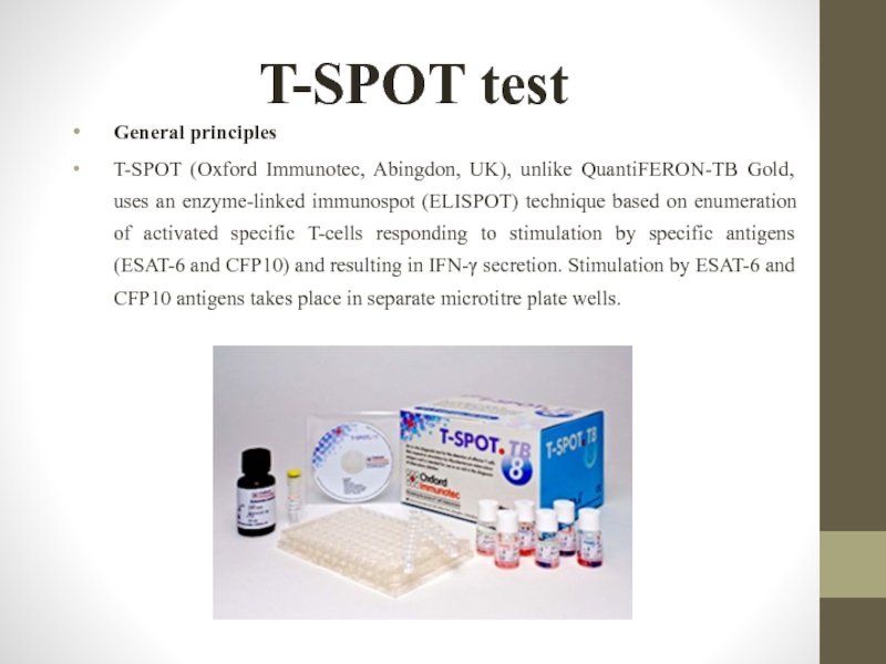
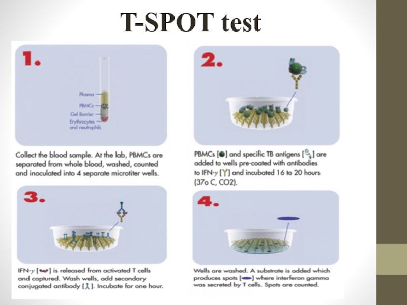
T-SPOT (Oxford Immunotec, Abingdon, UK), unlike QuantiFERON-TB Gold, uses
Слайд 42T-SPOT test
Baseline epidemiological data
As for the QuantiFERON-TB Gold assay, baseline epidemiological
Слайд 43T-SPOT test
- The test result is ‘positive’ if (Panel A minus
- The test result is ‘negative’ if both (Panel A minus nil control) and (Panel B minus nil control) ≤ 5 spots (this includes values less than zero), AND a nil control count <10 spots AND a positive control count >20 spots (or show saturation);
Слайд 44Screening fluorography
In Ukraine screening fluorography examination is conducted every two years
“The organized population” – employees of large companies, institutions and students in higher education. Planning preventive FG-examination and the number of contingents reported by companies medical and sanitary units institutions personnel department, district education departments and others. Their examination conducted by mobile x-ray stations.
“Employees of small businesses” – employees of agencies, enterprises conducting examination in district city clinics.
“Disorderly population” – housekeepers, don’t working pensioners, self-employed persons. They inspection conducted in clinics in the city of residence.
Слайд 45Screening fluorography
“Obligatory contingents” include:
Students of higher and specialized secondary educational institutions;
Persons
Employees of kindergartens and school children's institutions;
Employees of medical and pharmaceutical institutions;
Food industry workers who work in all phases of preparation and sale of food;
Domestic service workers;
Trade workers;
Employees of public transport;
Water utility workers;
Workers, working in hazardous occupational conditions with high air pollution. In rural areas these contingents also includes machine operators and cattle farms employees;
Mothers to their discharge from the hospital.
Слайд 46Screening fluorography
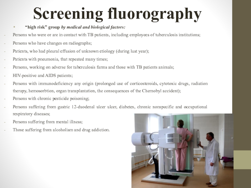
“high risk” group by medical and biological factors:
Persons who were
Persons who have changes on radiographs;
Patients, who had pleural effusion of unknown etiology (during lust year);
Patients with pneumonia, that repeated many times;
Persons, working on adverse for tuberculosis farms and those with TB patients animals;
HIV-positive and AIDS patients;
Persons with immunodeficiency any origin (prolonged use of corticosteroids, cytotoxic drugs, radiation therapy, hemosorbtion, organ transplantation, the consequences of the Chernobyl accident);
Persons with chronic pesticide poisoning;
Persons suffering from gastric 12-duodenal ulcer ulcer, diabetes, chronic nonspecific and occupational respiratory diseases;
Persons suffering from mental illness;
Those suffering from alcoholism and drug addiction.
Слайд 47Screening fluorography
“high risk” group by social factors:
Persons without permanent residence (refugees,
Persons, held in penitentiary system;
Persons, who have returned from the prison (for 3 years);
Persons, who got in remand centers and are there for a week or more;
Unemployed;
Persons, who are registered in the state employment as job seekers and the unemployed and those registered more than a year;
Members of low-income families who are registered in the Department of Labor and Social Protection;
Novices, monks;
Pilgrims, pilgrims upon arrival at place of pilgrimage;
Persons, who provide paid sex services.
Слайд 48Sputum samples collection
In order to collect diagnostic material shall be used
made from impact-resistant and transparent material that prevents leakage of fluid and allows to estimate the quantity and quality of samples collected without opening the cover;
can be easily marked and keeps it throughout the period of storage, transportation and carrying out research;
with the compaction screw tops (do not use bottles with closely corked cover, because at the opening of the container there is rarefied space that leads to the formation of aerosol, creating potential danger intra-laboratory contamination);
have the volume 30,0-50,0 ml;
have a wide hole for sputum collection (at least 30 mm in diameter) for the patient can easily separate the mucus inside the container without pollution exposing its outer surface.
Слайд 49Sputum samples collection
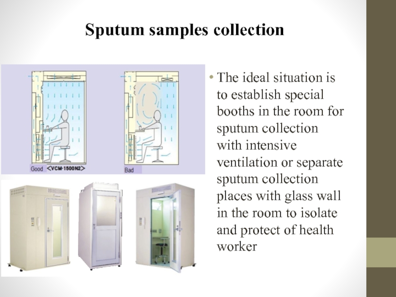
The ideal situation is to establish special booths in
Слайд 50The quantity and quality of collected sputum estimating
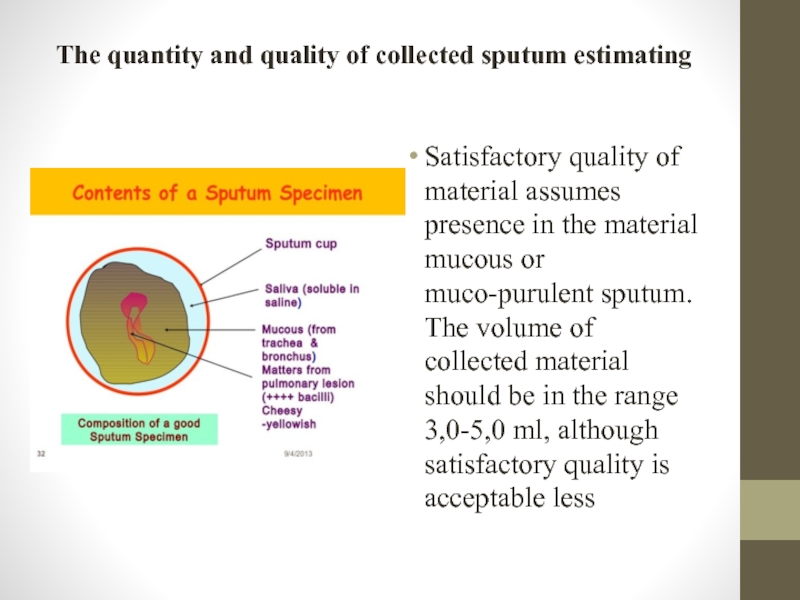
Satisfactory quality of material
Слайд 51Ziehl-Neelsen staining method
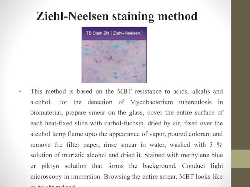
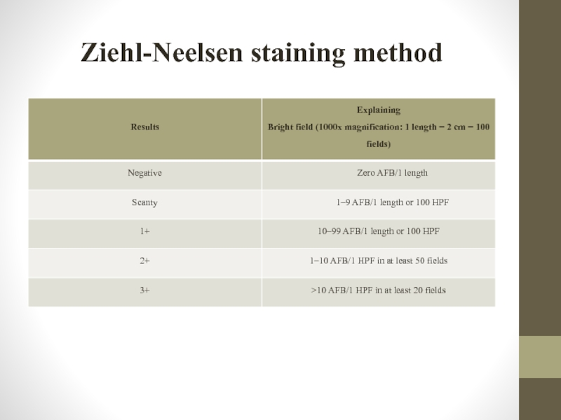
This method is based on the MBT resistance to
Слайд 53Storage of specimens
In order to increase the cultural method results period
Слайд 54Storage of specimens
Aseptic material should be sent to the laboratory immediately!
For
Listed solutions recommended, primarily, to preserve samples of sputum. If their application, material can be kept at room temperature. But conservants are toxic for mycobacteria, and their use can reduce the seeding of mycobacteria. To reduce the toxicity of conservants is recommended to keep samples in the refrigerator at a temperature of (+4 to +8) °C.
Diagnostic material can be frozen and in case it will not be subjected to repeated unfreezing and freeze viability of Mycobacterium will be kept.
Слайд 55Transportation of specimens
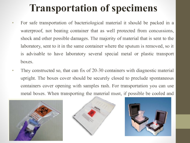
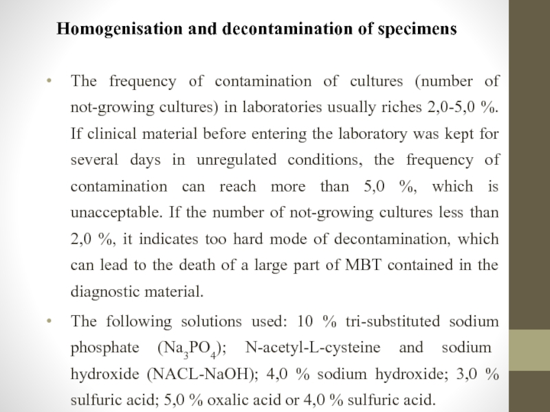
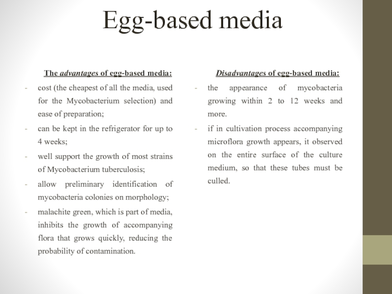
For safe transportation of bacteriological material it should be
They constructed so, that can fix of 20-30 containers with diagnostic material upright. The boxes cover should be securely closed to preclude spontaneous containers cover opening with samples rash. For transportation you can use metal boxes. When transporting the material must, if possible be cooled and not be in the sun.
Слайд 56Homogenisation and decontamination of specimens
The frequency of contamination of cultures (number
The following solutions used: 10 % tri-substituted sodium phosphate (Na3PO4); N-acetyl-L-cysteine and sodium hydroxide (NACL-NaOH); 4,0 % sodium hydroxide; 3,0 % sulfuric acid; 5,0 % oxalic acid or 4,0 % sulfuric acid.
Слайд 57CULTURE MEDIA
There are 4 main groups of various culture media for
- egg-based media: Löwenstein-Jensen (LJ) medium, Finn II and Ogawa medium;
- agar-based media: Middlebrook 7H10 and Middlebrook 7H11;
- liquid media: Middlebrook 7H9 broth;
- liquid synthetic and semi-synthetic nutrient media.
Слайд 58Egg-based media
The advantages of egg-based media:
cost (the cheapest of all
can be kept in the refrigerator for up to 4 weeks;
well support the growth of most strains of Mycobacterium tuberculosis;
allow preliminary identification of mycobacteria colonies on morphology;
malachite green, which is part of media, inhibits the growth of accompanying flora that grows quickly, reducing the probability of contamination.
Disadvantages of egg-based media:
the appearance of mycobacteria growing within 2 to 12 weeks and more.
if in cultivation process accompanying microflora growth appears, it observed on the entire surface of the culture medium, so that these tubes must be culled.
Слайд 59Agar-based media
These media are prepared in slant tubes or plates and
Слайд 60Liquid media
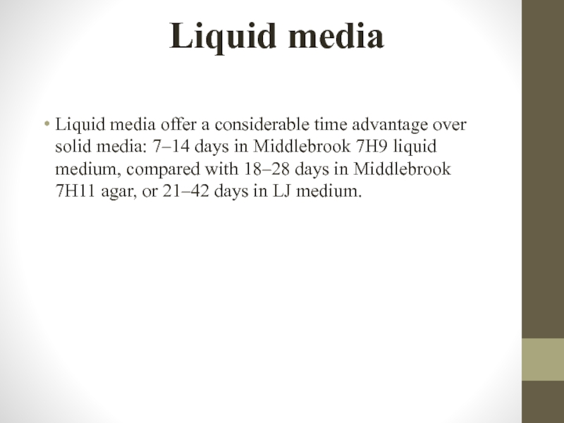
Liquid media offer a considerable time advantage over solid media:
Слайд 61Culture examination
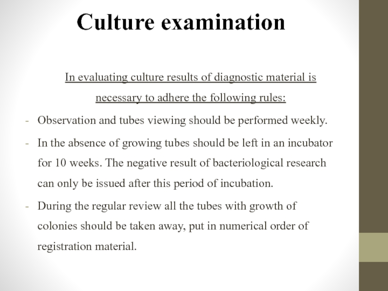
In evaluating culture results of diagnostic material is necessary to
Observation and tubes viewing should be performed weekly.
In the absence of growing tubes should be left in an incubator for 10 weeks. The negative result of bacteriological research can only be issued after this period of incubation.
During the regular review all the tubes with growth of colonies should be taken away, put in numerical order of registration material.
Слайд 62Culture examination
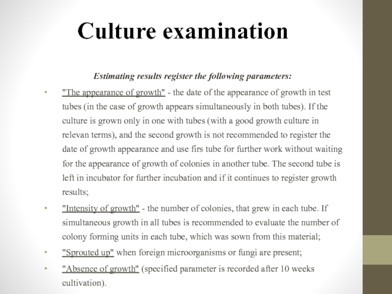
Estimating results register the following parameters:
"The appearance of growth" -
"Intensity of growth" - the number of colonies, that grew in each tube. If simultaneous growth in all tubes is recommended to evaluate the number of colony forming units in each tube, which was sown from this material;
"Sprouted up" when foreign microorganisms or fungi are present;
"Absence of growth" (specified parameter is recorded after 10 weeks cultivation).
Слайд 63Characterization of M. tuberculosis colonies
Cultures should be read within 5 to
Virulent Mycobacterium tuberculosis cultures typically grow on solid media in the form of R-colonies of various sizes and have a yellowish or slightly creamy shade (ivory color), a rough surface that resembles semolina or cauliflower. Colonies are usually dry, wrinkled, but in the case of dissociation they may be moist, lightly pigmented colonies, pink and yellow pigment which very different from the orange or yellow pigment or saprophytic some nontuberculous mycobacteria.
Слайд 65BACTEC MGIT 960 system
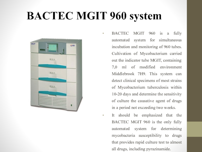
BACTEC MGIT 960 is a fully automated system
It should be emphasized that the BACTEC MGIT 960 is the only fully automated system for determining mycobacteria susceptibility to drugs that provides rapid culture test to almost all drugs, including pyrazinamide.
Слайд 66BACTEC MGIT 960 system
Advantages of the method: the receiving culture twice
The device weighs 351 kg, its size is small (92h135h85sm), not required special conditions for its placement in a laboratory. It consists of three sections that accommodate over 320 tubes each, so the maximal simultaneous loading device - 960 tubes. Control over included in the indicator tube material, carries a built-in a device computer. Liquid crystal display and custom indicators on each section give information about the presence of positive and negative results
Слайд 67BACTEC MGIT 960 system
An important component of the system is Mycobacteria
To accelerate growth and reduce contamination of Mycobacterium provided addition liquid nutritional supplements OADC and five lyophilized antibiotic PANTA added to 7H9, which contribute to the indicator tube before sowing. The device evaluates tube as positive if the number of living organisms in it reached 105-106 per 1,0 ml of medium.
The BACTEC MGIT 960 System was designed with simplicity in mind, ensuring maximum productivity with minimal staffing and training. Bar code scanning guides the simple 4-step operating procedure, eliminating potential errors
Слайд 68BACTEC MGIT 960 system
Firstly you should press «Tube enter» on BACTEC
The tubes, which are not fixed growth of Mycobacterium during 42 days, the system evaluated as negative. Negative result (no growth of mycobacteria) introduced as green signal of negative indicator on the relevant box and icon on the display.
Слайд 69Identification of Mycobacterium tuberculosis
Growth in different media
In LJ medium MBT
The absence of MBT growth in the medium with 500 mg / ml of salicylic acid-sodium or 500 mg / ml paranitro-benzoic acid (PNBK), and with 1000 mg / ml tioatsetazon (tibon).
Growth on the medium with 5,0 % NaCl
The method is based on the ability of nontuberculous mycobacteria of IV group grow on the medium with 5,0 % NaCl. Besides this group, in this environment grow only M. terrae complex (including M. triviale, M. terrae, M. nonchromogenicum (III group)) and M. flavescens (group II), as well as some mycobacteria from I groups (M. marinum ). All other mycobacteria, including M. tuberculosis and M. bovis, do not grow on this environment.
Слайд 70Identification of Mycobacterium tuberculosis
Detection of cord factor
Nontuberculous mycobacteria grow diffusely
Sensitivity to cycloserine
All strains of M. bovis-BCG observed resistance to 30,0-50,0 mg / ml of cycloserine. This biological feature of the BCG vaccine strain is an important diagnostic test to identify it.
Слайд 71Biochemical tests of identification
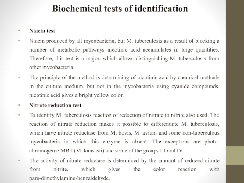
Niacin test
Niacin produced by all mycobacteria, but M.
The principle of the method is determining of nicotinic acid by chemical methods in the culture medium, but not in the mycobacteria using cyanide compounds, nicotinic acid gives a bright yellow color.
Nitrate reduction test
To identify M. tuberculosis reaction of reduction of nitrate to nitrite also used. The reaction of nitrate reduction makes it possible to differentiate M. tuberculosis, which have nitrate reductase from M. bovis, M. avium and some non-tuberculous mycobacteria in which this enzyme is absent. The exceptions are photo- chromogenic MBT (M. kansasii) and some of the groups III and IV.
The activity of nitrate reductase is determined by the amount of reduced nitrate from nitrite, which gives the color reaction with para-dimethylamino-benzaldehyde.
Слайд 72Biochemical tests of identification
Determining ability to growth in the medium with
M. tuberculosis is susceptible to nicotinamide. M. bovis vaccine strain has a natural resistance to nicotinamide. Differentiation is based on this features of tuberculosis complex.
Determination of catalase and peroxidase activity simultaneously
Principle of catalase reaction consists in disjoined of hydrogen peroxide by enzyme catalase to water and atomic oxygen, which is accompanied by of bubbles of oxygen and transition pyrogallol in purple-galin in the presence of hydrogen peroxide under the influence of peroxidase.
Слайд 73Biochemical tests of identification
Thermostability catalase
Catalase in MBT is different. In virulent
The reaction of hydrolysis of Tween-80
An important reaction for MBT identification in the second and third groups is the tvin-80 hydrolysis reaction. Tween-80 binding neutral red and the mixture reaction has straw-yellow color. Principle of reactions is in enzyme hydrolysis of tween-80. This releases a neutral color red from pink to red. The positive reaction observed in M. aquae (unlike M. scrofulaceum, in which the reaction is negative), on the group III it is positive only for M. terrae.
Слайд 74Drug susceptibility testing for Mycobacterium tuberculosis complex
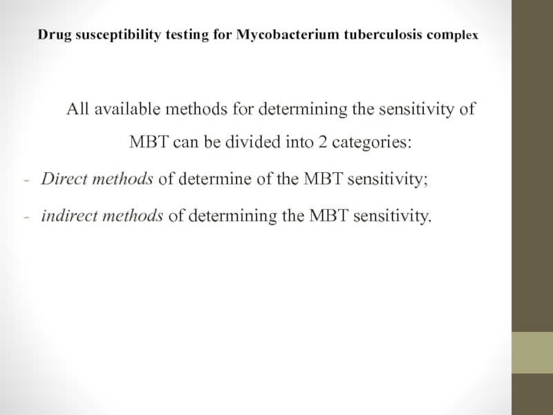
All available methods for determining
Direct methods of determine of the MBT sensitivity;
indirect methods of determining the MBT sensitivity.
Слайд 75Drug susceptibility testing for Mycobacterium tuberculosis complex
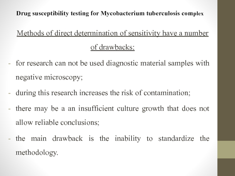
Methods of direct determination of
for research can not be used diagnostic material samples with negative microscopy;
during this research increases the risk of contamination;
there may be a an insufficient culture growth that does not allow reliable conclusions;
the main drawback is the inability to standardize the methodology.
Слайд 76Indirect methods of determining the sensitivity of MBT
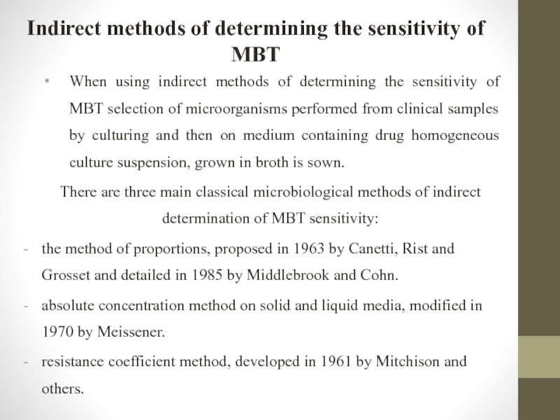
When using indirect
There are three main classical microbiological methods of indirect determination of MBT sensitivity:
the method of proportions, proposed in 1963 by Canetti, Rist and Grosset and detailed in 1985 by Middlebrook and Cohn.
absolute concentration method on solid and liquid media, modified in 1970 by Meissener.
resistance coefficient method, developed in 1961 by Mitchison and others.
Слайд 77The proportion method on Löwenstein-Jensen medium
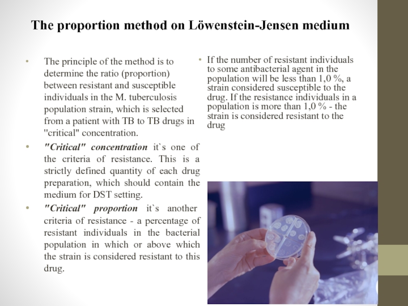
The principle of the method is
"Critical" concentration it`s one of the criteria of resistance. This is a strictly defined quantity of each drug preparation, which should contain the medium for DST setting.
"Critical" proportion it`s another criteria of resistance - a percentage of resistant individuals in the bacterial population in which or above which the strain is considered resistant to this drug.
If the number of resistant individuals to some antibacterial agent in the population will be less than 1,0 %, a strain considered susceptible to the drug. If the resistance individuals in a population is more than 1,0 % - the strain is considered resistant to the drug
Слайд 78Indirect absolute concentrations drug susceptibility testing
The method is performing by dosed
Usually "critical" concentration of drugs used, which is the criterion of resistance, inhibits the growth of all or almost all mycobacteria, defined as the presence of 20 or fewer colonies of the pathogen and allows to define the culture of MBT as sensitive or resistant to TB medication.
The evaluation results to determination resistance of mycobacteria to antituberculosis drugs conduct in 3 weeks of incubation in an incubator. If MBT do not grow on the control nutrient medium it should wait 1-2 weeks to get a pronounced growing in control, and then give the final answer.
When using the method of absolute concentrations MBT culture is considered resistant if on the culture medium with a certain drug grows more than 20 colonies with abundant microbial growth in vitro control (no drug).
Слайд 79Drug susceptibility testing in liquid media (MGIT 960)
The system BACTEC MGIT
A set of indicator tubes growing control (no drug) and containing TB drug is placed in a special medium with a bar code by which the device provides continuous monitoring of introduced to the culture tube.
Results are interpreted automatically, based on accounting multiplication of mycobacteria in vitro without drug at the time of control growth in 4-13 day after inoculation culture.
Set MGIT 960 SIRE Kit includes 4 bottles of major anti-TB drugs and 8 bottles of enriching liquid. Critical (low) drug concentrations achieved in nutrient broth after dilution. To determine the sensitivity of mycobacteria to pyrazinamide PZA) as control using MGIT tube with special pH = 5.9. Kit BACTEC MGIT 960 PZA includes 2 bottles with pyrazinamide lyophilized and six bottles with nutritional supplements.
Definition DST of isolated M. tuberculosis from cultures in newly diagnosed patients and relapsed patients with tuberculosis must be necessarily to spend DST to first-line drugs: isoniazid, rifampicin, ethambutol, pyrazinamide, streptomycin. In the case of drug resistance to these drugs or multidrug resistance is recommended to conduct DST to Ethionamidum, amikacin, capreomycin and fluoroquinolones (ofloxacin).
In previously treated patients (treatment failure and treatment after an interruption), and patients with chronic tuberculosis is necessary to determination DST of M. tuberculosis drugs to all immediately with the results of previous studies.
Слайд 80Investigation of the sensitivity of mycobacteria to medicinal preparations by the
The principle of method consists in determining the minimum inhibitory concentration of anti-TB drugs for clinical strains of Mycobacterium and minimum inhibitory concentration ratio of these drugs for deliberately sensitive laboratory strain of mycobacteria (typically, H37Rv). This is the most time consuming and expensive method because it requires the use of a large number of tubes with nutrient because it is used mainly for scientific research.
Слайд 81Absolute concentration method
DST results counted in 3- 4 weeks of incubation
To accelerate research direct method of absolute concentrations can be used. When setting this method performed direct seeding precipitate processed detergents diagnostic material simultaneously to control culture medium and environment with appropriate anti-tuberculosis drugs. Seeding of material to standard culture media is performed simultaneously (in order to obtain culture).
Culture of Mycobacterium considered as resistance, if in the indirect method of absolute concentrations grows more than 20 colonies.
However, the direct method of absolute concentrations can be used only for research material if bacterioscopic result is positive with massive bacterial excretion at least 2+. In this case, increases the risk of contamination. Also, necessary to consider that this method is not performed dosed seeding, which may complicate the interpretation of results. Therefore, in some cases, the results may be unreliable.
Слайд 82Nitrate reductase assay
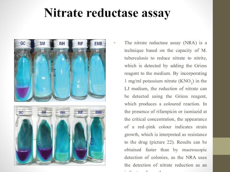
The nitrate reductase assay (NRA) is a technique based
Слайд 83GeneXpertMTB/RIF test
Test system GeneXpertMTB/RIF is recommended by WHO for use in
isolation and amplification is carried in the cartridge, pretreatment diagnostic material is reduced to a minimum of manipulation;
the possibility of contamination is greatly reduced;
it`s only determines the MBT resistance to rifampicin.
Test system GeneXpertMTB/RIF is a semi-nested PCR in real time in the cartridge that is conducted to identify:
M. tuberculosis DNA in sputum samples or concentrated sputum precipitates;
mutations in rpoB gene (resistance to rifampicin) in samples received from patients with a risk of resistance to this drug.
Слайд 84GeneXpertMTB/RIF test
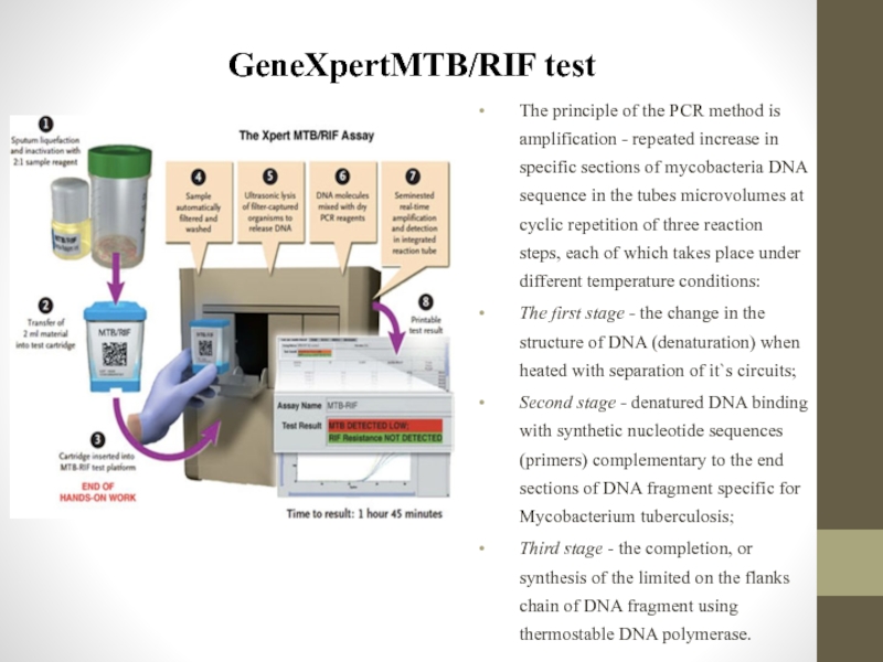
The principle of the PCR method is amplification - repeated
The first stage - the change in the structure of DNA (denaturation) when heated with separation of it`s circuits;
Second stage - denatured DNA binding with synthetic nucleotide sequences (primers) complementary to the end sections of DNA fragment specific for Mycobacterium tuberculosis;
Third stage - the completion, or synthesis of the limited on the flanks chain of DNA fragment using thermostable DNA polymerase.
Слайд 85GeneXpertMTB/RIF test
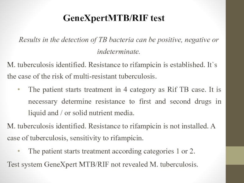
Results in the detection of TB bacteria can be positive,
M. tuberculosis identified. Resistance to rifampicin is established. It`s the case of the risk of multi-resistant tuberculosis.
The patient starts treatment in 4 category as Rif TB case. It is necessary determine resistance to first and second drugs in liquid and / or solid nutrient media.
M. tuberculosis identified. Resistance to rifampicin is not installed. A case of tuberculosis, sensitivity to rifampicin.
The patient starts treatment according categories 1 or 2.
Test system GeneXpert MTB/RIF not revealed M. tuberculosis.
Слайд 86GenoType test system
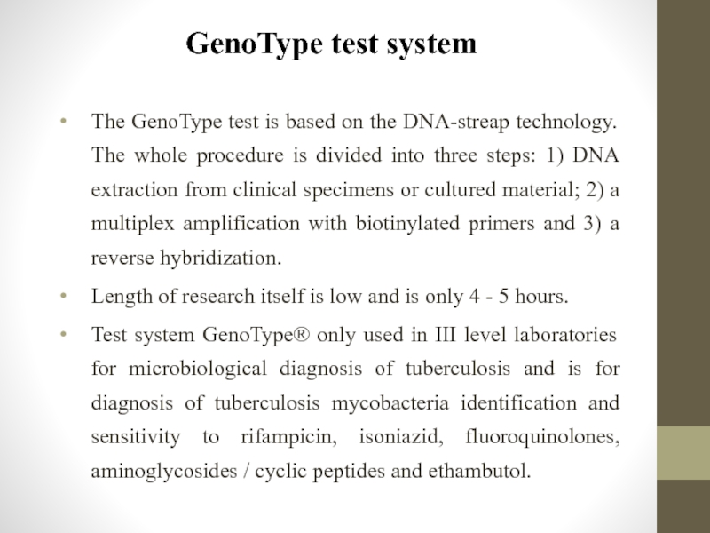
The GenoType test is based on the DNA-streap technology.
Length of research itself is low and is only 4 - 5 hours.
Test system GenoType® only used in III level laboratories for microbiological diagnosis of tuberculosis and is for diagnosis of tuberculosis mycobacteria identification and sensitivity to rifampicin, isoniazid, fluoroquinolones, aminoglycosides / cyclic peptides and ethambutol.
Слайд 87GenoType test system
Indications to GenoType test:
- HIV-infected patients with suspected TB.
-
• patients from MDR-TB contacts;
• patients, previously treated for tuberculosis;
• patients, who were born in a foreign country with a high TB incidence;
- Children or teenagers (0-17 years age group) with suspected TB.
Indications for patients with AFB in the sputum:
- TB patients with negative clinical and radiological dynamics and / or continuation or resumption of bacterial-excretion;
- Patients from social risk groups;
- Patients with newly diagnosed tuberculosis.
Слайд 88GenoType test system
Results interpretation.
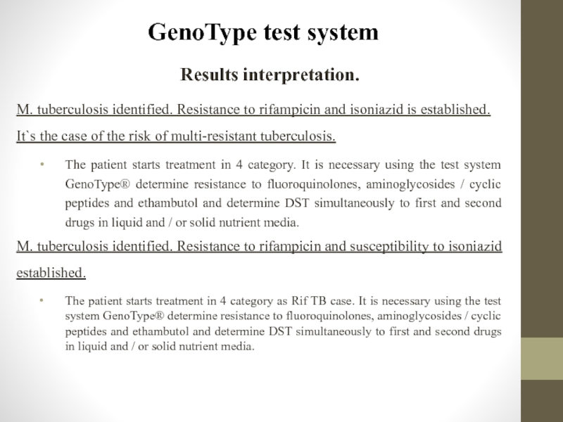
M. tuberculosis identified. Resistance to rifampicin and
The patient starts treatment in 4 category. It is necessary using the test system GenoType® determine resistance to fluoroquinolones, aminoglycosides / cyclic peptides and ethambutol and determine DST simultaneously to first and second drugs in liquid and / or solid nutrient media.
M. tuberculosis identified. Resistance to rifampicin and susceptibility to isoniazid established.
The patient starts treatment in 4 category as Rif TB case. It is necessary using the test system GenoType® determine resistance to fluoroquinolones, aminoglycosides / cyclic peptides and ethambutol and determine DST simultaneously to first and second drugs in liquid and / or solid nutrient media.
Слайд 89GenoType test system
Results interpretation.
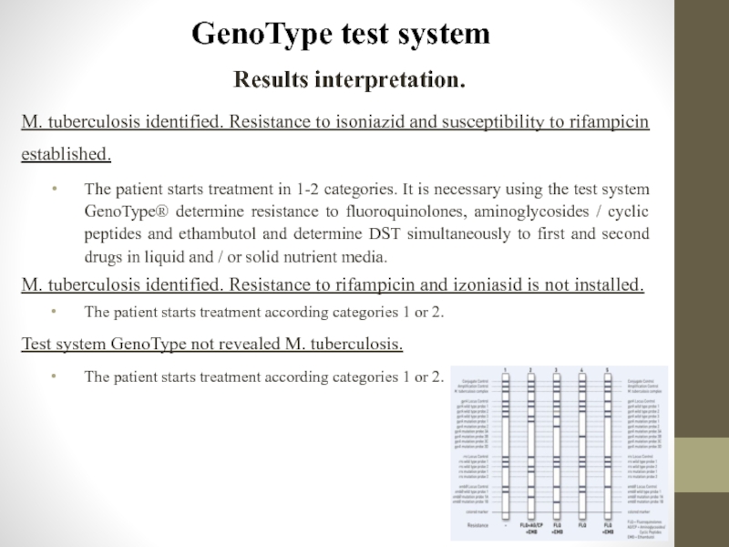
M. tuberculosis identified. Resistance to isoniazid and
The patient starts treatment in 1-2 categories. It is necessary using the test system GenoType® determine resistance to fluoroquinolones, aminoglycosides / cyclic peptides and ethambutol and determine DST simultaneously to first and second drugs in liquid and / or solid nutrient media.
M. tuberculosis identified. Resistance to rifampicin and izoniasid is not installed.
The patient starts treatment according categories 1 or 2.
Test system GenoType not revealed M. tuberculosis.
The patient starts treatment according categories 1 or 2.
