- Главная
- Разное
- Дизайн
- Бизнес и предпринимательство
- Аналитика
- Образование
- Развлечения
- Красота и здоровье
- Финансы
- Государство
- Путешествия
- Спорт
- Недвижимость
- Армия
- Графика
- Культурология
- Еда и кулинария
- Лингвистика
- Английский язык
- Астрономия
- Алгебра
- Биология
- География
- Детские презентации
- Информатика
- История
- Литература
- Маркетинг
- Математика
- Медицина
- Менеджмент
- Музыка
- МХК
- Немецкий язык
- ОБЖ
- Обществознание
- Окружающий мир
- Педагогика
- Русский язык
- Технология
- Физика
- Философия
- Химия
- Шаблоны, картинки для презентаций
- Экология
- Экономика
- Юриспруденция
The cardiac cycle and ECG презентация
Содержание
- 2. The cardiac cycle and ECG
- 3. Learning objectives 11.1.3.4 use an electrocardiogram
- 4. Success creteria 1.Investigate the electrical process
- 5. Terminology Atrium, ventricle, contract, systole, blood
- 7. Cardiac cycle Your heart beats around 70
- 8. Atrial systole The heart is filled
- 9. Ventricular systole About 0.1 seconds after the
- 10. Ventricular diastole Ventricular systole lasts for
- 13. Deoxygenated blood from body to RA through
- 15. Electrocardiograms (ECGs) It is relatively easy to
- 17. ECG teeth P- the sinoatrial node despoliation,

Слайд 3Learning objectives
11.1.3.4 use an electrocardiogram to describe the cardiac cycle
11.1.3.5
explain oxygen dissociation curves for hemoglobin and myoglobin in adult and embryo

Слайд 4Success creteria
1.Investigate the electrical process of in the heart .
2.Describe the structure of the heart and indicate the link between the structure of the heart muscles and its ability to automaticity.
3.Explain the mechanism of cardiac cycle.
4.Explain the essence of the Electrocardiography (ECG)

Слайд 5Terminology
Atrium, ventricle, contract, systole, blood pressure, valves, aorta, pulmonary vein
and arteria, relax, low and high pressure, diastole, vena cava, ECG – electrocardiogram, PQRST teeth, sinoatrial nodes and atrioventricular, atria, ventricles, Purkinje fibers, bundle branch block, septum, atrioventricular valves and semilunar valve, cardiac cycle, heart

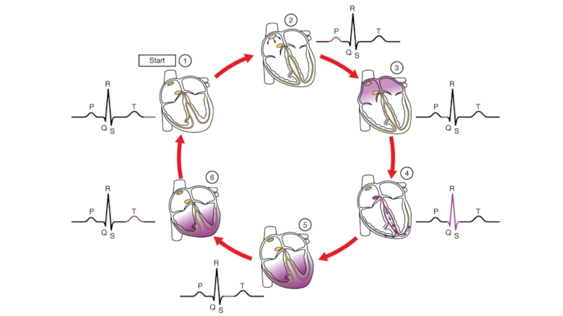
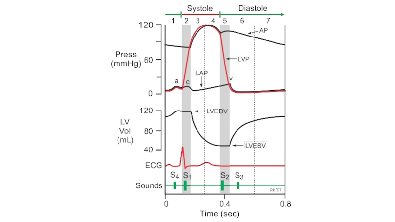
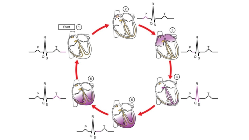
Слайд 7Cardiac cycle
Your heart beats around 70 times a minute. The cardiac
cycle is the sequence of events which makes up one heart beat.
Three stages in this cycle.
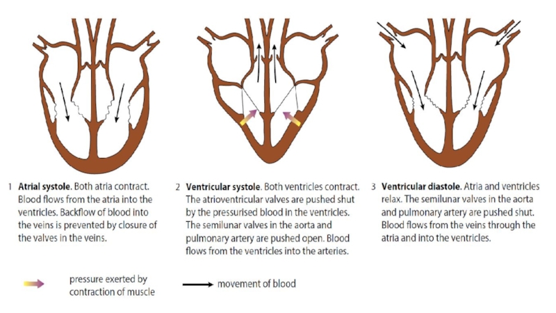
Atrial systole
Ventricular systole
Ventricular diastole
Three stages in this cycle.
Atrial systole
Ventricular systole
Ventricular diastole

Слайд 8Atrial systole
The heart is filled with blood and the muscle
in the atrial walls contracts. This stage is called atrial systole.
The pressure developed by this contraction is not very great, because the muscular walls of the atria are only thin, but it is enough to force the blood in the atria down through the atrioventricular valves into the ventricles. The blood from the atria does not go back into the pulmonary veins or the venae cavae, because these have semilunar valves to prevent backflow.
The pressure developed by this contraction is not very great, because the muscular walls of the atria are only thin, but it is enough to force the blood in the atria down through the atrioventricular valves into the ventricles. The blood from the atria does not go back into the pulmonary veins or the venae cavae, because these have semilunar valves to prevent backflow.

Слайд 9Ventricular systole
About 0.1 seconds after the atria contract, the ventricles contract.
This is called ventricular systole.
The thick, muscular walls of the ventricles squeeze inwards on the blood, increasing its pressure and pushing it out of the heart. As soon as the pressure in the ventricles becomes greater than the pressure in the atria, this pressure difference pushes the atrioventricular valves shut, preventing blood from going back into the atria.
Instead, the blood rushes upwards into the aorta and the pulmonary artery, pushing open the semilunar valves in these vessels as it does so.

Слайд 10Ventricular diastole
Ventricular systole lasts for about 0.3 seconds. The muscle
then relaxes, and the stage called ventricular diastole begins.
As the muscle relaxes, the pressure in the ventricles drops. The high-pressure blood which has just been pushed into the arteries would flow back into the ventricles but for the presence of the semilunar valves, which snap shut as the blood fills their cusps.
During diastole, as the whole of the heart muscle relaxes, blood from the veins flows into the two atria. The blood is at a very low pressure, but the thin walls of the atria are easily distended, providing very little resistance to the blood flow.

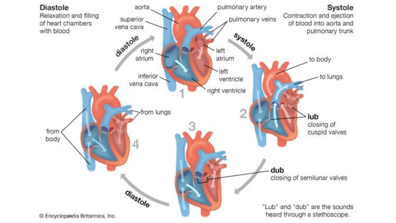
Слайд 13Deoxygenated blood from body to RA through vena cava
Blood from RA
to RV through tri-cuspid valve
Oxygenated blood to all the cells in the body via aorta
Deoxygenated from RV through pulmonary arteries to lungs to get oxygen
Oxygenated blood returns to LA via pulmonary veins.
Oxygenated blood to LV via the bi-cuspid valve.
The path of blood through the heart
Blood flow steps

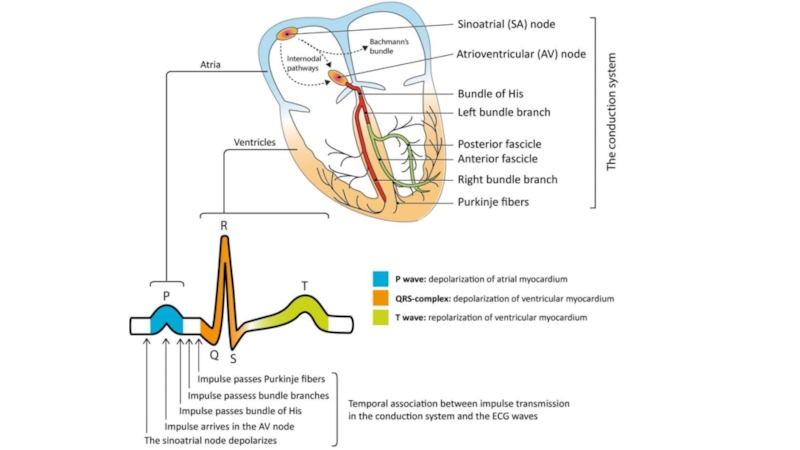
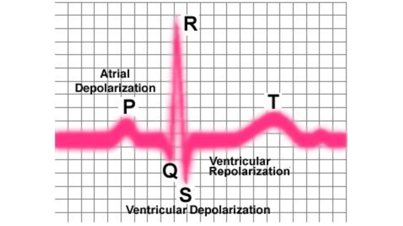
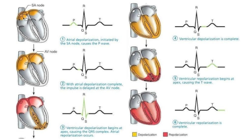
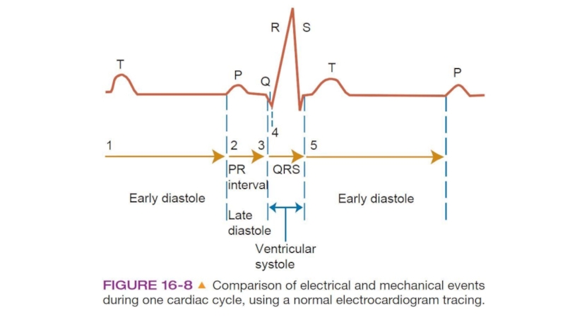
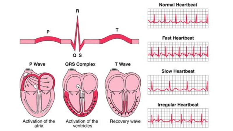
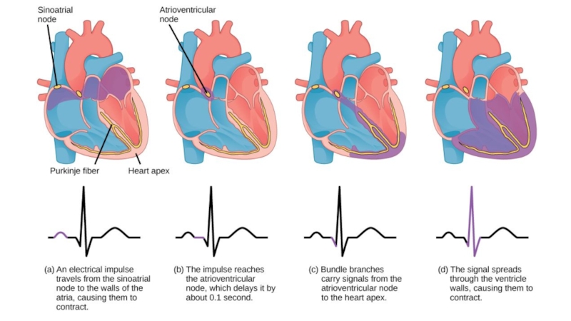
Слайд 15Electrocardiograms (ECGs)
It is relatively easy to detect and record the waves
of excitation flowing through heart muscle. Electrodes can be placed on the skin over opposite sides of the heart, and the electrical potentials generated recorded with time. The result, which is essentially a graph of voltage against time, is an electrocardiogram (ECG). The part labelled P represents the wave of excitation sweeping over the atrial walls. The parts labelled Q, R and S represent the wave of excitation in the ventricle walls. The T section indicates the recovery of the ventricle walls.
It is relatively easy to detect and record the waves of excitation flowing through")
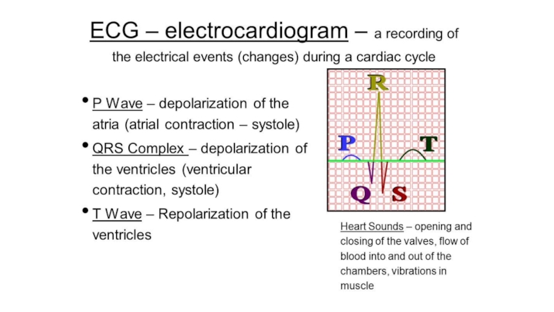
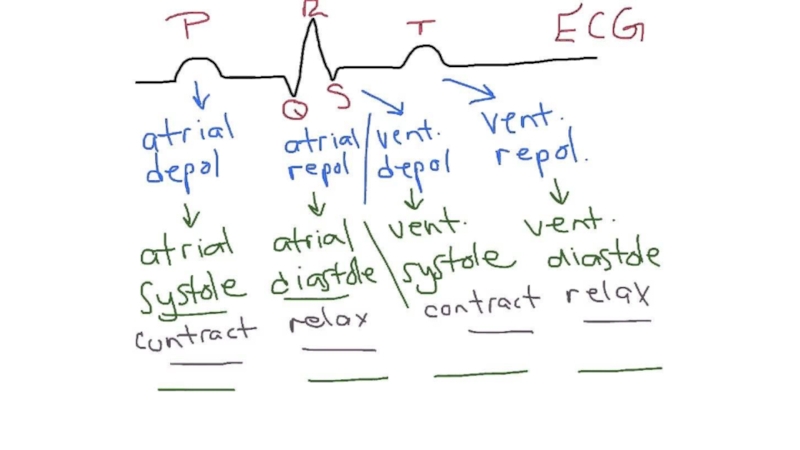
Слайд 17ECG teeth
P- the sinoatrial node despoliation, impulse arrive in the AV
node and impulse passes buddle of His
QRS complex - depolarization of ventricular myocardium
T- repolarization of ventricular myocardium
QRS complex - depolarization of ventricular myocardium
T- repolarization of ventricular myocardium