- Главная
- Разное
- Дизайн
- Бизнес и предпринимательство
- Аналитика
- Образование
- Развлечения
- Красота и здоровье
- Финансы
- Государство
- Путешествия
- Спорт
- Недвижимость
- Армия
- Графика
- Культурология
- Еда и кулинария
- Лингвистика
- Английский язык
- Астрономия
- Алгебра
- Биология
- География
- Детские презентации
- Информатика
- История
- Литература
- Маркетинг
- Математика
- Медицина
- Менеджмент
- Музыка
- МХК
- Немецкий язык
- ОБЖ
- Обществознание
- Окружающий мир
- Педагогика
- Русский язык
- Технология
- Физика
- Философия
- Химия
- Шаблоны, картинки для презентаций
- Экология
- Экономика
- Юриспруденция
Takayasu’s arteritis презентация
Содержание
- 1. Takayasu’s arteritis
- 2. EPIDEMIOLOGY More case reports from Japan ,India,
- 3. Age
- 5. Histopathology Idiopathic c/c infla arteritis of elastic
- 8. Wall thickening, Fibrosis, Stenosis, & Thrombus
- 9. Associated pathology-TB (LN)-55%
- 10. Clinical features Early pre pulseless/gen manif Fever,weight
- 12. Coronary involvement in TA Occurs in
- 13. Occular involvement-Amaurosis fugax, pain behind eye,
- 15. HTN is the most characteristic
- 16. Ishikawa clinical classification of Takayasu arteritis 1978
- 18. Cumulative survival 5years -91% (event free
- 21. Sharma BK, Jain S, Suri S,
- 23. nee
- 24. a/c phase-Axial T1-weighted image
- 25. Findings of TA on MRI
- 26. [18F]fluorodeoxyglucose PET for diagnosing Takayasu’s arteritis common
- 27. remission after treatment
- 28. Treatment of TA ・ Steroids
- 29. Medical treatment 0.7-1 mg/kg/day –prednisolone
- 30. Steroids → 50% response Methotrexate →further
- 31. Critical issue is in trying to
- 32. chronic phase-
- 33. Surgical treatment HTN with critical RAS Extremity
- 34. Surgical techniques Carry high morbidity &
- 35. Renal artery involvement Best treated by PTA
- 36. ostial stenosis of the right renal artery after deployment of a stent
- 37. Renal PTA - 33 stenoses (20 pts)
- 38. Aortoarteritic lesions Balloon dilation safe &
- 39. Left subclavian angiograms- 95% stenosis with
- 40. Joseph s et al, SCT J
- 41. Aortoplasty and Stenting PTA -desc thoracic and/or
- 42. long-segment diffuse stenotic involvement of the DTA after deployment of stents.
- 43. Treatment for cor A occulusion in TA
- 44. Percutaneous Management of Aneurysmal Lesions

Слайд 2EPIDEMIOLOGY
More case reports from Japan ,India, South-east Asia, Mexico
No geographic restriction
No
Incidence-2.6/million/year-N.America/Europe
The incidence in Asia is 1 case/1000-5000 women.

Слайд 3 Age
Mc-2nd & 3rd decade
May range
Indian studies-age 3- 50 yrs
Gender diff
Japan-F:M=8-9:1
India-F:M ratio varies from -1:1 - 3:1
( Padmavati S, Aurora AP, Kasliwal RR Aortoarteritis in India. J Assoc Physicians India 1987)
India=F:M- 6.4:1 (Panja et al, 1997 JACC)

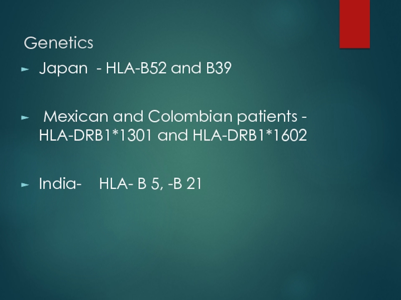
Слайд 4
Japan - HLA-B52 and B39
Mexican and Colombian patients - HLA-DRB1*1301 and HLA-DRB1*1602
India- HLA- B 5, -B 21
Слайд 5Histopathology
Idiopathic c/c infla arteritis of elastic arteries resulting in occlusive &/
Large vessels, esp, Aorta & its main branches (brachiocephalic, carotid, SCL, vertebral, RA)
+Coronary & PA
Ao valve –usually not beyond IMA
Multiple segs with dis & skipped nl areas
or diffuse involvement

Слайд 7
1)Gelatinous plaques-early
2)White plaques-collagen
3)Diffuse intimal thickening
Superficial– deep scarring
circumferential stenosis
4)Mural thrombus
5)2⁰ atheromatous changes
long standing,
HTN
Histology
Panarteritis-granulomatous lesion with giant cells
a/c phase diffuse infil-mono
granulomatous infil
2)c/c phase-coll rich fibrous tissue- adventitia thicker than media
3)Healed phase-no infl cells, vas media scarred
Gelatinous plaques-early2)White plaques-collagen3)Diffuse intimal thickening Superficial– deep scarring circumferential")
Слайд 8
Wall thickening, Fibrosis, Stenosis, & Thrombus formation →end organ ischaemia
More a/c
Stenotic lesions predominate & tend to be B/L
Nearly all pts with aneurysms also have stenoses

Слайд 9Associated pathology-TB (LN)-55%
Bazins disease(eryt induratum)
churg strauss synd
reteroperitoneal fib
PAN,UC,CD etc
-55% Erthema multiforme")
Слайд 10Clinical features
Early pre pulseless/gen manif
Fever,weight loss,headache, fatigue,malaise,night sweats, arthralgia
+/_ splenomegaly/ cervical,
Disappear partly/ completely in 3 months
50% -no h/o acute phase
Late ischemic phase
Sequel of occl of Ao arch/br
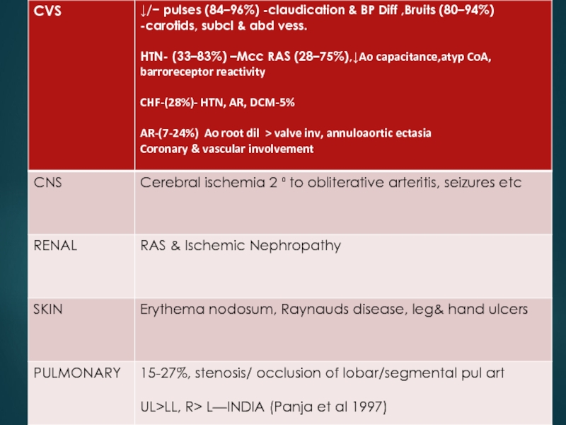
Diminished/absent pulses (84–96%)
Bruits (80–94%)
Hypertension (33–83% )
RAS(28–75%) &
CCF(28%)

Слайд 12Coronary involvement in TA
Occurs in 10~30%
Often fatal
Classified into 3 types
Type1:stenosis or
Type2:diffuse or focal coronary arteritis
Type3:coronary aneurysm

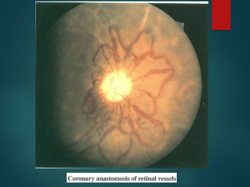
Слайд 13Occular involvement-Amaurosis fugax, pain behind eye,
Hypertensive retinopathy
Commonest
Arteriosclerotic –art narrowing, av nipping,silver wiring
Neuroretinopathy-exudates and papilloedema
Direct opthalmoscopy
Nonhypertensive retinopathy
UYAMA & ASAYAMA CLASS
stage 1- Dil of small vessels
stage 2- Microaneurysm
stage 3- Art-ven anastomoses
stage 4- Ocular complications
Mild -stage 1
Moderate -stage 2
Severe -stages 3 & 4
Flourescien angio sensitive

Слайд 15
HTN is the most characteristic manifestation in Indian patients,suggesting a high

Слайд 16Ishikawa clinical classification of Takayasu arteritis 1978
4 Complications
Retinopathy, Secondary HTN,

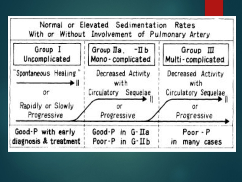
Слайд 18Cumulative survival
5years -91% (event free survival -74.9%)
10 years -84% (event
Single mild complication or no complication
5 year event free survival 97%
Single severe or multiple complications
5 year event free survival 59.7%
No deaths in groups I and IIA
19.6% mortality in groups IIB and III (CVA,CCF)
Subramanyan R, Joy J, Balakrishnan KG, et al.SCT. Natural
history of aortoarteritis (Takayasu’s arteritis). Circulation
1989; 80: 429-37.
10 years -84% (event free survival -64%)Single mild")
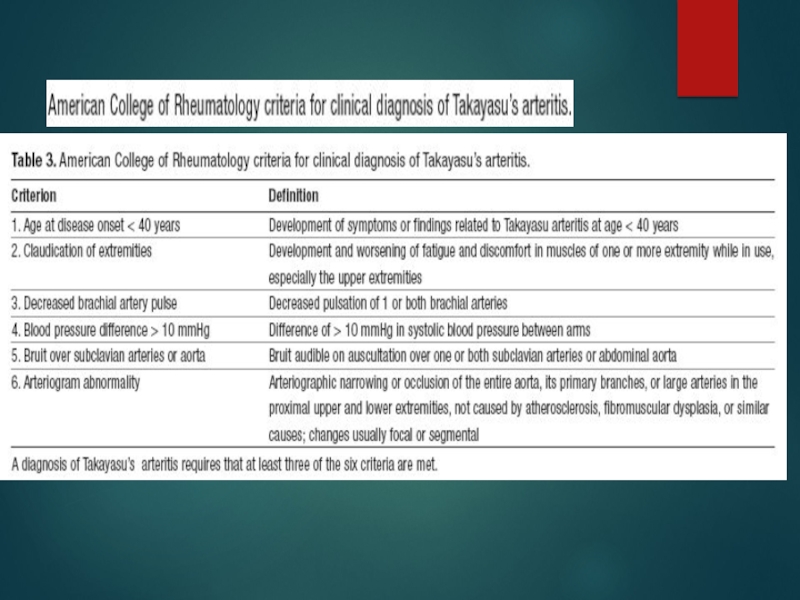
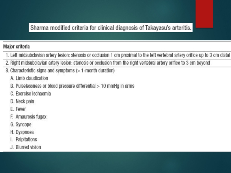
Слайд 21
Sharma BK, Jain S, Suri S, Numano F. Diagnostic criteria for
Takayasu


Слайд 24
a/c phase-Axial T1-weighted image
wall thickening of As aorta and
Axial T1-weighted image- improvement of wall thickening of As Ao and PA after steroid therapy

Слайд 25
Findings of TA on MRI
mural thrombi
signal alterations within
vascular dilation
thickened aortic valvular cusps
multifocal stenoses
concentric thickening of the aortic wall
Disadvantages
difficulty in visualizing small branch vessels and poor visualization of vascular calcification
may falsely accentuate the degree of vascular stenoses (renal & subclavian)

Слайд 26[18F]fluorodeoxyglucose PET for diagnosing
Takayasu’s arteritis
common [18F]FDG uptake pattern TA
late phase-patchy rather than continuous ,linear
shown to identify more affected vascular regions than morphologic imaging with MRI
does not provide any information about changes in the wall structure or luminal blood flow
sensitivities of 83% and specificity 100%
( Meller Jet al. Value of F-18 FDG hybrid camera PET and MRI in earlyTakayasu aortitis. Eur Radiol 2003)
Sensitivity of 92%, specificity of 100% and a diagnostic accuracy of 94%
( Webb M et al. The role of 18F-FDG PET in characterising disease activity in Takayasu arteritis. Eur J Nucl Med Imaging 2004


Слайд 28Treatment of TA
・
Steroids
immunosuppressants:
Cyclosporine,Cyclophosphamide,
Mtx,Mycophenolate mofetil
Anti-platelet therapy(low-dose Aspirin)
angioplasty/surgery
If uncontrolled
Control of vasculitis
Symptomatic occlusion
thrombosis
angioplasty/surgeryIf uncontrolledControl of vasculitisSymptomatic occlusionthrombosis")
Слайд 29Medical treatment
0.7-1 mg/kg/day –prednisolone for 1-3 months
common tapering regimen once
↓ pred by 5 mg/week → 20 mg/day.
Thereafter, ↓by 2.5 mg/week → 10 mg/day
↓1 mg/day each week, as long as disease does not become more active
Pulse iv corticosteroids - CNS symptoms- no data to support

Слайд 30
Steroids → 50% response
Methotrexate →further 50% respond
25% with active disease will
resistant to steroids/ recurrent disease once corticosteroids are tapered
cyclophosphamide (1-2 mg/kg/day),
azathioprine (1-2mg/kg/day), or
methotrexate (0.3 mg/kg/week)
Mycophenolate mofetil/ anti TNF α agents- infliximab

Слайд 31
Critical issue is in trying to determine whether or not disease
During Rx- regular clinical examination and ESR+ C-RP initially - every few days
CT or MR angio - 3 to 12 months - (active phase of Rx), and annually thereafter
Criteria for active disease

Слайд 32
chronic phase- persistent inflammation
steroids should be continued
<1.0 mg/dL of s.C-RP and 20 mm/h of ESR

Слайд 33Surgical treatment
HTN with critical RAS
Extremity claudication limiting daily activities
Cerebrovascular ischaemia or
Moderate AR
Cardiac ischaemia with confirmed coronary involvement
Aneurysms
Recommended at quiescent state-avoids compli
(restenosis, anastamotic failure, thrombosis, haemorrhage, & infection)

Слайд 34
Surgical techniques
Carry high morbidity & mortality
Steno /aneurysm -anastomotic points
Progressive nature
Diffuse nature of TA

Слайд 35Renal artery involvement
Best treated by PTA
Stent placement following PTA
Ostial lesions
Long segment
Incomplete relief of stenoses
Dissection


Слайд 37Renal PTA - 33 stenoses (20 pts)
Indi-sev HTN,angio 70% stenosis
nl-ESR
Tech success -28 lesions (85%) clin success-14(82%)
Failures - Coexistent abd Ao disease & tight, prox RAS
Tech diffi - tough, noncompliant stenoses, difficult to cross & resisted repeated, prolonged balloon inflations - backache & ↓SBP during balloon inflation
Follow-up –mean (8/12) -restenosis in 6 (21%)
Renal PTA in TA -tech difficulties; Short-term results - good, Complication rate-acceptable
Sharma s et al, AIIMS
Am J Roentgenol. 1992 Feb;158(2):417-22
 Indi-sev HTN,angio 70% stenosis with pr grad 20mm,")
Слайд 38Aortoarteritic lesions
Balloon dilation
safe & reasonably effective
Can be performed repeatedly without
Balloon dilation diff from atherosclerotic lesions
Minimal intimal involvement –permits easy wiring and balloon crossing
Resistance to dilation – high fibrotic element in the stenotic lesion
restenosis> frequent in TA - diffuse and long stenotic lesions

Слайд 39
Left subclavian angiograms- 95% stenosis with extensive collaterals
Post angioplasty and

Слайд 40
Joseph s et al, SCT
J Vasc Interv Radiol 1994;5:573–580
PTA- Scl A
24 pts →26 Scl A VB insufficiency, UL claudication, or both
Aortography → (focal-14 ,< 3 cm,extensive-12)
Initial tech & clinical success – 81% (17 /19 steno,4/7occlu)
Follow-up → mean26 months → ISR -6 ( all ext)
Cumu patency –S/L-100/50%
Long-term results -excellent in focal lesions ,less durable extensive disease
Tyagi s et al, GB Pant
Cardiovasc Intervent Radiol. 1998 May219-24
To compare PTA- Scl A in TA & athero
61 Scl A PTA (TA = 32 & athero = 23)
PTA succ in 52 stenotis,3 occl
TA -Higher balloon inflation P
TA -more residual stenosis
TA –restenosis more
restnosis could be effectively redilated
TA -Subclavian PTA - Safe, can be performed as effectively as in athero, good long-term results

Слайд 41Aortoplasty and Stenting
PTA -desc thoracic and/or abd Ao (TA) stenosis
16 pts
Aortography – stenosis→ DTA-5, abd Ao-10, Both -1
Initial tech & clinical success -100%
patency rate of 67% in a 52-month follow-up
Follow-up (mean 21months)- Restenosis -3
PTA has a definite role in TA management
residual gradient < 20 mm -criterion for successful aortoplasty
long-segment disease, dissection or persistence of a grad > 20 mm Hg after PTBA- aortic stenting
Rao AS et al, SCT
Radiology. 1993 Oct;189(1):173-9
 stenosis16 pts (12+4)- HTN/severe b/l- LL")

Слайд 43Treatment for cor A occulusion in TA
Surgery (CABG)- often not
・IMA can’t be used often
occlu of Innomi A / Scl A
calcification of aorta
High incidence of restenosis:36%
Angioplasty(PTCA)
・alternative to surgery
Very high incidence of restenosis:78%
DES-effectiveness ?
- often not indicated・IMA can’t be used oftenocclu")
Слайд 44 Percutaneous Management of Aneurysmal Lesions
Aneurysmal dilatation- isolation or together
fusiform or saccular
one of the major complications related to the prognosis in TA
Incidence of aneurysm rupture -low
Management - mainly surgical.
Covered stent-grafts may be useful