- Главная
- Разное
- Дизайн
- Бизнес и предпринимательство
- Аналитика
- Образование
- Развлечения
- Красота и здоровье
- Финансы
- Государство
- Путешествия
- Спорт
- Недвижимость
- Армия
- Графика
- Культурология
- Еда и кулинария
- Лингвистика
- Английский язык
- Астрономия
- Алгебра
- Биология
- География
- Детские презентации
- Информатика
- История
- Литература
- Маркетинг
- Математика
- Медицина
- Менеджмент
- Музыка
- МХК
- Немецкий язык
- ОБЖ
- Обществознание
- Окружающий мир
- Педагогика
- Русский язык
- Технология
- Физика
- Философия
- Химия
- Шаблоны, картинки для презентаций
- Экология
- Экономика
- Юриспруденция
Sarcoma of soft tissue презентация
Содержание
- 1. Sarcoma of soft tissue
- 2. Soft Tissue Sarcomas:Definition Sarcomas are malignant tumors
- 3. Soft Tissue Sarcomas: Statistic Rare and
- 4. Soft Tissue Sarcomas: Histology
- 5. Soft Tissue Sarcomas: Histology Histopathology is determined
- 6. Kaposi’s sarcoma PNST
- 7. Sarcomas: Age as factor in Histology Childhood:
- 8. STS-Grade The biologic behavior of sarcomas is
- 9. Low-grade sarcomas Fibromyxoid sarcoma Better differentiated, less
- 10. High grade-sarcoma Highly cellular, poorly differentiated, mesenchymal
- 11. STS-Genetic risk factors Neurofibromatosis-Von Recklinghausen’s disease Li-Fraumeni
- 12. STS- risk factors Radiation Exposure Lymphedema
- 13. STS-Diagnosis Physical examination: assessment of the size
- 14. STS-Diagnosis Usually incisional or core biopsy preferred
- 15. STS-Diagnosis Imaging MRI preferred Enhances the
- 16. STS-Workup Evaluation for sites of potential metastasis:
- 17. STS-Workup Extremity-STS: MRI of
- 18. STS staging
- 19. STS staging
- 20. STS-treatment: Surgical excision The only hope for
- 21. STS-treatment The best excision with
- 22. STS- Radiotherapy Brachytherapy “seeds of iridium-192 External-beam therapy Standardized fields
- 23. STS- Radiotherapy Indications: high grade of the
- 24. STS- Radiotherapy For survival: Limb conserving+
- 25. STS-chemotherapy Adjuvant chemotherapy-controversial Meta-analysis: improved PFS (15%)
- 26. STS- Recurrent disease Local extremity rec.: if
- 27. STS- Resection of pulmonary metastasis Conditions: primary
- 28. STS-chemotherapy for metastatic disease Palliative, not curative
- 29. STS-chemotherapy for metastatic disease Every STS :
- 30. Stojadinovic et al. J Clin Oncol 2002;
- 31. GastroIntestinal Stromal Tumors (GIST): A Brief Overview
- 32. 10-20 cases per million. Similar incidence in
- 33. GIST: A Brief Overview Clinical Presentation Abdominal
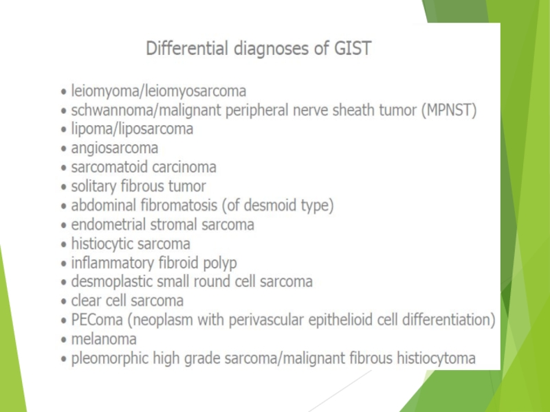
- 35. Desmoid
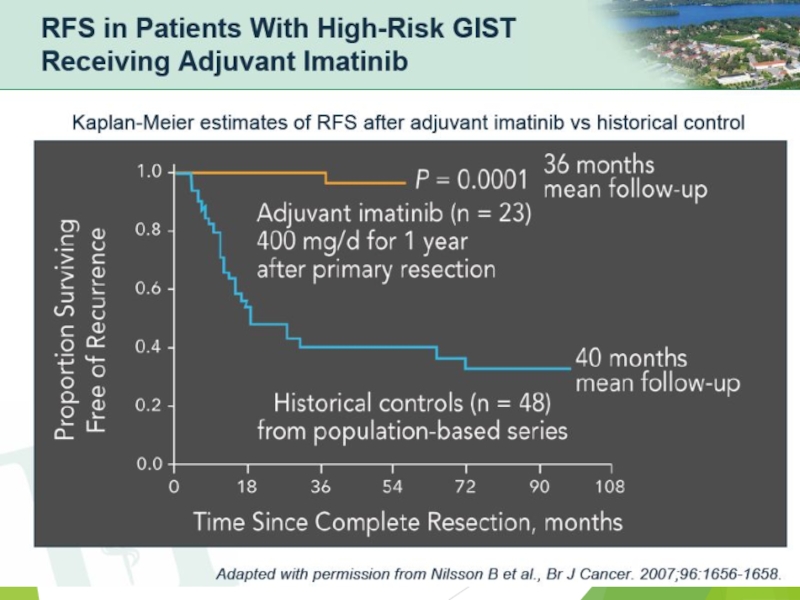
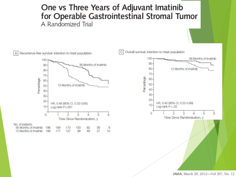
- 36. Can we prevent recurrence of high risk GIST?
- 38. IMATINIB
- 39. Imatinib Mesylate: Mechanism of Action Imatinib
- 42. Imatinib Mesylate in metastatic GIST
- 43. Overall survival of EORTC trial
- 45. Pediatric Sarcomas Ewing’s Sarcoma Rhabdomyosarcoma Osteosarcoma
- 46. Osteogenic Sarcoma The most common bone tumor
- 52. Treatment of Osteogenic Sarcoma Chemotherapy (every sarcoma
- 54. Ewing Sarcoma The second most common bone
- 56. “Onion skin” sign («луковая шелуха»)
- 58. Ewing Sarcoma Molecular biology methods of diagnosis:
- 59. Ewing Sarcoma – Treatment considerations Biopsy and
- 60. Malignant bone tumors Osteosarcoma During growth spurt

Слайд 2Soft Tissue Sarcomas:Definition
Sarcomas are malignant tumors that arise from skeletal and
extraskeletal connective tissues (mesenchymal cells).
Including:
Adipose tissue
Bone
Cartilage
Smooth muscle
Skeletal muscle
Including:
Adipose tissue
Bone
Cartilage
Smooth muscle
Skeletal muscle

Слайд 3Soft Tissue Sarcomas: Statistic
Rare and unusual cancer.
About 1% of adults
human cancers
15% of pediatric malignancies
Most commonly occur in the extremities (50%)
Other sites: Abdominal cavity/ retroperitoneum, Trunk/ thoracic region and head and neck.
15% of pediatric malignancies
Most commonly occur in the extremities (50%)
Other sites: Abdominal cavity/ retroperitoneum, Trunk/ thoracic region and head and neck.


Слайд 5Soft Tissue Sarcomas: Histology
Histopathology is determined by anatomic site. Common:
Extremity:
Malignant fibrous histocytoma liposarcoma
Retroperitoneal:
liposarcoma
leiomyosarcoma
Visceral: GIST
Retroperitoneal:
liposarcoma
leiomyosarcoma
Visceral: GIST


Слайд 7Sarcomas: Age as factor in Histology
Childhood: embryonal rhabdomyosarcoma
Bone: Ewing’s
sarcoma, osteosarcoma
Synovial sarcoma is more likely to be seen in young adults (<35 years old)
Liposarcoma, MFH are the predominant types in the oldest population
Synovial sarcoma is more likely to be seen in young adults (<35 years old)
Liposarcoma, MFH are the predominant types in the oldest population

Слайд 8STS-Grade
The biologic behavior of sarcomas is extremely variable
Histologic grade is a
major prognostic factor
Based on degree of mitosis, cellularity, presence of necrosis,
Differentiation, stromal content
Based on degree of mitosis, cellularity, presence of necrosis,
Differentiation, stromal content

Слайд 9Low-grade sarcomas
Fibromyxoid sarcoma
Better differentiated, less cellular, tend to resemble the tissue
of origin in some extent, mitotic rate is low
Grow slower, low risk of metastasis, a high risk of local recurrence after surgical removal
Grow slower, low risk of metastasis, a high risk of local recurrence after surgical removal

Слайд 10High grade-sarcoma
Highly cellular, poorly differentiated, mesenchymal cells with marked nuclear abnormality,
high mitotic rate, anaplasia
Grow rapidly, show extensive local invasion, metastasize early through bloodstream
Grow rapidly, show extensive local invasion, metastasize early through bloodstream
Leiomyosarcoma

Слайд 11STS-Genetic risk factors
Neurofibromatosis-Von Recklinghausen’s disease
Li-Fraumeni syndrome
Retinoblastoma
Gardner’s syndrome
Inhibition of p53
60% of sarcomas
Phosphorylation
of RB
50% of sarcomas
50% of sarcomas

Слайд 12STS- risk factors
Radiation Exposure
Lymphedema
Post-surgical
Post-irradiation
Parasitic infection (filariasis)
Trauma
Chemical:
2,3,7,8-Tetrachlorodibenzodioxin
Polyvinyl chloride
Hemachromatosis
Arsenic
Hemachromatosis
Arsenic
Angiosarcoma
TraumaChemical: 2,3,7,8-Tetrachlorodibenzodioxin Polyvinyl chloride Hemachromatosis ArsenicAngiosarcoma")
Слайд 13STS-Diagnosis
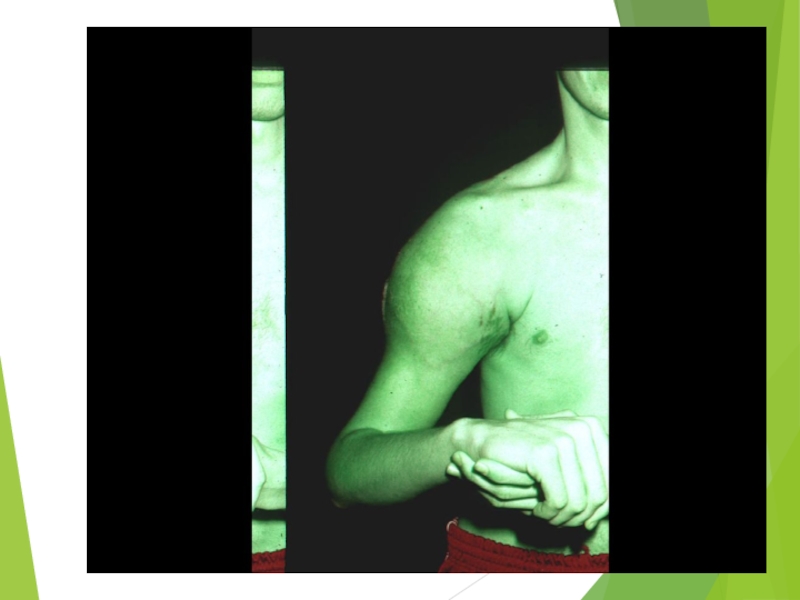
Physical examination: assessment of the size of the mass and its
relationship to neurovascular and bony structures
Extremity sarcomas usually present as painless mass.
Biopsy: any soft tissue mass that is symptomatic or enlarging or any new mass that persists beyond 4 weeks should be sampled.
Extremity sarcomas usually present as painless mass.
Biopsy: any soft tissue mass that is symptomatic or enlarging or any new mass that persists beyond 4 weeks should be sampled.

Слайд 14STS-Diagnosis
Usually incisional or core biopsy preferred
The incision should be centered over
the mass in its most superficial location.

Слайд 15STS-Diagnosis
Imaging
MRI preferred
Enhances the contrast between tumor and adjacent structures
Provides excellent
3-dimensional definition of fascial plans
Combination of CT and MR images did not significantly improve accuracy
Combination of CT and MR images did not significantly improve accuracy

Слайд 16STS-Workup
Evaluation for sites of potential metastasis:
LN mets. Occur in less than
3% of adults STS.
For extremity lesions, lungs is the principal site for mets.
For visceral lesions the liver is the principal site.
Low grade STS, the risk for mets.<15%
High grade STS the risk for mets. >50%
For extremity lesions, lungs is the principal site for mets.
For visceral lesions the liver is the principal site.
Low grade STS, the risk for mets.<15%
High grade STS the risk for mets. >50%

Слайд 17
STS-Workup
Extremity-STS:
MRI of the lesion
CT chest,bone scan
Visceral-STS:
MRI if needed
CT chest and
abdomen
Childhood sarcomas:
PET-CT
MRI of the primary site
Bone scan if needed
BMB
Childhood sarcomas:
PET-CT
MRI of the primary site
Bone scan if needed
BMB


Слайд 20STS-treatment: Surgical excision
The only hope for cure
The goal is complete removal
of the tumor with negative margins and maximal preservation of function.
Limb sparing procedures should be preformed, when possible.
Less radical procedure do not adversely affect local control or outcome
Limb sparing procedures should be preformed, when possible.
Less radical procedure do not adversely affect local control or outcome

Слайд 21STS-treatment
The best excision with 2-3cm margins.
The centrifugal
growth creates pseudo-capsule, malignant cells penetrate this capsule.
90% recur with only removal of visible tumor.
30% recur after excision of tumor bed, without radiotherapy

Слайд 22STS- Radiotherapy
Brachytherapy “seeds of iridium-192
External-beam therapy
Standardized fields

Слайд 23STS- Radiotherapy
Indications:
high grade of the limbs
intermediate grade of the limbs with
close or positive margins
Little role in low grade, should be considered for a recurrence
Little role in low grade, should be considered for a recurrence

Слайд 24STS- Radiotherapy
For survival: Limb conserving+ adj. Radiotherapy= amputation
Preoperative 50Gy dose.
Postoperative
60-70Gy dose.
Pre. Vs. Post: doubling the wound complications, slightly better functional outcome
Pre. Vs. Post: doubling the wound complications, slightly better functional outcome

Слайд 25STS-chemotherapy
Adjuvant chemotherapy-controversial
Meta-analysis: improved PFS (15%) but not overall survival (4% n.s.)
Doxorubicin base.
ESFT (childhood-round cell tumors)
Initial chemo. Improved survival from 10% to 60%.
Necrosis of 90% confers better outcome
High dose chemo. With salvage autologous PBPC for recurrence.
ESFT (childhood-round cell tumors)
Initial chemo. Improved survival from 10% to 60%.
Necrosis of 90% confers better outcome
High dose chemo. With salvage autologous PBPC for recurrence.
 but not overall survival (4% n.s.) Doxorubicin base.ESFT (childhood-round cell")
Слайд 26STS- Recurrent disease
Local extremity rec.: if isolated should undergo resection and
adj. Radiotherapy if feasible- 2/3 long term survival
Distant metastasis:Lungs are the first metastatic site in 73% of rec.
If possible- metastectomy is the best option
Distant metastasis:Lungs are the first metastatic site in 73% of rec.
If possible- metastectomy is the best option

Слайд 27STS- Resection of pulmonary metastasis
Conditions: primary tumor controlled
No extrathoracic disease
Complete resection
of all lung disease appears possible
20%-30% 3 years survival after complete resection

Слайд 28STS-chemotherapy for metastatic disease
Palliative, not curative therapy
For unresectable pulmonary mets.
Extrapulmonary mets.
In more than one site.
Poor prognosis
Median survival less than 1 year

Слайд 29STS-chemotherapy for metastatic disease
Every STS : adriamycin, ifosfamide, decarbazin as single
or combination 20-40% response rate
Leiomyosarcoma (maybe MFH): docotaxel with gemcitabine
Angiosarcoma: paclitaxel, doxil
New chemotherapy: trabectidin (yondelis) product from marine tunicate Ecteinascidia tubinata (4% response but high stable dis.)
Leiomyosarcoma (maybe MFH): docotaxel with gemcitabine
Angiosarcoma: paclitaxel, doxil
New chemotherapy: trabectidin (yondelis) product from marine tunicate Ecteinascidia tubinata (4% response but high stable dis.)

Слайд 30Stojadinovic et al. J Clin Oncol 2002; 20: 4344–52
STS: 5-year Survival
Rates
5-year overall survival, %

Слайд 31GastroIntestinal Stromal Tumors (GIST): A Brief Overview
Definition
Rare soft tissue tumor of
the GI tract, mesentery, and omentum
Histologic subtypes include spindle, epitheliod, mixed
Originate from Cajal cells.
Histologic subtypes include spindle, epitheliod, mixed
Originate from Cajal cells.
: A Brief OverviewDefinitionRare soft tissue tumor of the GI tract, mesentery,")
Слайд 3210-20 cases per million.
Similar incidence in males and females.
Only 0.2% of
all GI tumors, 80% of GI sarcomas.
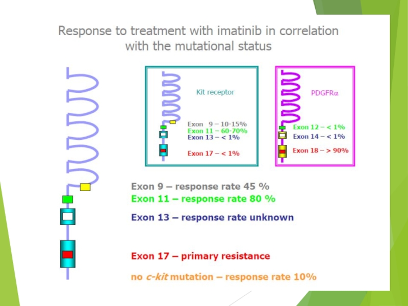
>90% positive for C-KIT.
Origin:
stomach 40-70%
Small intestine 20-40%
Colon and rectum 5-15%
Esophagus <5%
>90% positive for C-KIT.
Origin:
stomach 40-70%
Small intestine 20-40%
Colon and rectum 5-15%
Esophagus <5%

Слайд 33GIST: A Brief Overview
Clinical Presentation
Abdominal Pain, GI Bleeding, Mass, Obstruction
Primary tumor
only (46%), Metastatic disease (47%)
Prognostic Factors
No uniform prognostic guidelines, poor Px associated with
increasing tumor size
metastatic disease at presentation
high grade (high mitotic index)
Primary Treatment = Surgery
~67% primary tumors resectable,
However, 40-90% recur (most often: intra-abdominal, liver)
Prognostic Factors
No uniform prognostic guidelines, poor Px associated with
increasing tumor size
metastatic disease at presentation
high grade (high mitotic index)
Primary Treatment = Surgery
~67% primary tumors resectable,
However, 40-90% recur (most often: intra-abdominal, liver)
, Metastatic disease")


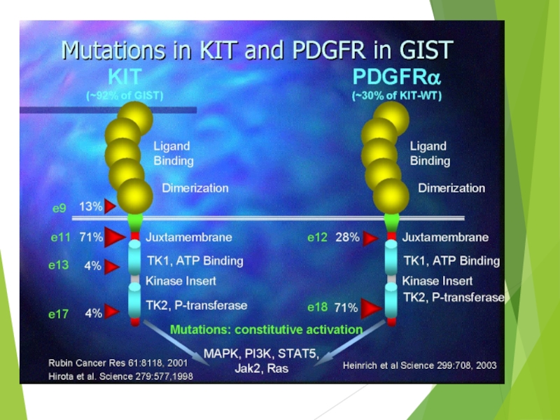

Слайд 39Imatinib Mesylate:
Mechanism of Action
Imatinib mesylate occupies the ATP binding pocket
of the kit kinase domain
This prevents substrate phosphorylation and signaling
A lack of signaling inhibits proliferation and survival
This prevents substrate phosphorylation and signaling
A lack of signaling inhibits proliferation and survival
Savage and Antman. N Engl J Med. 2002;346:683.


Слайд 45Pediatric Sarcomas
Ewing’s Sarcoma Rhabdomyosarcoma
Osteosarcoma
Multimodality approach: Chemotherapy, Radiation and Surgery
Curative Therapy
for majority of patients with localized disease

Слайд 46Osteogenic Sarcoma
The most common bone tumor
Peak incidence: second decade of
life
Females earlier than males
May be primary or secondary (radiation-induced and as a part of Li-Fraumeni syndrome)
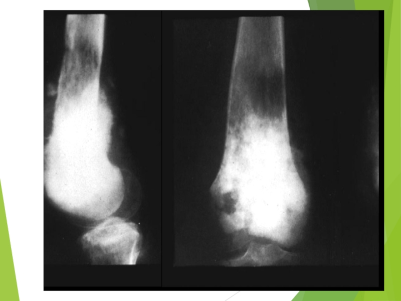
Most commonly located in methaphyses of long bones, especially around the knee
The most common sites of mets: lungs, bones (20% of all children with OS have macroscopic lung mets in lungs at the time of initial diagnosis)
Females earlier than males
May be primary or secondary (radiation-induced and as a part of Li-Fraumeni syndrome)
Most commonly located in methaphyses of long bones, especially around the knee
The most common sites of mets: lungs, bones (20% of all children with OS have macroscopic lung mets in lungs at the time of initial diagnosis)

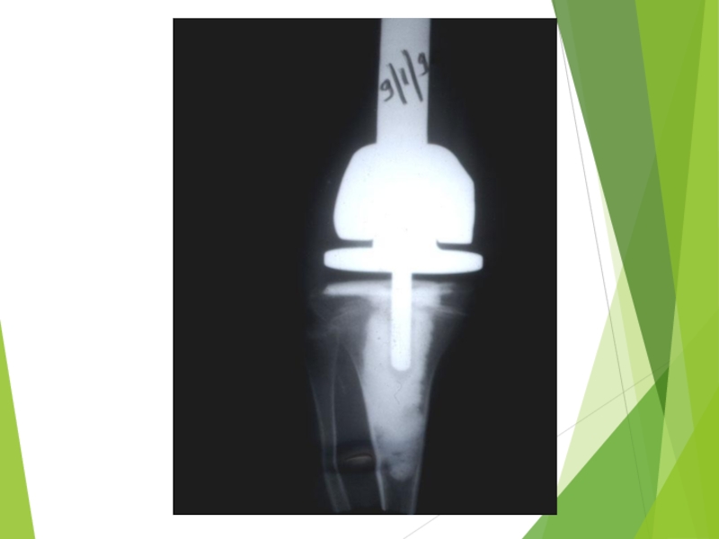
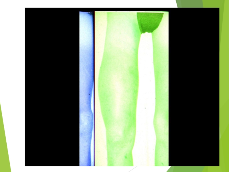
Слайд 52Treatment of Osteogenic Sarcoma
Chemotherapy (every sarcoma in children is systemic disease
– before era of chemotherapy 80% of pts developed distant metastases despite excellent local control)
Surgery (limb-sparing with endoprothesis)
Resection selected lung mets
Chemotherapy
OS is not sufficiently radiosensitive, at least 6000 cGy
5-y DFS in non-metastatic pts: 60-75%
5-y DFS in metastatic to lungs pts: 20-25%
Surgery (limb-sparing with endoprothesis)
Resection selected lung mets
Chemotherapy
OS is not sufficiently radiosensitive, at least 6000 cGy
5-y DFS in non-metastatic pts: 60-75%
5-y DFS in metastatic to lungs pts: 20-25%

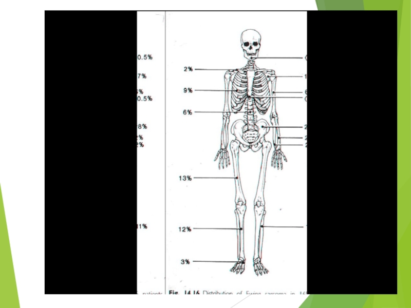
Слайд 54Ewing Sarcoma
The second most common bone tumor
The peak incidence is
appeared to be earlier than OS
The most common location: diaphyses of long bones, frequently bones of pelvis
The most common sites of mets: lungs and bones (20% of all pts have lung mets at the time of initial diagnosis), may be in bone marrow
ES is one of small round blue cells tumors (others are neuroblastoma, rhabdomyosarcoma, and lymphoma)

")
Слайд 58Ewing Sarcoma
Molecular biology methods of diagnosis: t (11,22) and t (21,22)
in approximately 95% of cases
PCR for t (11,22) in tumorous tissue, peripheral blood, and bone marrow
Prognosis of pts with PCR positive in peripheral blood and/or bone marrow approaches that of pts with overt metastatic disease
PCR for t (11,22) in tumorous tissue, peripheral blood, and bone marrow
Prognosis of pts with PCR positive in peripheral blood and/or bone marrow approaches that of pts with overt metastatic disease
 and t (21,22) in approximately 95% of")
Слайд 59Ewing Sarcoma – Treatment considerations
Biopsy and definitive diagnosis
Neoadjuvant chemotherapy
Surgery ± radiotherapy
(5500 cGy)
Continuation of chemotherapy
Percentage of necrosis (> or < 90%) have prognostic implications
5-y DFS in non-metastatic pts with more 90% necrosis after neoadjuvant chemotherapy is about 75%
Continuation of chemotherapy
Percentage of necrosis (> or < 90%) have prognostic implications
5-y DFS in non-metastatic pts with more 90% necrosis after neoadjuvant chemotherapy is about 75%
Continuation of chemotherapyPercentage")
Слайд 60Malignant bone tumors
Osteosarcoma
During growth spurt (12-18 years)
Metaphysis
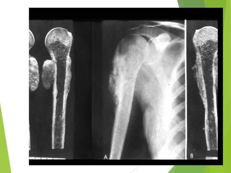
Distal femur>proximal tibia>proximal humerus
No known
chromosomal abberations
No radiosensitive
No really efficacious second-line chemotherapy
No radiosensitive
No really efficacious second-line chemotherapy
Ewing sarcoma
Much younger patients (2y – 20 y)
Diaphysis
Pelvic bones>femur>chest wall
EWS/FLI1; t(11;22)
Radiosensitive
There is second-line chemotherapy
MetaphysisDistal femur>proximal tibia>proximal humerusNo known chromosomal abberationsNo radiosensitiveNo really")