- Главная
- Разное
- Дизайн
- Бизнес и предпринимательство
- Аналитика
- Образование
- Развлечения
- Красота и здоровье
- Финансы
- Государство
- Путешествия
- Спорт
- Недвижимость
- Армия
- Графика
- Культурология
- Еда и кулинария
- Лингвистика
- Английский язык
- Астрономия
- Алгебра
- Биология
- География
- Детские презентации
- Информатика
- История
- Литература
- Маркетинг
- Математика
- Медицина
- Менеджмент
- Музыка
- МХК
- Немецкий язык
- ОБЖ
- Обществознание
- Окружающий мир
- Педагогика
- Русский язык
- Технология
- Физика
- Философия
- Химия
- Шаблоны, картинки для презентаций
- Экология
- Экономика
- Юриспруденция
Radiology and imaging the biliary, liver and pancreas презентация
Содержание
- 1. Radiology and imaging the biliary, liver and pancreas
- 2. The biliary tract These include: 1.
- 3. Gall Bladder The normal gall bladder is
- 4. Smooth muscle fibers are found in
- 5. The gall bladder concentrates this bile and
- 8. Cholecystitis
- 9. Chronic cholecystitis Chronic cholecystitis may follow acute
- 10. Cholecystography In cholecystography we
- 11. Technique After a light fat-free dinner the
- 12. Occasionally nausea may be complained or mild
- 13. The amount of iodine in bile
- 14. Cause of Non- visualization of Gall Bladder
- 15. There may be faulty absorption from the
- 16. Normal Cholecystography Gall bladder is visualized
- 18. Non Functioning Gall bladder Common cause for
- 19. When gall bladder is sub normally functional
- 20. Simple X-ray of the biliary tract Opaque
- 21. Non-opaque gall-stones, for instance large cholesterol stones,
- 22. Differentiating renal calculus from gall bladder calculus
- 28. Gas in the biliary tract Usually in
- 29. It is important to remember that both
- 30. Cholesterols [strawberry gall bladder] There is diffuse
- 31. Cholesterosis are: small round translucent
- 32. A cholesterol polyp is not a tumor
- 36. Calcification of walls of Gall Bladder
- 40. Milk of Calcium Gall Bladder Milk of
- 41. Tumors of Gall Bladder Small papillomas are
- 42. Adenoma of gall bladder is rear. It
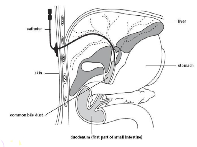
- 45. Percutaneous Cholangiography Indications are: • To differentiate
- 46. Contraindications are: • Hemorrhagic diathesis
- 47. Interpretation of Percutaneous Cholangiography •
- 48. • Dilatation of duct with an
- 51. The same duct, following removal of the stone through the drainage catheter
- 52. Ultrasound This view is widely used as
- 53. Gall stones characteristically produce high density echoes
- 54. normal
- 57. stones
- 58. Isotope scanning 99 Tc HIDA (a derivative
- 59. NIDA is also a most valuable screening
- 60. normal
- 62. Operative and post - operative cholangiography It
- 63. Many surgeons now perform operative cholangiography as
- 64. Operative cholangiography Operative cholangiography, if skillfully
- 65. Post - operative cholangiography Post -
- 66. Operative and T-tube cholangiogram
- 67. post operative study
- 68. Endoscopic retrograde cholangio pancreatography (ERCP) Under radiological
- 69. Biliary obstruction due to stone or neoplasm
- 70. For this examination the patient
- 72. endoscope
- 76. Stricture of the cystic duct
- 77. stones
- 78. The Liver The main indication or the
- 79. The techniques available include: • Simple X ray
- 80. Simple X ray provides little information apart
- 81. Indications are: the liver cysts
- 82. Tumors usually show as rounded areas with
- 83. tumor
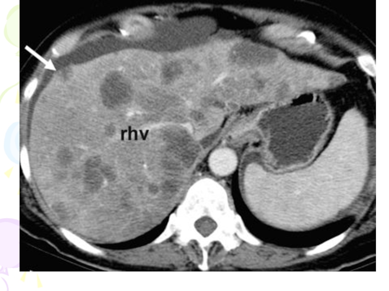
- 87. CT scanning CT shows the liver in
- 88. cyst
- 92. mts
- 94. stone
- 95. Necrotic gall bladder
- 96. MRI MRI is similar to CT
- 98. 3-D image
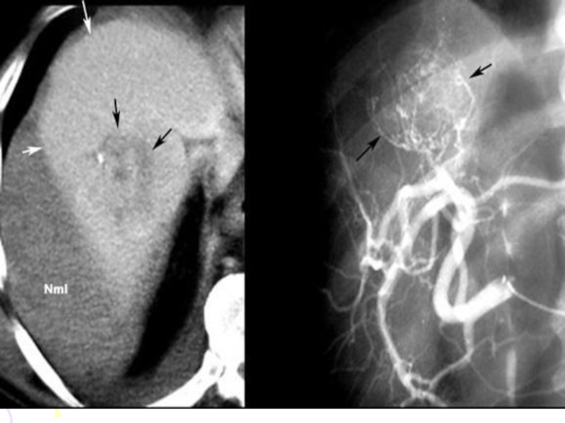
- 99. Angiography Hepatic angiography is performed by
- 100. The technique uses: for the diagnosis
- 101. liver tumor during embolizations
- 103. hemangioma
- 104. Isotope scanning The study is usually
- 105. Following are the indications of liver imaging:
- 107. CHOICE OF EXAMINATION IN BILIARY AND LIVER
- 108. CT and MRI will also perform this
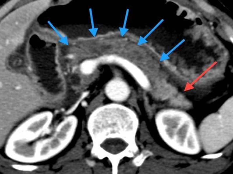
- 109. In suspected obstructive jaundice, ultrasound is the
- 110. PANCREAS Pancreas lies in the posterior abdominal
- 113. Simple X ray will occasionally show extensive
- 116. Barium studies will show: distortion of
- 119. Ultrasound: the normal pancreas may be
- 120. In acute pancreatitis, the whole organ
- 121. Rupture of pancreas
- 122. CT scan CT is now the method
- 123. Small tumors such as islet cell
- 124. cyst
- 126. MRI MRI can also show the
- 127. ERCP ERCP is the method of
- 128. These include: 1. Stone extraction from pancreatic
- 129. Choice of examination CT is probably the
- 130. Ultrasound in skilled hands is almost reliable,

Слайд 2The biliary tract
These include:
1. Simple X-ray
2. Oral Cholecystography
3. Intravenous Cholangiography
4.
Operative and postoperative Cholangiography
5. Percutaneous transhepatic cholangiography (PTC)
6. Endoscopic retrograde cholangio pancreatography (ERCP)
7. Ultrasound
8. Isotope scanning
9. Computed tomography (CT)
10. Magnetic resonance imaging (MRJ)
5. Percutaneous transhepatic cholangiography (PTC)
6. Endoscopic retrograde cholangio pancreatography (ERCP)
7. Ultrasound
8. Isotope scanning
9. Computed tomography (CT)
10. Magnetic resonance imaging (MRJ)

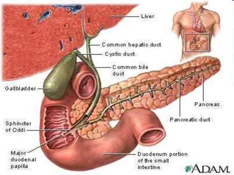
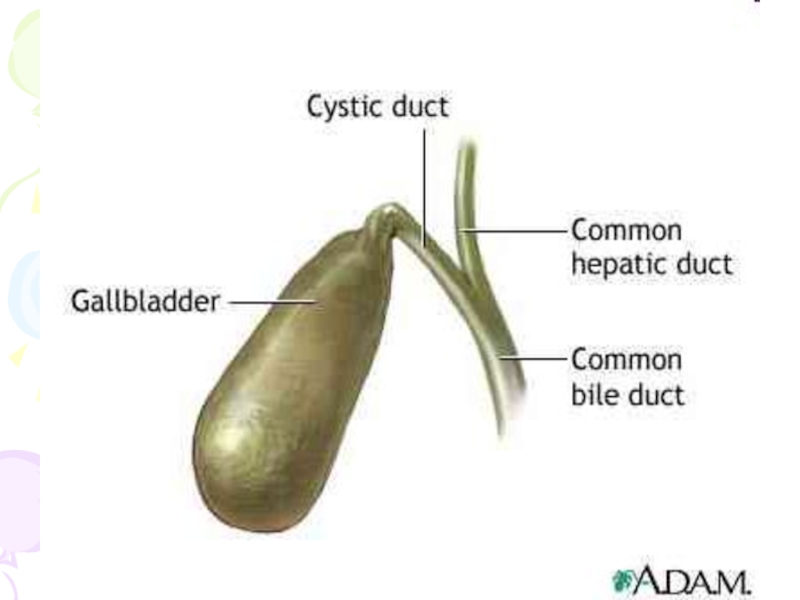
Слайд 3Gall Bladder
The normal gall bladder is composed of four parts:
fundus
body
infundibulum and
a neck

Слайд 4 Smooth muscle fibers are found in the wall of the
fundus and infundibulum but are completely absent in the body. The liver is constantly secreting thin watery bile which flows into the bile ducts where it is diluted by addition of mucus.

Слайд 5The gall bladder concentrates this bile and when the pressure in
gall bladder falls below that in bile ducts more watery bile runs in. This process continues till the gall bladder is full of concentrated bile. Concentration of bile in gall bladder is carried out by absorption of water. Bilirubin is concentrated about 20 times and cholesterol, bile salts and calcium about 5 to 10 times.

Слайд 8Cholecystitis
Acute Cholecystitis
In acute cholecystitis cholecystography is contraindication because:
opaque medium may aggravate the inflammation and
there is failure to concentrate the dye
Only plain x-ray may be of use in revealing opaque calculi.
opaque medium may aggravate the inflammation and
there is failure to concentrate the dye
Only plain x-ray may be of use in revealing opaque calculi.

Слайд 9Chronic cholecystitis
Chronic cholecystitis may follow acute cholecystitis with or without stones.
Chronically inflamed gall bladder is usually distended with mucus, its walls are thickened and its mucosa is thrown into heavy folds. In case where cystic ducts are blocked by stone the gall bladder becomes permanently distended, its wall undergoes atrophy and later on becomes thin sheet of fibrous tissue-like parchment.

Слайд 10Cholecystography
In cholecystography we see the following three points
by which we can estimate the biliary function
i. The rate of filling of gall bladder.
ii. Capability of gall bladder mucosa to concentrate bile as evidenced by the intensity of the shadow.
iii. The rate of emptying of gall bladder.
i. The rate of filling of gall bladder.
ii. Capability of gall bladder mucosa to concentrate bile as evidenced by the intensity of the shadow.
iii. The rate of emptying of gall bladder.

Слайд 11Technique
After a light fat-free dinner the patient is given 3 or
6gm of telepaque; next morning after 12-14 hours supine and erect x-rays are taken.
Patient is then given fatty meal (egg, butter, etc.) and x-ray is taken after 1 hour. This completes the examination in most cases.
Patient is then given fatty meal (egg, butter, etc.) and x-ray is taken after 1 hour. This completes the examination in most cases.

Слайд 12Occasionally nausea may be complained or mild diarrhea may develop as
side reaction. Sensitivity to iodine is a contraindication for the procedure. Use of telepaque is restricted in obstructive jaundice, vomiting and diarrhea.
Iodine is absorbed from the gastrointestinal tract is secreted by liver. The bile as it comes from the liver ordinarily does not contain enough of the drug to be radio-opaque. The gall bladder receiving the bile does not contain enough of the drug to be radio-opaque. The gall bladder concentrates it by removing water.
Iodine is absorbed from the gastrointestinal tract is secreted by liver. The bile as it comes from the liver ordinarily does not contain enough of the drug to be radio-opaque. The gall bladder receiving the bile does not contain enough of the drug to be radio-opaque. The gall bladder concentrates it by removing water.

Слайд 13 The amount of iodine in bile then becomes sufficient to
make it radio-opaque and a dense shadow of gall bladder. To visualize all this mucosa of gall bladder, it must be intact and functioning well. If gall bladder is diseased, as in chronic cholecystitis, the concentrating ability of the mucosa is impaired or lost and gall bladder visualization becomes a failure.

Слайд 14Cause of Non- visualization of Gall Bladder
The patient could
not take drug or could not retain it.
Obstruction at the cardia or pylorus may prevent the material reaching to small intestine.
Liver function maybe impaired to the point where a sufficient amount of the material is not secreted. Results are satisfactory if obstructive jaundice is present
Obstruction at the cardia or pylorus may prevent the material reaching to small intestine.
Liver function maybe impaired to the point where a sufficient amount of the material is not secreted. Results are satisfactory if obstructive jaundice is present

Слайд 15There may be faulty absorption from the intestine
Obstruction of cystic duct
may prevent entrance of bile into the gall bladder.
Previously removed gall bladder.
Previously removed gall bladder.

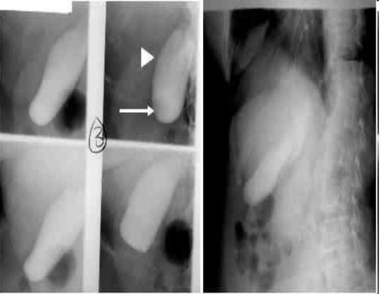
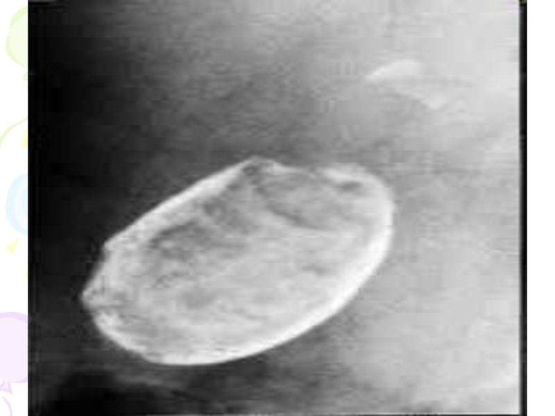
Слайд 16Normal Cholecystography
Gall bladder is visualized with uniform density
There is
no filling defect suggestive of radio-opaque stones
After fatty meal there is sufficient contraction of gall bladder. Contraction of the gall bladder in response to fatty is the result of production of a hormone known as cholecystokinin by mucous membrane of intestine.
Absence of contraction of gall bladder after fatty meal is suggestive of cholecystitis.

Слайд 18Non Functioning Gall bladder
Common cause for non-functioning gall bladder is chronic
cholecystitis with stones. Multiple doses of telepaque 3gm each day for 3 or 4 days (after each meal) is advised due to which at times the margins of stone may become opaque and seen.

Слайд 19When gall bladder is sub normally functional a shadow of the
gall bladder is visible but faint. Density may be so low that the presence or absence may not be determined under the conditions and examination may be repeated after giving double dose of telepaque.

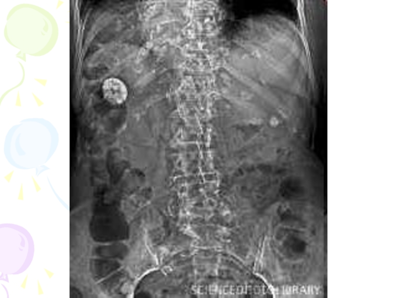
Слайд 20Simple X-ray of the biliary tract
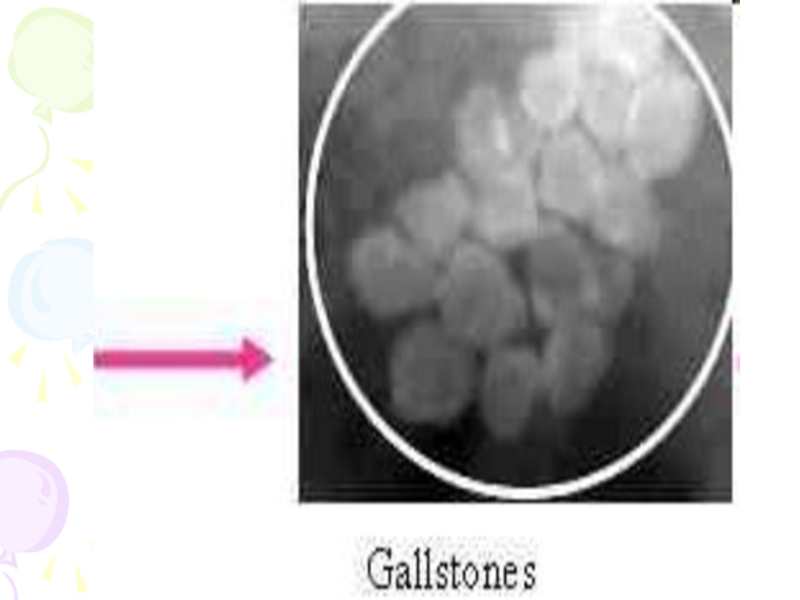
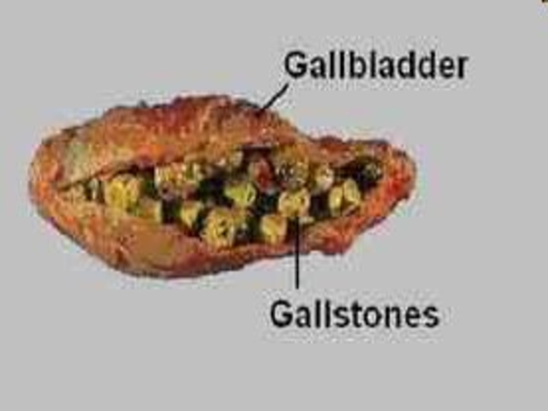
Opaque gall stones will be readily
shown. These vary in type. They may be large laminated structures, which are usually solitary or few in number. On the other hand small calculi may be multiple and numerous. An opaque stone in the cystic or common bile duct can be diagnosed by its position relative to the normal gall-bladder. This is easy if there are also opaque stones in the gall-bladder.

Слайд 21Non-opaque gall-stones, for instance large cholesterol stones, will not be diagnosed
by plain x-rays and will require contrast studies, or ultrasound for their demonstration. Multiple (minute) calculi may form sediment in the gall bladder giving rise to so-called "milky bile". This will outline the whole gall-bladder in the supine patient and will sediment to form a horizontal level in the gall-bladder in the erect film.


Слайд 28Gas in the biliary tract
Usually in the hepatic ducts, is only
occasionally noted at plain x-ray. This implies either a fistula between the bowel and biliary react, or an incompetent sphincter Oddi. The latter condition may be seen following passage of a large calculus or following operative intervention and exploration.

Слайд 29It is important to remember that both oral and intravenous cholecystography
are unlikely to be successful in the presence of obstructive jaundice. This is because with biliary obstruction the excretion of contrast from the liver is impaired.

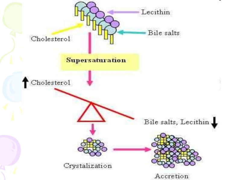
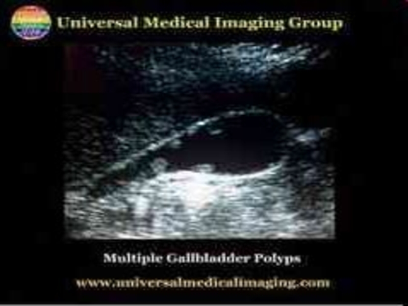
Слайд 30Cholesterols [strawberry gall bladder]
There is diffuse deposition of cholesterol on the
mucosa of the gall bladder. The gall bladder shadow appears normal and concentrates normally. When cholesterol deposits measures 2mm or more, they can be detectable. Multiplicity of fixed mural defects is absolutely polyps are extremely rare.

Слайд 31Cholesterosis are:
small round translucent defects attached to the gall bladder
wall
these vary in size from 1 to 2mm
their attachment to the wall is determined by lack of movement in different position
these vary in size from 1 to 2mm
their attachment to the wall is determined by lack of movement in different position

Слайд 32A cholesterol polyp is not a tumor but consists of small
collection of cholesterol crystals. It may form a sessile mass or be attached to the wall by a thin delicate stalk. It has also been called "lipid gall bladder" or "strawberry gall bladder" because of the gross appearance of multiple tiny collections of cholesterol on the surface of reddened mucosa resembling strawberry seeds.


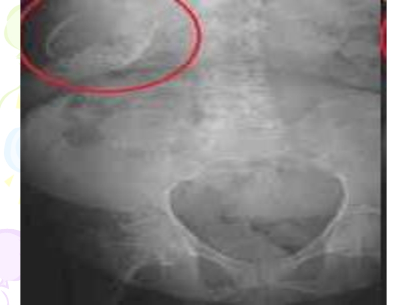
Слайд 36Calcification of walls of Gall Bladder
This is a rare condition.
It cannot occur without existing fibrosis and therefore is a sequel to a chronic cholecystitis. It is known as porcelain gall bladder. Calcium is laid down symmetrically and the usual ovoid outline of the gall bladder is visible. Calcified gall bladder either completely fails to concentrate the contrast medium of does it feebly and slowly.

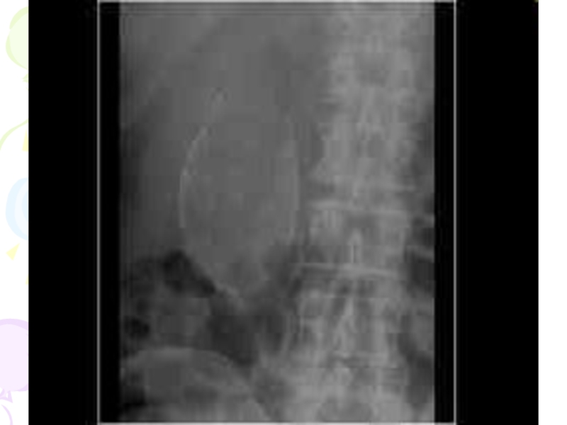
Слайд 40Milk of Calcium Gall Bladder
Milk of calcium bile is a condition
in which the gall bladder becomes filled with an accumulation of bile containing a high percentage of calcium carbonate due to which gall bladder is visualized in pain abdomen film without administering telepaque to the patient. It follows obstruction of cystic duct.

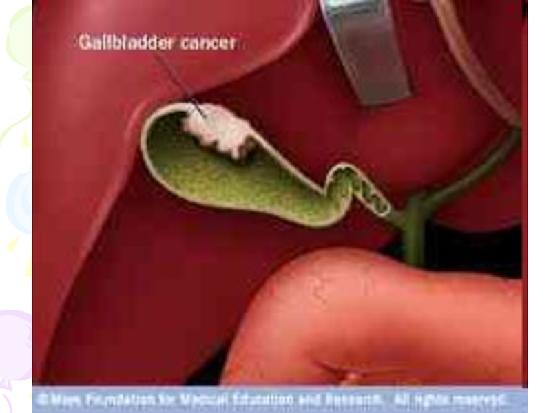
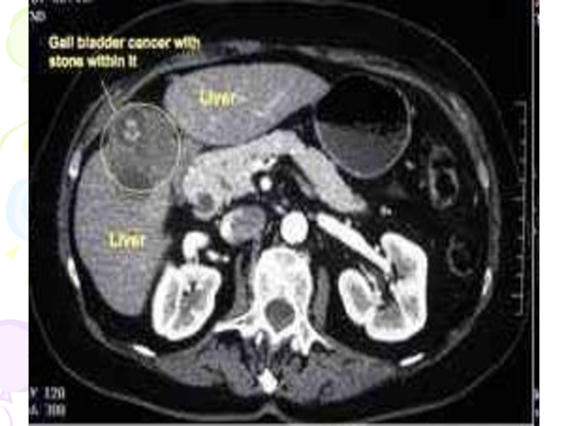
Слайд 41Tumors of Gall Bladder
Small papillomas are most frequent. These are seen
as small translucent defects usually on the lateral walls of the gall bladder. Their average size is about 0.5cm and not longer than 1cm. They are found in multiple. Gall bladders concentrate the dye well. These are fixed and never changes position. In the filled gall bladder these resembles stones or small pocket of duodenum gas.

Слайд 42Adenoma of gall bladder is rear. It looks like single small
semi -circular or circular translucent defect in the fundus of a well defined gall bladder. It never alters its position and it is best seen when gall bladder has evacuated half of its content.

Слайд 45Percutaneous Cholangiography
Indications are:
• To differentiate between obstructive and non obstructive jaundice.
•
To determine the presence and site of carcinoma of billiary system.
• To demonstrate the number of calculi in billiary system.
• To decompress the billiary tree prior to surgery. The procedure is restricted to patients schedule for surgery and it is performed an hour or two before it.
• To demonstrate the number of calculi in billiary system.
• To decompress the billiary tree prior to surgery. The procedure is restricted to patients schedule for surgery and it is performed an hour or two before it.

Слайд 46Contraindications are:
• Hemorrhagic diathesis
• Vitamin K resistant hypoprothrombinemia
•
Febrile cholangitis

Слайд 47Interpretation of Percutaneous Cholangiography
• Calculi producing filling defects.
•
Obstructed duct is usually due to calculi.
• Smooth narrowness of a short segment indicates stricture.
• Rigid and irregular duct indicates carcinoma
• Smooth narrowness of a short segment indicates stricture.
• Rigid and irregular duct indicates carcinoma

Слайд 48• Dilatation of duct with an uneven and ragged obstruction
pattern indicates carcinoma in the ampulla of pancreas.
• Smooth, flat, shallow obstruction with dilation of the duct indicates carcinoma.
• Tortuous and marked dilatations of ducts indicate pancreatic carcinoma obstruction and may be rounded, bulbous, tapered or notched.
• Detection of metastasis
• Smooth, flat, shallow obstruction with dilation of the duct indicates carcinoma.
• Tortuous and marked dilatations of ducts indicate pancreatic carcinoma obstruction and may be rounded, bulbous, tapered or notched.
• Detection of metastasis


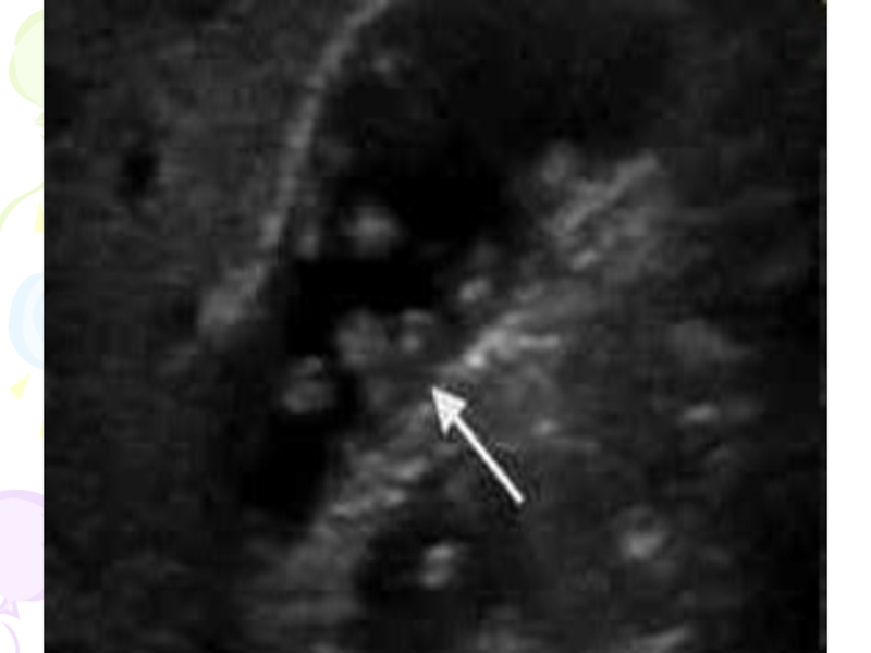
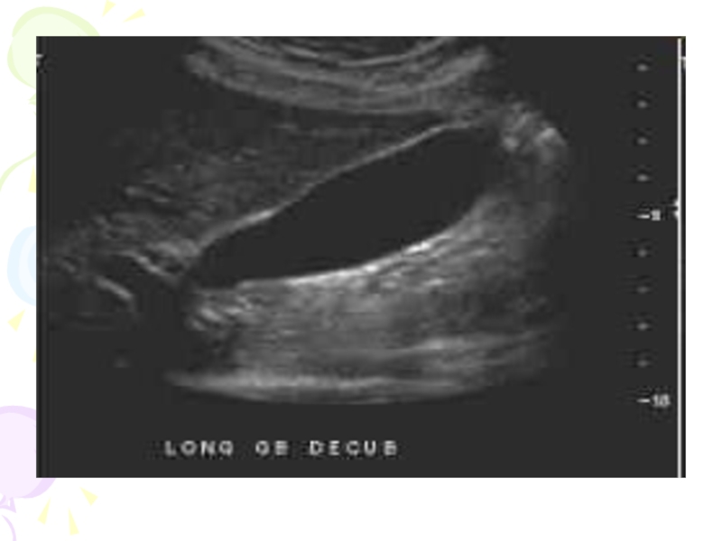
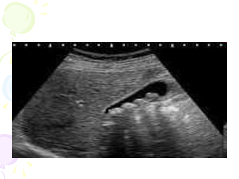
Слайд 52Ultrasound
This view is widely used as the preliminary examination in suspected
gall bladder or billiary tract diseases and has the added advantage that the liver and pancreas can be assessed at the same examination. It is highly accurate in the diagnosis of gall stones including the non-opaque stones visible at the simple radiography.

Слайд 53Gall stones characteristically produce high density echoes and cast acoustic shadows
appearing dark bands.
Ultrasound will also demonstrate the dilated intra hepatic ducts or common bile duct and thus will help to differentiate obstructive from non -obstructive jaundice.
Ultrasound will also demonstrate the dilated intra hepatic ducts or common bile duct and thus will help to differentiate obstructive from non -obstructive jaundice.


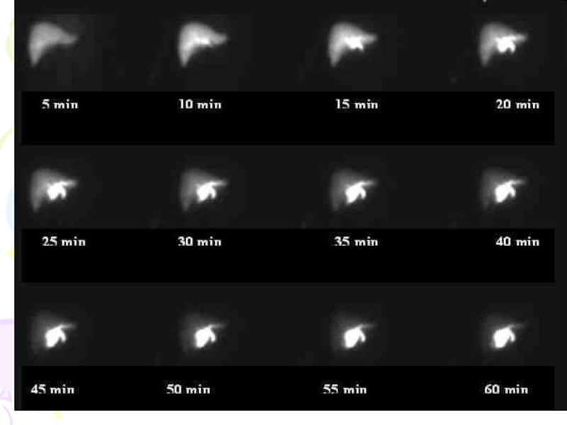
Слайд 58Isotope scanning
99 Tc HIDA (a derivative of iminodiacetic acid (IDA)) is
a drug which is concentrated by the hepatocytes and excreted in the bile. Serial gamma camera pictures taken at 10 minutes interval after administration show the normal gall bladder and billiary tract at 30 min and drainage into the gut at 60 min. In billiary obstruction, there is no evidence of gut activity even on delayed films at 24 hr.
) is a drug which is")
Слайд 59NIDA is also a most valuable screening test for acute cholecystitis
when the gall bladder will fail to fill despite the gut activity. Similar appearances are seen if the cystic duct or Hartmann's pouch are obstructed by calculi.


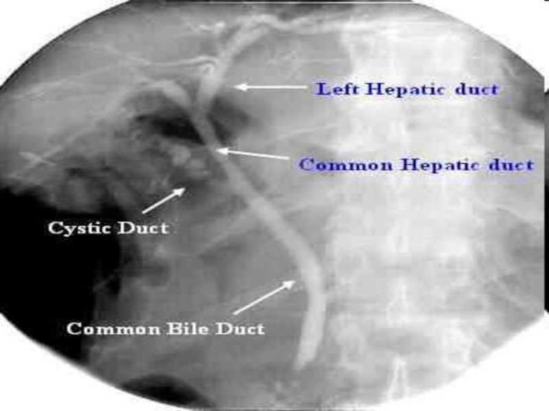
Слайд 62Operative and post - operative cholangiography
It is well known that in
calculous billiary diseases cholecystectomy alone will leave a proportion of patients with residual stones in the billiary ducts. On the other hand the exploration of the ducts adds considerably to the risk of the operation and many miss stones which are free to move about in the ducts or are soft consistency ("putty stones").

Слайд 63Many surgeons now perform operative cholangiography as a routine at operations
for billiary stones.
A small tube is inserted into the cystic duct and the bile ducts are filled with contrast medium. Films are obtained and examined during the operation and should demonstrate most removal calculi in the ducts.
A small tube is inserted into the cystic duct and the bile ducts are filled with contrast medium. Films are obtained and examined during the operation and should demonstrate most removal calculi in the ducts.

Слайд 64Operative cholangiography
Operative cholangiography, if skillfully performed, adds little to the
operative time and will ensure against leaving stones in the duct which may require a second operation. The best results are obtained when there is direct co - operation between surgeon and radiologist and apparatus permitting serial film is used. The possibility of artefact due to gas bubbles must be borne in mind and guarded against.

Слайд 65Post - operative cholangiography
Post - operative cholangiography is carried out
in the immediate post operative period by injecting the T tube drain in the common bile duct with contrast medium. This method will also show residual calculi which have been missed at the operation. These can be removed with a catheter with a catheter Snare passed through the T tube tract under image intensifier control.


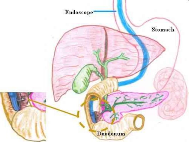
Слайд 68Endoscopic retrograde cholangio pancreatography (ERCP)
Under radiological control, the ampulla of Vater
is cannulated and the common bile duct or the pancreatic duct can be entered. Contrast medium can then be injected and the biliary ducts or the pancreatic ducts can be shown.
Under radiological control, the ampulla of Vater is cannulated and the")
Слайд 69Biliary obstruction due to stone or neoplasm can be visualized or
alternatively a normal biliary tree may be shown.
In skilled hands this is probably the method of choice for the investigation of obstructive jaundice.
It is also of great value in the investigation of the pancreatic diseases.
Not only can the pancreatic duct be injected and shown to be normal, abnormal or obstructed but pancreatic juice can be directly collected and analyzed.

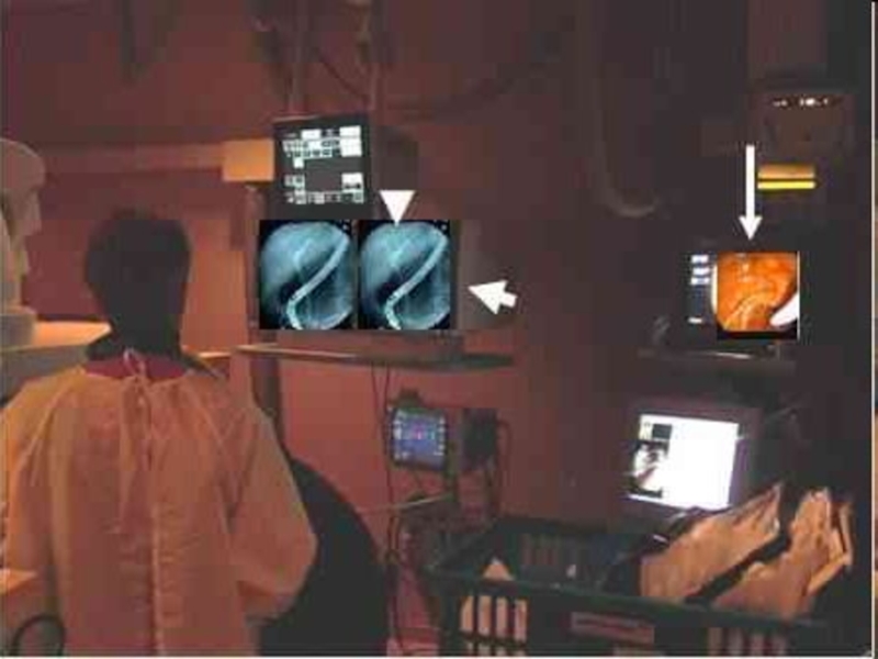
Слайд 70
For this examination the patient is usually fasting and lightly sedated.
A side viewing duodenoscope or other fibre - scope is passed to the duodenum. The ampulla of Vater is identified and a small Teflon catheter is passed from the endoscope into the apex of the papilla under visual control.


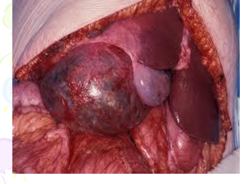
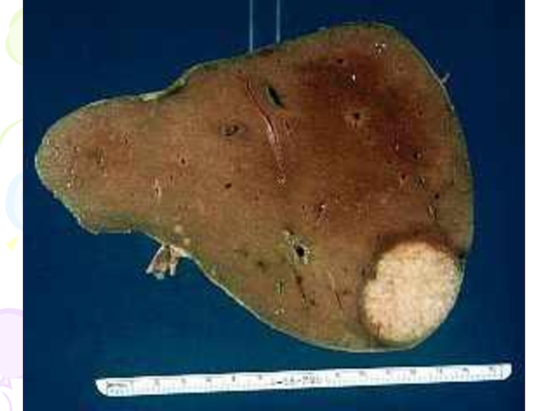
Слайд 78The Liver
The main indication or the investigation of the liver by
imaging are the diagnosis or excision of tumors, primary or secondary, of cysts and of inflammatory lesions. Other indications are the investigation of hepatomegaly, of cirrhosis, and of portal hypertension.

Слайд 79The techniques available include:
• Simple X ray
• Ultra sound
• CT
• Isotope scanning
• MRI
• Hepatic angiography
• Splenic and arterial
portography

Слайд 80Simple X ray provides little information apart from the confirming enlargement
of the liver and showing the occasional calcified lesion as hydatid cysts.
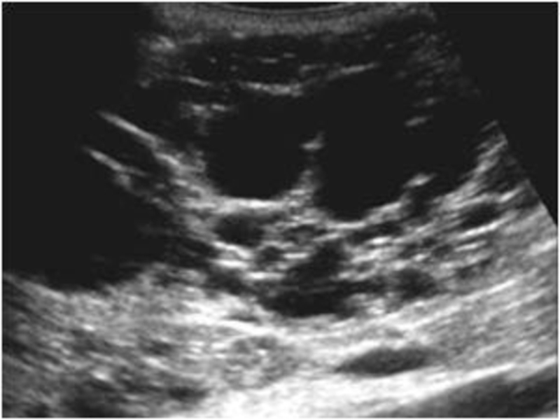
Ultrasound
Ultrasound is widely used in the investigation of the liver and biliary system.
Ultrasound
Ultrasound is widely used in the investigation of the liver and biliary system.

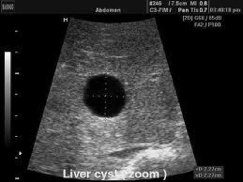
Слайд 81Indications are:
the liver cysts
abscess
Hematomas
and neoplasm both primary and
secondary are readily identified

Слайд 82Tumors usually show as rounded areas with diminished echoes.
Cysts are
completely transonic.
As already noted above dilated bile ducts are indefinable and are characteristic of obstructive jaundice.
The gall bladder and gall stones are readily shown.
As already noted above dilated bile ducts are indefinable and are characteristic of obstructive jaundice.
The gall bladder and gall stones are readily shown.


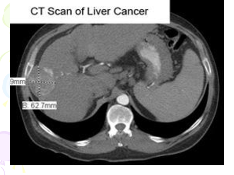
Слайд 87CT scanning
CT shows the liver in axial sections with high resolution.
Primary
and secondary neoplasm can be demonstrated and differentiated from the cysts.
Adjacent organs are also shown on the scans including abdominal nodes thus making CT invaluable in the staging of tumors.
Contrast enhancement following IV injection is frequently under taken to highlight focal lesions. Dynamic scans involving rapid serial images after contrast injections may assist in diagnosing vascular lesions such as haemangiomas and some tumors.


Слайд 96MRI
MRI is similar to CT in the accuracy of showing focal
lesions in the liver. It has the advantage of easy imaging in the coronal and sagittal planes but is still relatively more costly than CT.


Слайд 99Angiography
Hepatic angiography is performed by percutaneous transfemoral catheterization of the coeliac
axis or superselective catheterization of the hepatic artery followed by injection of a bolus of a contrast medium.

Слайд 100The technique uses:
for the diagnosis of tumors
angiomas
aneurysms
and other vascular lesions
for the
transcatheter embolisation of vascular lesions as well as tumor
for the treatment of tumors by local chemotherapy drug infusion
for the treatment of tumors by local chemotherapy drug infusion


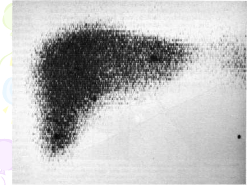
Слайд 104Isotope scanning
The study is usually performed 15-30 minutes after injection with
radiopharmaceutical substent since 10-15 min are required for complete clearing of the colloid from the blood stream in the normal object

Слайд 105Following are the indications of liver imaging:
• Evaluation of the
liver size, shape and position.
• Evaluation of the abdominal masses.
• Detection: abscess, hematoma, tumor and cyst.
• Pre and post treatment evaluation of the hepatic metastases.
• Evaluate diffuse hepatic disease such as cirrhosis, hepatitis or metabolic disease.
• Evaluation of the abdominal masses.
• Detection: abscess, hematoma, tumor and cyst.
• Pre and post treatment evaluation of the hepatic metastases.
• Evaluate diffuse hepatic disease such as cirrhosis, hepatitis or metabolic disease.

Слайд 107CHOICE OF EXAMINATION IN BILIARY AND LIVER DISEASE
Suspected liver masses (tumors,
primary or secondary, cysts or abscess) should be examined in the 1st place by isotope scanning or ultrasound. Both these methods will usually demonstrate liver masses quite well. Ultrasound has the advantage that it will differentiate cysts and abscess from solid masses.

Слайд 108CT and MRI will also perform this function but are more
expansive investigations and
CT involves radiation. They are used for more precise diagnosis; as a prelude to surgery; and for tumor staging.
CT involves radiation. They are used for more precise diagnosis; as a prelude to surgery; and for tumor staging.

Слайд 109In suspected obstructive jaundice, ultrasound is the primary investigation of choice,
though biliary isotope scanning will show moderately dilated ducts, as will CT. Transhepatic cholangiography or ERCP may be required to define the point of obstruction, and percutaneous biliary drainage may be used in treatment.

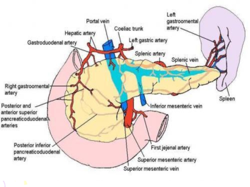
Слайд 110PANCREAS
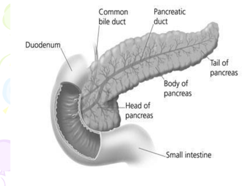
Pancreas lies in the posterior abdominal wall in the epigastrium and
left hypochondrium. Head is surrounded on three sides by duodenal ring and prepyloric areas of stomach. Head is covered anteriorly by distal end of the stomach, duodenum and transverse colon.

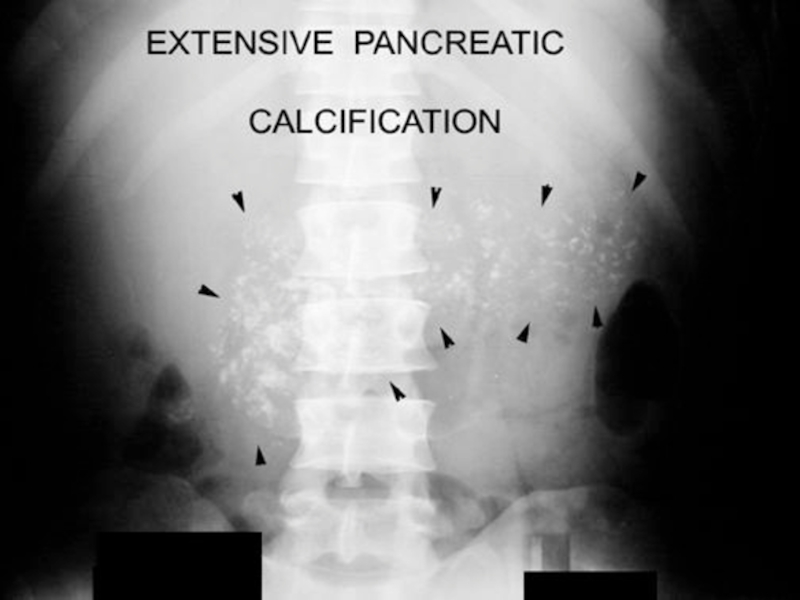
Слайд 113Simple X ray will occasionally show extensive nodules of calcification in
the pancreas associated with chronic pancreatitis but is otherwise little help.

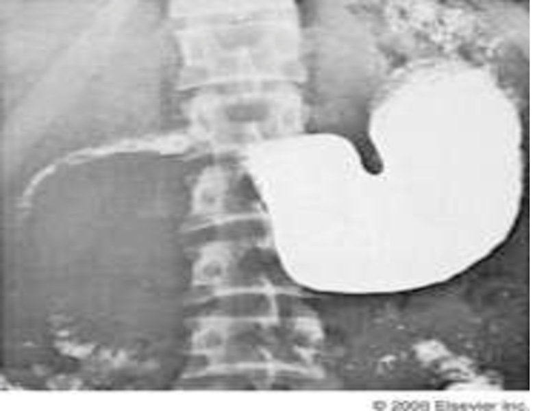
Слайд 116Barium studies will show:
distortion of the duodenal loop by masses in
the head of the pancreas
large pancreatic masses or cysts may displace the stomach.
large pancreatic masses or cysts may displace the stomach.


Слайд 119
Ultrasound: the normal pancreas may be partly obscured by bowel gas
but when well seen appears as 1 to 3 cm diameter band arching over the aorta at the level of superior mesenteric artery and of slightly higher echogenicity than normal liver.

Слайд 120 In acute pancreatitis, the whole organ is enlarged and edematous
appearing more transonic than normal.
Abscess and cysts appear as well defined rounded transonic areas.
Tumors appear as local masses enlarging the pancreas.
Abscess and cysts appear as well defined rounded transonic areas.
Tumors appear as local masses enlarging the pancreas.


Слайд 122CT scan
CT is now the method most widely used for the
demonstration of the pancreatic morphology.
The pancreas and its relationships to the adjacent organs are clearly identified.
Cysts and mass lesions greater than 2 cm in diameter are easily identified.
The pancreas and its relationships to the adjacent organs are clearly identified.
Cysts and mass lesions greater than 2 cm in diameter are easily identified.

Слайд 123 Small tumors such as islet cell adenomas are more difficult
to diagnose.
Acute pancreatitis may be clearly demonstrated at CT as diffuse swelling and edema of the whole organ, though some cases, particularly in first 24 hours, will not show diagnostic appearances.
Dilation and irregularity of the main pancreatic duct can also be seen at CT.
Acute pancreatitis may be clearly demonstrated at CT as diffuse swelling and edema of the whole organ, though some cases, particularly in first 24 hours, will not show diagnostic appearances.
Dilation and irregularity of the main pancreatic duct can also be seen at CT.


Слайд 126MRI
MRI can also show the pancreas well but has no advantage
over CT, which is much cheaper and more readily available.

Слайд 127ERCP
ERCP is the method of choice for demonstrating the pancreatic
duct and its obstruction or stenosis by calculi or tumor. The bile ducts can be demonstrated by at the same time and variety of diagnostic and therapeutic procedures performed.

Слайд 128These include:
1. Stone extraction from pancreatic and bile ducts
2. Sphincterectomy
3. Biopsy of ampulla
4. Cytology of
pancreatic juice
5. Balloon dilation of benign structures
6. Biliary stem insertion.
7. Pancreatic cyst drainage
5. Balloon dilation of benign structures
6. Biliary stem insertion.
7. Pancreatic cyst drainage

Слайд 129Choice of examination
CT is probably the best method for speedy demonstration
of the pancreas. The normal pancreas is usually well shown as are masses and cysts. Small tumors whether carcinomas or islet cell adenomas are more difficult to define.

Слайд 130Ultrasound in skilled hands is almost reliable, but is to some
extent operator dependent.
Isotope scanning is less reliable than CT or ultrasound and is no longer used for pancreatic lesions. Neither CT nor ultrasound will differentiate an inflammatory pancreatic mass from a tumor lesion.
However fine needle biopsy guided by CT or ultrasound will permit a definitive diagnosis.
Isotope scanning is less reliable than CT or ultrasound and is no longer used for pancreatic lesions. Neither CT nor ultrasound will differentiate an inflammatory pancreatic mass from a tumor lesion.
However fine needle biopsy guided by CT or ultrasound will permit a definitive diagnosis.