Слайд 1Semey State Medical University
SIW
Topic: Properties of heart and vessels in children
Prepared
by: Mohammad Aquib, GMF 340
Checked by: Shchetinina Nataliya
Semey,2015
Слайд 2Plan
1.Features of embryonic cardiovascular system.
2. Morphological and histological features of the
heart.
3. Characteristics of the functions of the circulatory system.
4. The morphology and function of the cardiovascular system in children.
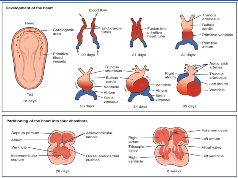
Слайд 4Stawage the heart (the end of the 2nd week of fetal
development)
The separation of the heart into left and right half (the end of the third week of embryonic development), education and the formation of atrial foramen ovale
The formation of the interventricular septum (the fifth week of fetal development)
Education septum that separates the bulb on the mouth of the pulmonary artery and the aorta (the fourth week of intrauterinedevelopment)
Слайд 5Forming a third septum that combines atrium and venous sinus (4-5
week)
Internal formation(trabecular) (3 Week 4) and the outer layer of the myocardium (4-5 week)
The formation of the fibrous ring atrioventricular openings
(2nd month of intrauterine debelopment)
Слайд 6Stages of development of the fetal circulation:
gistiotrofny type of food (first
two weeks) - the circulatory system is absent; Nutrients come from the yolk sac;
yolk circulation period (from 3 weeks to 2 months of fetal development);
placental circulation period (end of 2 nd - beginning of the 3 months of fetal development) - fetal blood is separated from the mother's blood placental membrane.
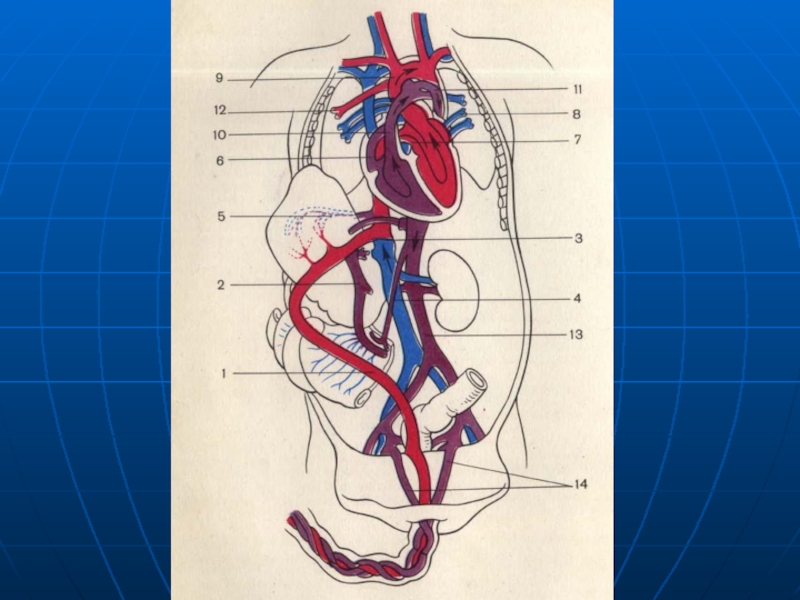
Слайд 8Features newborn circulation
oxygen saturation occurs in the placenta, where it flows
through the umbilical vein to the liver of the fetus and through the venous (Arantsiev) duct empties into the vena cava;
pulmonary circulation is not functioning, the principal amount of blood through the patent ductus arteriosus enters the aorta;
Слайд 9all organs (except liver) get mixed blood;
the blood returns to the
placenta through the umbilical artery,
functioning shunts: Arantsiev duct, an oval hole, Botalov flow;
a synchronous contraction of the ventricles, rather than sequentially.
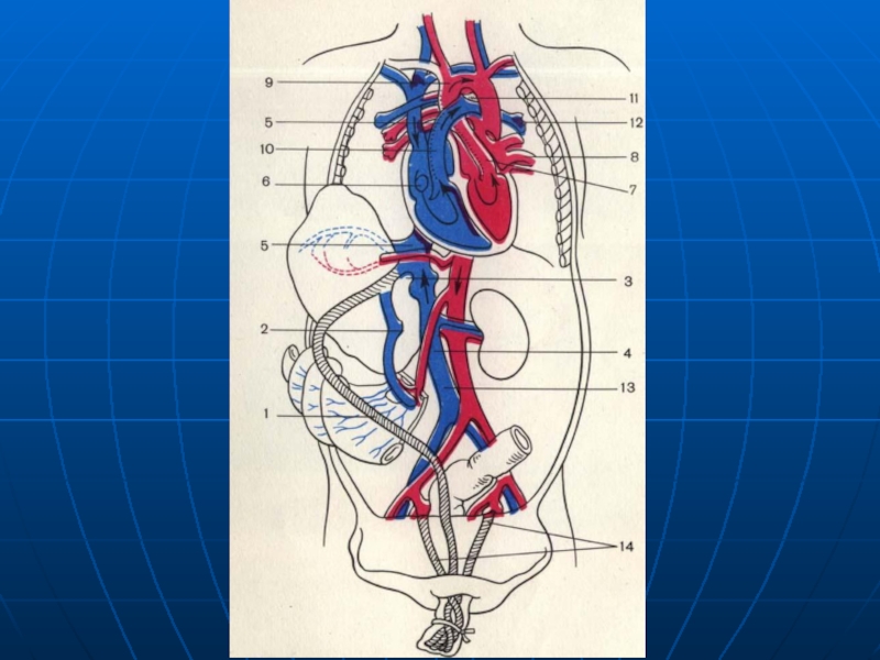
Слайд 11Features newborn circulation:
cease to function 6 main structures: 4 (umbilical Vienna,
ductus venosus and the two umbilical arteries) that provide blood flow and placental 2 (foramen ovale and ductus arteriosus), which shunted blood from the pulmonary circulation into the aorta;
starts functioning pulmonary circulation
Слайд 12The morphological features of the heart
1. The relatively large mass of
the heart (the newborn corresponds to 0.8% of the adult body weight - 0.4%).
2. Features of the form heart of a conditioned aspect ratio of the cavity.
3. The ratio of heart weight to body weight increases unevenly.
Слайд 13closes Arantsiev duct committed spasm and subsequent obliteration of blood (Botallova)
flow;
a relatively wide lumen of the arteries and veins of the same caliber.
4. The most intensive growth of the heart in the 1st year of life in pre - and pubertal periods (10 - 14 years).
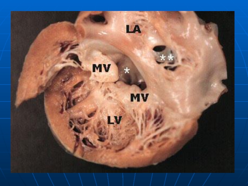
Слайд 145. The walls of the heart macroscopically not have a clear
differentiation flap valves are formed enough, capillary (papillary), the muscles are underdeveloped; capillary muscle tendon thread 2 times shorter than that of adults.
6. subepicardial department little adipose tissue, its amount increases markedly after 7 years
Слайд 15Динамика массы сердца
До года масса сердца удваивается;
До 2 – 3 лет
утраивается;
До 15 –16 лет увеличивается в 15 – 16 раз.
Слайд 16Соотношение правого и левого желудочков.
К 1-му году составляет 1 : 1,5
;
К 5-ти годам – 1 : 2;
К 14-ти годам – 1 : 2,76.
Толщина стенки левого желудочка возрастает в 3 раза за период роста ребенка, правого – на 1/3.
Слайд 17Heart weight (relative to body weight)
5 - 6 months. - 0.38%
At
8 years old boys - 0.44%
In 12 years in girls - 0.48%
Слайд 19Properties neonatal cardiac muscle:
a) muscle fibers are thin, are located close
to one another;
b) have a large number of large nuclei;
c) poorly developed interstitial, and elastic connective tissue, a well-developed net of blood vessels;
g) Soft flap valves and epicardium.
Слайд 20The duration of the cardiac cycle in children of all ages:
Newborn
- to 0,40-0,50
10 years - 0.70
adults - to 0,77-0,80
The duration of ventricular diastole:
infants - 0.23
adults - 0.48 to
The physiological importance of: increasing blood filling of the ventricles
Слайд 21Differences vessels Child and adult:
Arteries are relatively wider
Artery wider than veins
Vienna
grow rather than artery
In 16 years in the lumen of the vein 2 times wider than the lumen of the arteries
Thin-walled blood vessels of newborn, they are not sufficiently developed muscle and elastic fibers
Слайд 22With age comes the differentiation of the vascular wall, increasing the
number of elastic and muscle fibers
The development of blood vessels ends up to 12-13 years
Children have a well-developed capillary network
Capillaries intestine, kidney, skin, lung, relatively and absolutely larger than in adults


 The separation of")
Internal formation(trabecular) (3 Week")
 - the")

 get mixed blood;the blood returns to the placenta through the umbilical")


 flow;a relatively wide lumen")


5 - 6 months. - 0.38%At 8 years old boys")
 muscle fibers are thin, are located close to one another;b) have")