ICCU
9.2017
- Главная
- Разное
- Дизайн
- Бизнес и предпринимательство
- Аналитика
- Образование
- Развлечения
- Красота и здоровье
- Финансы
- Государство
- Путешествия
- Спорт
- Недвижимость
- Армия
- Графика
- Культурология
- Еда и кулинария
- Лингвистика
- Английский язык
- Астрономия
- Алгебра
- Биология
- География
- Детские презентации
- Информатика
- История
- Литература
- Маркетинг
- Математика
- Медицина
- Менеджмент
- Музыка
- МХК
- Немецкий язык
- ОБЖ
- Обществознание
- Окружающий мир
- Педагогика
- Русский язык
- Технология
- Физика
- Философия
- Химия
- Шаблоны, картинки для презентаций
- Экология
- Экономика
- Юриспруденция
Pericardial diseases презентация
Содержание
- 1. Pericardial diseases
- 2. Pericard : anatomical and physyological considerations Outer
- 3. Pericardial fluid In normal hearts there is
- 4. Most common forms of pericardial syndromes Acute
- 6. Etiology
- 7. Etiology
- 8. ESC guidelines 2004
- 12. Acute pericarditis
- 14. Acute pericarditis Most common form of pericardial
- 15. Acute pericarditis: etiology 80-95% of cases -
- 16. Acute pericarditis: etiology (cont’d) Developed countries:
- 17. Acute pericarditis: etiology (cont’d) Developing countries:
- 18. Acute pericarditis: diagnosis Typical chest pain (pleuritic
- 19. Acute pericarditis: diagnosis Basic diagnostic evaluation
- 20. ECG in acute pericarditis
- 21. ECG in acute pericarditis
- 22. ECG in acute pericarditis
- 23. Acute pericarditis: diagnosis Basic diagnostic evaluation
- 24. Indications for pericardiocentesis Cardiac tamponade Large
- 25. Acute pericarditis: diagnostic studies of pericardial fluid
- 26. Acute pericarditis: diagnostic studies of pericardial fluid
- 27. Acute pericarditis: other diagnostic modalities Pericardial biopsy
- 28. Management of pericarditis
- 29. Acute pericarditis: risk stratification
- 30. Acute pericarditis: risk stratification At least one
- 31. Acute pericarditis: therapy Targets toward specific etiology
- 32. Acute pericarditis: therapy
- 34. NEJM 2013, Sep 1
- 35. ICAP trial Colchicine 0.5 mg x
- 36. ICAP trial
- 37. ICAP trial
- 38. ICAP trial
- 39. ICAP trial
- 40. ICAP trial
- 42. Acute pericarditis: therapy Corticosteroids increase risk of
- 43. Acute pericarditis: therapy
- 44. Acute pericarditis: therapy
- 45. Acute pericarditis: therapy (cont’d) Rest and avoidance
- 46. Acute pericarditis: therapy (cont’d) Athlets. Return
- 47. Acute pericarditis: prognosis Recurrence is most common
- 48. Recurrent pericarditis
- 49. Recurrent pericarditis
- 50. Recurrent pericarditis: therapy
- 51. Pericardial effusion
- 52. Echo (4-chamber view) in pt with large pericardial effusion and cardiac tamponade PE PE
- 54. Pericardial effusion Large idiopatic chronic pericardial effusion
- 55. Pericardial effusion Pericardiectomy is recommended in a
- 56. Pericardial effusion: etiology Pericardial effusion without evidence
- 57. Pericardial effusion: etiology
- 58. Pericardial effusion: management
- 59. Pericardial effusion: management
- 60. Pericardial effusion: management
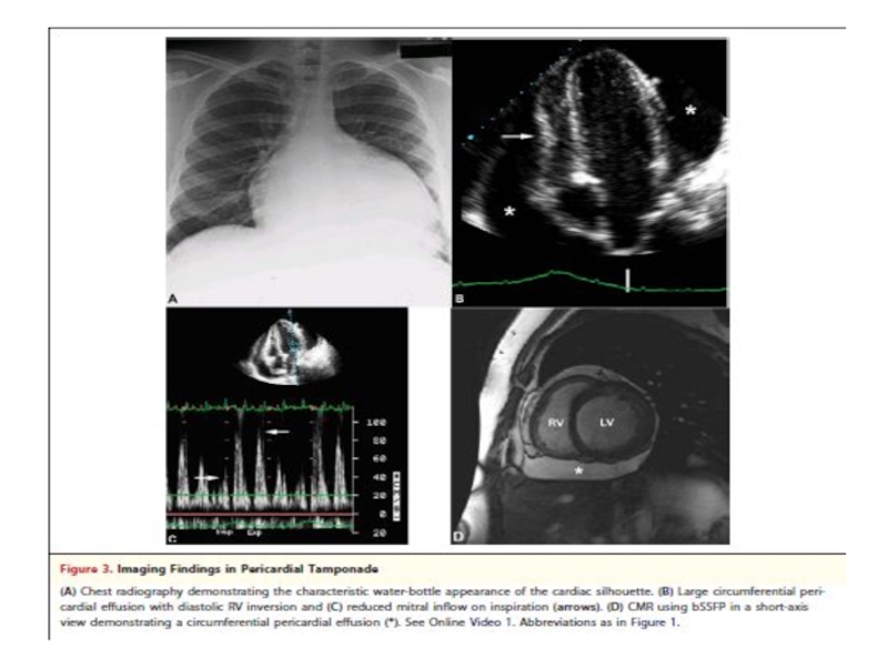
- 61. Cardiac tamponade
- 63. Cardiac tamponade Clinical signs Beck’s triad: hypotension,
- 64. Cardiac tamponade Electrocardiographic signs -
- 66. Cardiac tamponade
- 67. Approaches for pericardiocentesis parasternal apical subxyphoid / subcostal
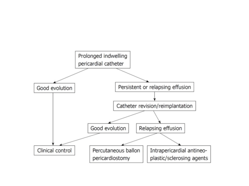
- 69. Recommendations for management of neoplastic involvement of the pericardium
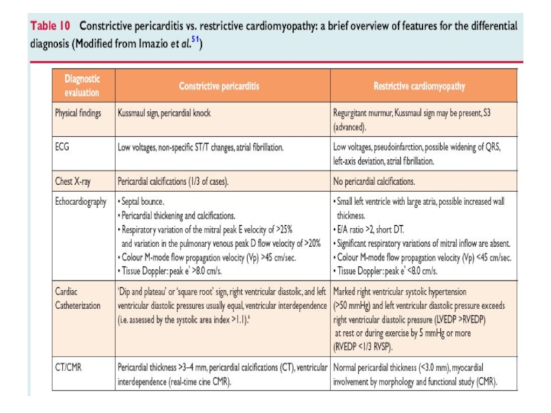
- 70. Constrictive pericarditis
- 71. Constrictive pericarditis
- 73. Constrictive pericarditis Fibrotic pericardium impedes normal diastolic
- 74. Constrictive pericarditis: etiology Idiopathic or viral -
- 76. 500 patients Mean FU – 72
- 77. Circulation 2011; 124: 1270
- 78. Constrictive pericarditis: symptoms Right heart failure:
- 79. Constrictive pericarditis Pericardial constriction should
- 81. Transient constrictive pericarditis 10-20% of cases during
- 82. Effusive constrictive pericarditis In 8% of patients
- 83. Constrictive pericarditis: treatment
- 84. Thank you for attention
- 85. Backup slides
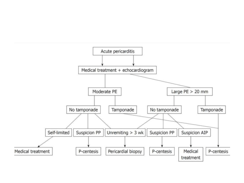
- 94. Triage of patients with acute pericarditis Imazio et al. JACC 2004; 43:1042-6
- 95. Causes of pericardial effusion Inflammation Infection Noninfectious
- 96. Etiology of pericarditis Infectious pericarditis Pericarditis in
- 97. Acute pericarditis: therapy (cont’d)
- 98. COPPS trial Am Heart J 2011; 62:527-32
- 99. COPPS trial
- 100. COPPS trial
- 101. COPPS trial
- 102. Rx of acute pericarditis in children
- 103. Rx of acute pericarditis in children
Слайд 1Pericardial diseases
Dr. Michael Kapeliovich MD, PhD
Director Emergency Cardiology Service
Deputy Director

Слайд 2Pericard : anatomical and physyological considerations
Outer layer - fibrous pericardium
Inner layer - serous or visceral pericardium (epicardium)
Proximal portion of aorta and pulmonary artery are enclosed in pericardial sac
Functions of pericardium:
- prevents friction between the heart and surrounding
structures
- acts as mechanical and immunological barrier
- limits distention of the heart

Слайд 3Pericardial fluid
In normal hearts there is a small amount of pericardial
fluid (25-50 ml)
Produced by visceral pericardium
Produced by visceral pericardium
increased production of fluid
pericardial effusion
 Produced")
Слайд 4Most common forms of pericardial syndromes
Acute and recurrent pericarditis
Pericardial effusion
Cardiac
tamponade
Constrictive pericarditis
Constrictive pericarditis


Слайд 14Acute pericarditis
Most common form of pericardial disease
~5% of presentations to ED
for non-ischemic chest pain
Incidence of acute pericarditis in a prospective study 28/ 100 000 of the population per year in an urban area in Italy
Incidence of acute pericarditis in a prospective study 28/ 100 000 of the population per year in an urban area in Italy

Слайд 15Acute pericarditis: etiology
80-95% of cases - idiopathic ( in Western Europe
and in North America )
Such cases are generally presumed to be viral
Major non-idiopathic etiologies:
- tuberculosis
- neoplasia
- systemic (generally autoimmune disease)
Such cases are generally presumed to be viral
Major non-idiopathic etiologies:
- tuberculosis
- neoplasia
- systemic (generally autoimmune disease)

Слайд 16Acute pericarditis: etiology (cont’d)
Developed countries:
emerging cases of pericarditis –
iatrogenic posttraumatic, following cardiac surgery, PCI, pacemaker insertion, catheter ablation.
In these cases pathogenesis is determined by combination of:
- direct pericardial trauma
- pericardial bleeding
- individual predisposition
In these cases pathogenesis is determined by combination of:
- direct pericardial trauma
- pericardial bleeding
- individual predisposition
Developed countries: emerging cases of pericarditis – iatrogenic posttraumatic, following cardiac surgery,")
Слайд 17Acute pericarditis: etiology (cont’d)
Developing countries:
high prevalence of tuberculosis-related pericarditis
(70-80%) in Sub-Saharian Africa,
in ~90% the disease associated with HIV infection
in ~90% the disease associated with HIV infection
Developing countries: high prevalence of tuberculosis-related pericarditis (70-80%) in Sub-Saharian Africa,")
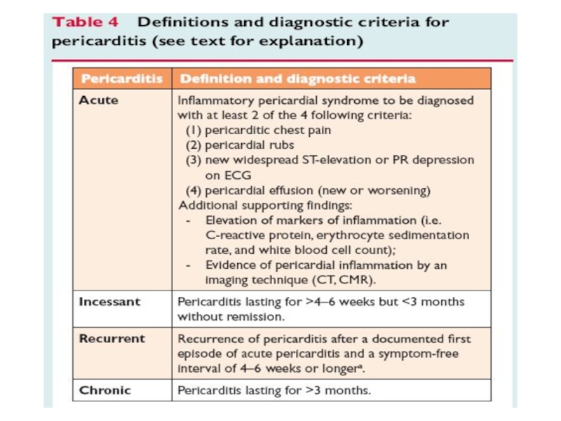
Слайд 18Acute pericarditis: diagnosis
Typical chest pain (pleuritic CP)
Pericarial friction rub
Widespread ST-segment elevation
and PR depression
Pericardial effusion
Pericardial effusion
At least 2 of 4 criteria should be present for Dx of acute pericarditis
Pericarial friction rubWidespread ST-segment elevation and PR depressionPericardial effusion")
Слайд 19Acute pericarditis: diagnosis
Basic diagnostic evaluation
Physical examination – auscultation
ECG
Trans-thoracic echocardiography (TTE)
Chest
x-ray
Blood tests
- routine blood tests
- markers of inflammation (C-reactive protein [CRP],
erythrocyte sedimentation rate [ESR])
- markers of myocardial damage (CK, Tn)
Blood tests
- routine blood tests
- markers of inflammation (C-reactive protein [CRP],
erythrocyte sedimentation rate [ESR])
- markers of myocardial damage (CK, Tn)
Chest x-rayBlood tests - routine")


Слайд 23Acute pericarditis: diagnosis
Basic diagnostic evaluation
The need for routine etiology search in
all cases of pericarditis is controversial and in low risk patients is not considered necessary

Слайд 24Indications for pericardiocentesis
Cardiac tamponade
Large or symptomatic pericardial effusion despite medical therapy
Highly
suspected tuberculous, purulent, or neoplastic etiology
ESC guidelines, 2004

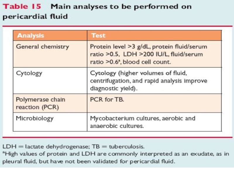
Слайд 25Acute pericarditis: diagnostic studies of pericardial fluid
Protein
LDH
Glucose
Cell count
Less useful for diagnosis
of specific etiology but are warranted to distinguish exudate from transudate

Слайд 26Acute pericarditis: diagnostic studies of pericardial fluid
Adenosin deaminase measurement for TB
Tumor
marker measurement ( carcino-embryonic antigen [CEA], cytokeratin 19 fragment )
Cytology
Culture and polymerase chain reactions for infections
Cytology
Culture and polymerase chain reactions for infections

Слайд 27Acute pericarditis: other diagnostic
modalities
Pericardial biopsy (during surgical drainage)
-
if cardiac tamponade relapsed after pericardiocentesis
- in patients without definite diagnosis whose illness lasted
for > 3 weeks
Pericardioscopy with target biopsy
Thoracic and abdominal CT
- in patients without definite diagnosis whose illness lasted
for > 3 weeks
Pericardioscopy with target biopsy
Thoracic and abdominal CT
 - if cardiac tamponade relapsed")


Слайд 30Acute pericarditis: risk stratification
At least one predictor of poor prognosis is
sufficient to identify a high risk cases
Cases of moderate risk – cases without negative prognostic predictors but incomplete or lacking response to NSAID therapy
Low risk cases – those without negative prognostic predictors and good response to anti-inflammatory therapy
Cases of moderate risk – cases without negative prognostic predictors but incomplete or lacking response to NSAID therapy
Low risk cases – those without negative prognostic predictors and good response to anti-inflammatory therapy

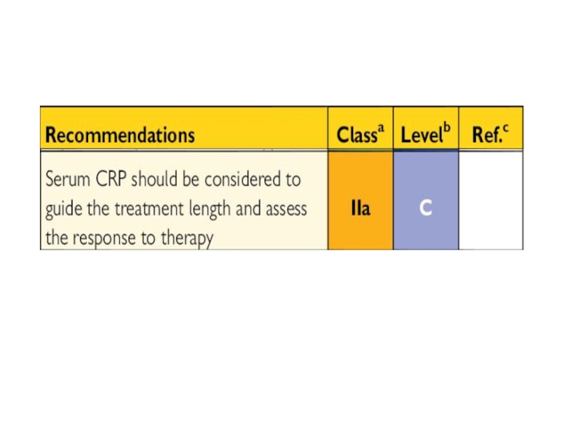
Слайд 31Acute pericarditis: therapy
Targets toward specific etiology if known
Empirical therapy for most
cases (idiopathic or presumed to be viral)
Rx until inflammatory marker (CRP, ESR) normalize (~7-14 days), than gradual tapering of the drug can be considered
Rx until inflammatory marker (CRP, ESR) normalize (~7-14 days), than gradual tapering of the drug can be considered


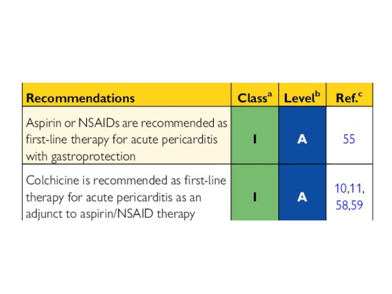

Слайд 35ICAP trial
Colchicine 0.5 mg x 2/d for 3 months
(for patients < 70 kg 0.5 mg x 1/d) vs placebo
In addition to conventional antiinflammatory therapy with Aspirin or Ibuprofen
In addition to conventional antiinflammatory therapy with Aspirin or Ibuprofen


Слайд 42Acute pericarditis: therapy
Corticosteroids increase risk of pericaditis recurrence
Indications:
- contraindication
for aspirin and NSAID
- failure of treatment with aspirin and at least another NSAID
- need for treatment of concomitant systemic condition
- failure of treatment with aspirin and at least another NSAID
- need for treatment of concomitant systemic condition


Слайд 45Acute pericarditis: therapy (cont’d)
Rest and avoidance of physical activity are useful
adjunctive measures until active disease is no longer evident (absence of pericardial effusion, normalization of inflammatory markers)
For athlets return to competitive sports not earlier than 6 months after episode of pericarditis particularly with myopericarditis
For athlets return to competitive sports not earlier than 6 months after episode of pericarditis particularly with myopericarditis
Rest and avoidance of physical activity are useful adjunctive measures until active")
Слайд 46Acute pericarditis: therapy (cont’d)
Athlets. Return to competitive sports only if:
asymptomatic
achieve
normalization of ECG abnormalities
achieve normalization of markers of inflammation
achieve normalization of LV function, wall motion
abnormalities and cardiac dimentions
no evidence of clinically relevant arrhythmias on Holter
monitoring and exercise tolerance test
achieve normalization of markers of inflammation
achieve normalization of LV function, wall motion
abnormalities and cardiac dimentions
no evidence of clinically relevant arrhythmias on Holter
monitoring and exercise tolerance test
 Athlets. Return to competitive sports only if:asymptomaticachieve normalization of ECG abnormalitiesachieve")
Слайд 47Acute pericarditis: prognosis
Recurrence is most common complication
Incidence ~30%
Autoimmune pathogenetic mechanism
is most
probable
probable


 in pt with large pericardial effusion and cardiac tamponadePEPE")
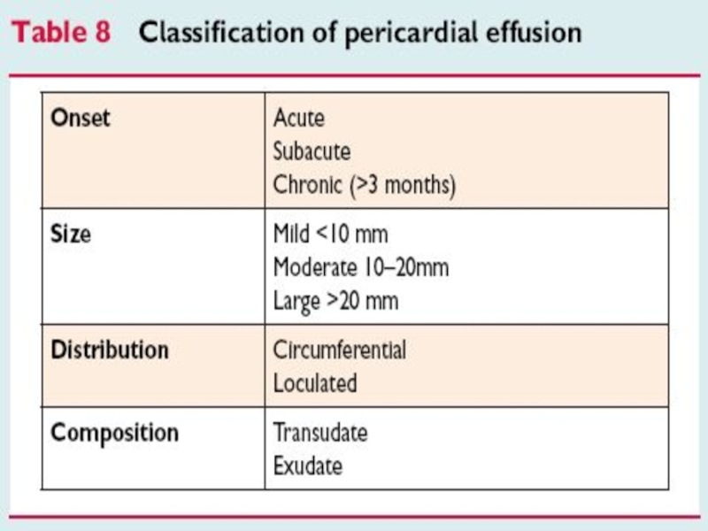
Слайд 54Pericardial effusion
Large idiopatic chronic pericardial effusion defined as collection of pericardial
fluid that persists for >3 months and has no apparent cause
Risk of progression to cardiac tamponade ~30%
Drainage of large pericardial effusion is recommended after 6-8 weeks of Rx
Risk of progression to cardiac tamponade ~30%
Drainage of large pericardial effusion is recommended after 6-8 weeks of Rx

Слайд 55Pericardial effusion
Pericardiectomy is recommended in a case of large effusion after
pericardiocentesis
No medical therapy have been proven effective for reduction of an isolated pericardial effusion in the absence of inflammation
No medical therapy have been proven effective for reduction of an isolated pericardial effusion in the absence of inflammation

Слайд 56Pericardial effusion: etiology
Pericardial effusion without evidence of inflammation and pericarditis is
often a clinical dilema
The presence of inflammatory signs (elevated CPR
and/or ESR) favor diagnose of pericarditis
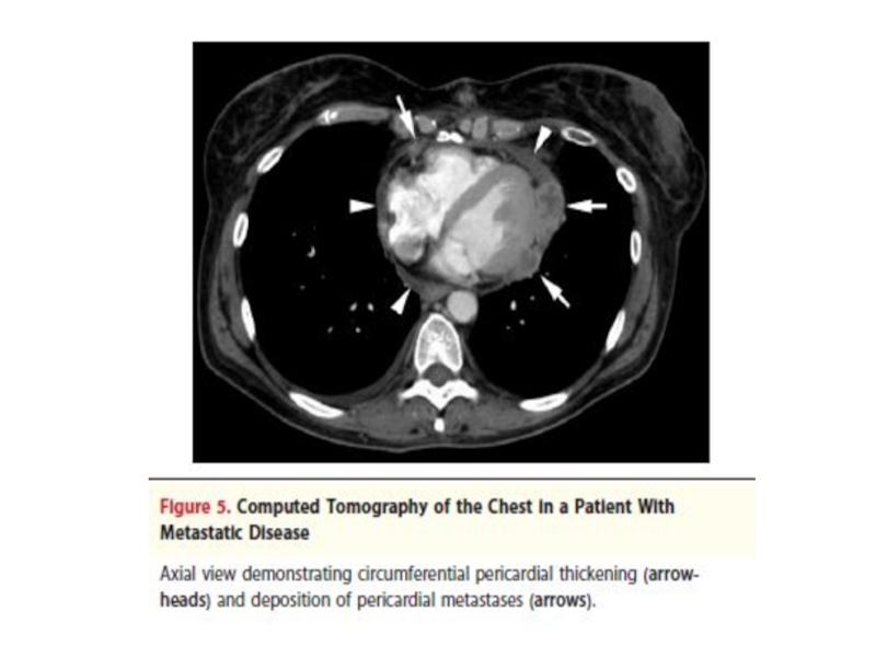
Large effusion and cardiac tamponade without
inflammatory signs are often associated with
neoplastic etiology
The presence of inflammatory signs (elevated CPR
and/or ESR) favor diagnose of pericarditis
Large effusion and cardiac tamponade without
inflammatory signs are often associated with
neoplastic etiology


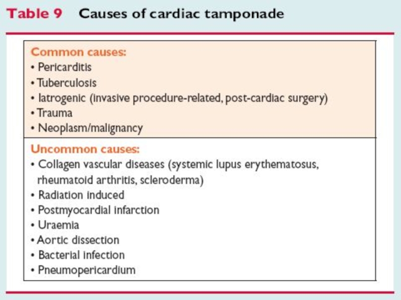
Слайд 63Cardiac tamponade
Clinical signs
Beck’s triad: hypotension, muffled heart sounds,
elevated
jugular venous pressure
pulsus paradoxus >10 mm Hg: difference between
the pressure at which Korotkoff sounds first appear
and that at which they are present with
each heart beat
pulsus paradoxus >10 mm Hg: difference between
the pressure at which Korotkoff sounds first appear
and that at which they are present with
each heart beat

Слайд 64Cardiac tamponade
Electrocardiographic signs
- reduced voltage
- electrical alternance
Echocardiographic
signs
- large peicardial effusion (most often)
- “swinging” motion
- repriratory changes in trans-mitral and trans-aortic flow
- large peicardial effusion (most often)
- “swinging” motion
- repriratory changes in trans-mitral and trans-aortic flow


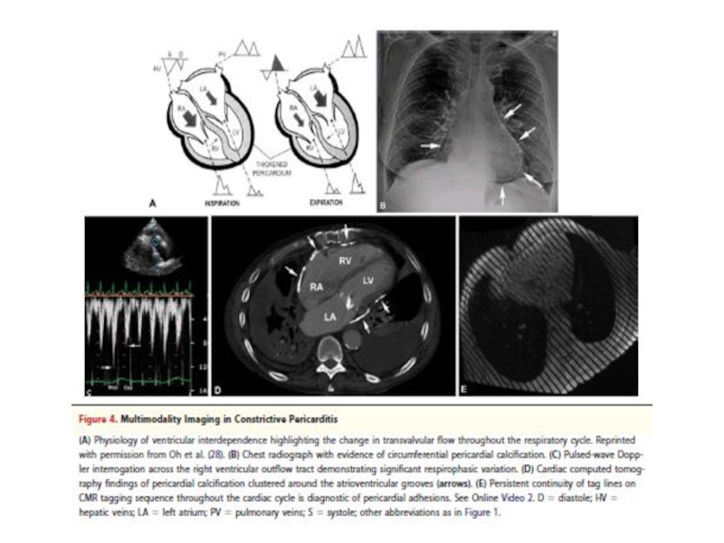
Слайд 73Constrictive pericarditis
Fibrotic pericardium impedes normal diastolic filling because of loss of
elasticity
Usually pericardium is considerably thickened but in ~20% of cases can be of normal thickness
Types of constrictive pericarditis:
- chronic (usually)
- subacute transient
- occult constriction
Usually pericardium is considerably thickened but in ~20% of cases can be of normal thickness
Types of constrictive pericarditis:
- chronic (usually)
- subacute transient
- occult constriction

Слайд 74Constrictive pericarditis: etiology
Idiopathic or viral - 42-49%
Cardiac surgery - 11-37%
Radiation Rx
- 9-31% (mostly for Hodgkin disease or breast cancer)
Connective tissue disorders (3-7%)
Infection 3-6% (TB or purulent pericarditis)
Connective tissue disorders (3-7%)
Infection 3-6% (TB or purulent pericarditis)

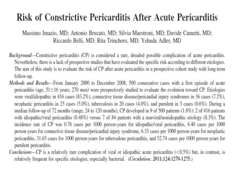
Слайд 76
500 patients
Mean FU – 72 months
Constrictive pericarditis – 1.8%
Idiopathic/Viral (2 of
416 pts) – 0.48%
Nonviral/Nonidiopathic (7 of 84 pts) – 8.3%
Nonviral/Nonidiopathic (7 of 84 pts) – 8.3%
Circulation 2011; 124: 1270
 – 0.48%Nonviral/Nonidiopathic")

Слайд 78Constrictive pericarditis: symptoms
Right heart failure: range from periferal edema to anasarca
No pulmonary congestion
Usually normal heart size
Fatigability and dyspnea related to diminished
cardiac output (CO) response to exertion

Слайд 79Constrictive pericarditis
Pericardial constriction should be considered in any patient
with unexplained elevation of jugular venous pressure, particularly with history of cardiac surgery, radiation therapy, or bacterial pericarditis

Слайд 81Transient constrictive pericarditis
10-20% of cases during resolution of pericardial inflammation
Patients with
newly diagnosed constrictive pericarditis who are hemodynamically stable, can be managed conservatively for 2-3 months period with empiric anti-inflammation therapy, before pericardiectomy is recommended

Слайд 82Effusive constrictive pericarditis
In 8% of patients with cardiac tamponade who underwent
pericardiocentesis and cardiac catheterization
Diagnostic characteristics of effusive-constrictive pericarditis: failure of right atrial (RA) pressure to fall by 50% or to level below 10 mm Hg after pericardiocentesis
Usually present with clinical signs of pericardial effusion, constrictive pericarditis, or both
Diagnostic characteristics of effusive-constrictive pericarditis: failure of right atrial (RA) pressure to fall by 50% or to level below 10 mm Hg after pericardiocentesis
Usually present with clinical signs of pericardial effusion, constrictive pericarditis, or both


Слайд 95Causes of pericardial effusion
Inflammation
Infection
Noninfectious etiology
-------------------------------------------------------------------------
Chronic inflammation + fibrosis + calcification
Thickened and
calcified pericardium
Constriction

Слайд 96Etiology of pericarditis
Infectious pericarditis
Pericarditis in systemic autoimmune diseases
Type 2 (auto)immune process
Pericarditis
and pericardial effusion in diseases of surrounding organs
Pericarditis in metabolic disorders
Neoplastic
Idiopathic
Pericarditis in metabolic disorders
Neoplastic
Idiopathic
immune processPericarditis and pericardial effusion in")
")