ZAPORIZHZHIAN STATE MEDICAL UNIVERSITY
The department of pathological anatomy and forensic medicine with basis of law
- Главная
- Разное
- Дизайн
- Бизнес и предпринимательство
- Аналитика
- Образование
- Развлечения
- Красота и здоровье
- Финансы
- Государство
- Путешествия
- Спорт
- Недвижимость
- Армия
- Графика
- Культурология
- Еда и кулинария
- Лингвистика
- Английский язык
- Астрономия
- Алгебра
- Биология
- География
- Детские презентации
- Информатика
- История
- Литература
- Маркетинг
- Математика
- Медицина
- Менеджмент
- Музыка
- МХК
- Немецкий язык
- ОБЖ
- Обществознание
- Окружающий мир
- Педагогика
- Русский язык
- Технология
- Физика
- Философия
- Химия
- Шаблоны, картинки для презентаций
- Экология
- Экономика
- Юриспруденция
Pathology of pregnancy and delivery презентация
Содержание
- 1. Pathology of pregnancy and delivery
- 2. Physiology of menstrual cycle: During reproductive
- 3. Ovulation releases an egg from an ovarian
- 4. Pregnancy 1. In endometrium: under the progestational
- 5. Pregnancy is divided into trimesters 1. The
- 6. Pregnancy Periods of normal births (delivery): 1.
- 7. Abortion - it is miscarriage of pregnancy.
- 8. Abortion A spontaneous abortion is a loss
- 9. Abortion Definitions are as follows: Inevitable abortion—no
- 10. PATHOLOGY OF IMPLATATION (Ectopic Pregnancy) 1. Placenta
- 11. PATHOLOGY OF IMPLATATION (Ectopic Pregnancy) 1. Placenta
- 12. PATHOLOGY OF IMPLATATION (Ectopic Pregnancy) 2. Deep
- 13. 3. Extra-uterine pregnancy Reasons of development:
- 14. PATHOLOGY OF IMPLATATION (Ectopic Pregnancy) Diagnostics:
- 15. PATHOLOGY OF DELIVERY 1. Amniotic embolism –
- 16. PATHOLOGY OF DELIVERY Danger: an amniotic
- 17. PATHOLOGY OF DELIVERY 2. Delivery infection of
- 18. Trophoblastic Disease 1. Hydatidiform Mole - tumor
- 19. Trophoblastic Disease 1. Hydatidiform Mole - tumor
- 20. Trophoblastic Disease 1. Hydatidiform Mole - tumor
- 22. Histologically: , the hydatidiform mole has large
- 23. Trophoblastic Disease 2. Choriocarcinoma This is cancer
- 24. The pathologist determines the histologic diagnosis of
Слайд 1PATHOLOGY OF PREGNANCY AND DELIVERY
Lecture on pathological anatomy
for the
3-rd year students

Слайд 2Physiology of menstrual cycle:
During reproductive life, the endometrium goes through a
monthly cycle. Under influence of realizing-factors of hypothalamus there is beginning of menstrual cycle from removing of functional layer of the endometrium.
Phases of menstrual cycle:
1. proliferative phase - begins with menstruation.
2. secretory phase - begins at ovulation, and should be 14 days, with less variability. At that time the producing of progesterone begins. The endometrium is ready to receive an ovule (ovum). If impregnation is not coming, from the 28th day the regression of corpus luteum begins, the secretion of progesterone decreases, the desquamation of function layer begins. If there is impregnation the corpus luteum of pregnancy develops, so pregnancy begins, that leads - 280 days (or 40 weeks, 9 months ).

Слайд 3 Ovulation releases an egg from an ovarian follicle. The egg is
swept into the fallopian tube and begins to descend. Spermatozoa begin ascending. Fertilization of the egg by a single sperm occurs in the ampullary portion of the fallopian tube about a day after ovulation. The fertilized egg begins to develop into the blastocyte and descend into the endometrial cavity, where implantation occurs on the wall of the fundus about a week after ovulation. In norm, the blastocyte implantates on the posterior uterus wall.
Pregnancy

Слайд 4Pregnancy
1. In endometrium: under the progestational influence of the corpus luteum
of pregnancy, the endometrial stroma undergoes a decidual change. The endometrial glands become hypersecretory. Even if the pregnancy is ectopic or trophoblastic disease is present, an exaggerated
hypersecretory response may occur. The changes can persist for up to 8 weeks after delivery.
2. The nourish of blastocyte takes place in the functional
layer of endometrium. Trophoblast forms the primary chorionic villi, endometrium atrophies above blastocytes, and veritable chorionic villi are formed under blastocytes, consisting of cytotrophoblast, syncytiotrophoblast and vessels.
3. Underlying endometrium form decidua basalis and decidua fetalis that form placenta. Placenta is formed to the 3-d
month of pregnancy.

Слайд 5Pregnancy is divided into trimesters
1. The first trimester lasts until 12
weeks but is also defined as up to 14 weeks of gestational age
2. The second trimester from 12 -14 until 24-28 weeks of gestational age
3. The third trimester from 24-28 weeks until delivery.
An infant delivered prior to 24 weeks is considered to be previable,
from 24 to 37 weeks is considered pre-term,
from 37 to 42 weeks is considered term.
A pregnancy carried beyond 42 weeks is considered postdate or post-term.

Слайд 6Pregnancy
Periods of normal births (delivery):
1. Fights – un-rhythmic and weak reductions
of myometrium.
2. Labour (muscular contraction) – rhythmic, increasing on force, reductions of pregnant uterus. Opening of delivery ways, dissection of front fetus bubble and pour out of pericarp waters begins.
3. Period of banishment and birth of fetus.
4. Separation and birth of placenta – the physiological bleeding at the separation of placenta is 400 ml, blood clots are formed in the micro-vessels of the wound.
:1. Fights – un-rhythmic and weak reductions of myometrium.2. Labour (muscular")
Слайд 7Abortion - it is miscarriage of pregnancy.
Classification:
1. According to the development
a) Artificial (therapeutic) – medical or criminal
b) Spontaneous
2. According to the time of development:
a) early – up to 14 weeks,
b) late - 14-22 (20) weeks (a baby is not viable),
c) premature births – 22(20)-37 weeks (a baby is a viable).
Artificial delivery is introduction of liquid between a fetus bubble and endometrium arising up of uterus capacity of constriction, because of volume arising up in uterus cavity.
 Artificial (therapeutic) –")
Слайд 8Abortion
A spontaneous abortion is a loss of the child before 20
weeks gestation.
The type of spontaneous abortion is defined by whether any or all of the products of conception have passed and whether or not the cervix is dilated.
Definitions are as follows:
Abortus—fetus lost before 20 weeks gestation, less than 500g, or less than 25cm.
Complete abortion—complete expulsion of all products of conception before 20 weeks gestation.
Incomplete abortion—partial expulsion of some but not all products of conception before 20 weeks gestation.

Слайд 9Abortion
Definitions are as follows:
Inevitable abortion—no expulsion of products, but bleeding and
dilation of the cervix such that a viable pregnancy is unlikely.
Threatened abortion—any intrauterine bleeding before 20 weeks, without dilation of the cervix or expulsion of any products of conception.
Missed abortion—death of the embryon or fetus before 20 weeks with complete retention of products of conception
Threatened abortion—any intrauterine bleeding before 20 weeks, without dilation of the cervix or expulsion of any products of conception.
Missed abortion—death of the embryon or fetus before 20 weeks with complete retention of products of conception

Слайд 10PATHOLOGY OF IMPLATATION (Ectopic Pregnancy)
1. Placenta previa
2. Deep implatation of
blastocyst
3. Extra-uterine pregnancy
3. Extra-uterine pregnancy
 1. Placenta previa 2. Deep implatation of blastocyst 3. Extra-uterine pregnancy")
Слайд 11PATHOLOGY OF IMPLATATION (Ectopic Pregnancy)
1. Placenta previa. It is anomalous attachment
of placenta in a lower segment of uterus:
incomplete (partial, marginal, low implantation) – only the edge of an internal part of endocervix is covered by placenta
complete
A fetus develops normally, but innate pathology is arising up in 5 times.
Complications:
air embolism through the opened vessels,
amniotic embolism (97% to lethality),
massive bleeding, that leads to hemorrhagic shock.
Treatment: Cesarean Section
1. Placenta previa. It is anomalous attachment of placenta in a")
Слайд 12PATHOLOGY OF IMPLATATION (Ectopic Pregnancy)
2. Deep implatation of blastocyst. In norm
blastocyst is implantated not deeper than middle of endometrium. During deep implantation, chorionic villi grow into myometrium. Pregnancy and first half of delivery is going normally. Complications develop in the 3d period of delivery. Part of placenta, at its separation, remains the uterus fastened on a bottom (placenta accreta) > an uterus is not abbreviated > bleeding from the cavity of uterus (obstetric) occurs > violation of process of haemostasis.
Treatment:
hand inspection of uterus cavity and separation of placenta,
vacuum-extraction of placenta tailings (parts),
amputation of uterus.
A placenta polypus it is an organized fragment of placenta, which remained in the cavity of uterus.
Treatment:
hand inspection of uterus cavity and separation of placenta,
vacuum-extraction of placenta tailings (parts),
amputation of uterus.
A placenta polypus it is an organized fragment of placenta, which remained in the cavity of uterus.
 2. Deep implatation of blastocyst. In norm blastocyst is implantated not")
Слайд 133. Extra-uterine pregnancy
Reasons of development:
scars in endometrium after: inflammatory processes
or abortion,
scars or joints in fallopian tube,
joints in small pelvis,
bends of fallopian tubes,
violation of reductions rhythm of fallopian tube (hormonal adjusting).
According to the localization it is divide:
1. ovarian – fetus does not develop, the rudimentary organs of embryon remain in small pelvis and petrificated (litopedion),
2. intra-abdominal – embryon is in the abdominal cavity,
3. intra-ligamental – between fallopian tube and ovary,
4. tubal pregnancy –
a) the ampullar pregnancy - tend to be of longer duration,
b) interstitial - in the corner of uterus (in the intrauterine part of tube),
c) isthmus pregnancy.
Sometimes blood clot and chorionic villi are recovered outside of the tube following rupture of an ectopic pregnancy.

Слайд 14PATHOLOGY OF IMPLATATION (Ectopic Pregnancy)
Diagnostics:
A positive pregnancy test (presence of
human chorionic gonadotropin)
Ultrasound investigation
Culdocentesis (needle aspiration) with presence of blood
Treatment:
amputation of fallopian tube (at histological research of remote fallopian tube it is find out chorionic villi in the tubes wall)
coagulation of the torn fallopian tube
Ultrasound investigation
Culdocentesis (needle aspiration) with presence of blood
Treatment:
amputation of fallopian tube (at histological research of remote fallopian tube it is find out chorionic villi in the tubes wall)
coagulation of the torn fallopian tube
Diagnostics: A positive pregnancy test (presence of human chorionic gonadotropin)Ultrasound investigationCuldocentesis")
Слайд 15PATHOLOGY OF DELIVERY
1. Amniotic embolism – an amniotic liquid gets into
the blood
Reasons of development:
a) An acute rise of pressure of amniotic liquid, as compared to pressure in the venous vessels of uterus, which develops at:
quick (fast) delivery,
postmature pregnancy,
large fetus with time-lagged delivery
b) an amniotic liquid gets into the vessels of uterus at:
the premature removing of placenta,
placenta percreta (the placenta invades through the full thickness of the uterine wall, and attaches to adjacent organs in the abdomen)
during the Cesarean Section.
")
Слайд 16PATHOLOGY OF DELIVERY
Danger:
an amniotic liquid is rich in with vaso-active
molecules and fragments of vital functions of fetus (squamosus epithelium, hair). At embolism by round-fetus waters in the vessels of woman lungs microscopy it is found out the scales of epithelium, hairs, they do not participate in thanatogenesis, but they are the diagnostic criterion of amniotic embolism.
Reasons of death:
a) massive and rapid reception of large volume of biologically-active liquids > penetration into lungs > spasm of bronchiols and arterioles > cardio-pulmonal shock > fibrillation of heart;
b) fractional amniotic embolism.
Reasons of death:
a) massive and rapid reception of large volume of biologically-active liquids > penetration into lungs > spasm of bronchiols and arterioles > cardio-pulmonal shock > fibrillation of heart;
b) fractional amniotic embolism.

Слайд 17PATHOLOGY OF DELIVERY
2. Delivery infection of uterus (Postpartum endometritis) - A
placenta ground takes off in the 3d period of delivery, so bacteriemia can be observed through the damaged of vessels during or after delivery.
By nature:
bacterial
endogenous infecting in 3 trimesters at intensifying of chronic inflammatory process.
Bacteria intensively propagate oneself and quickly enter the blood stream > festering endometritis > bacteriemia > sepsis
By nature:
bacterial
endogenous infecting in 3 trimesters at intensifying of chronic inflammatory process.
Bacteria intensively propagate oneself and quickly enter the blood stream > festering endometritis > bacteriemia > sepsis
 - A placenta ground takes off")
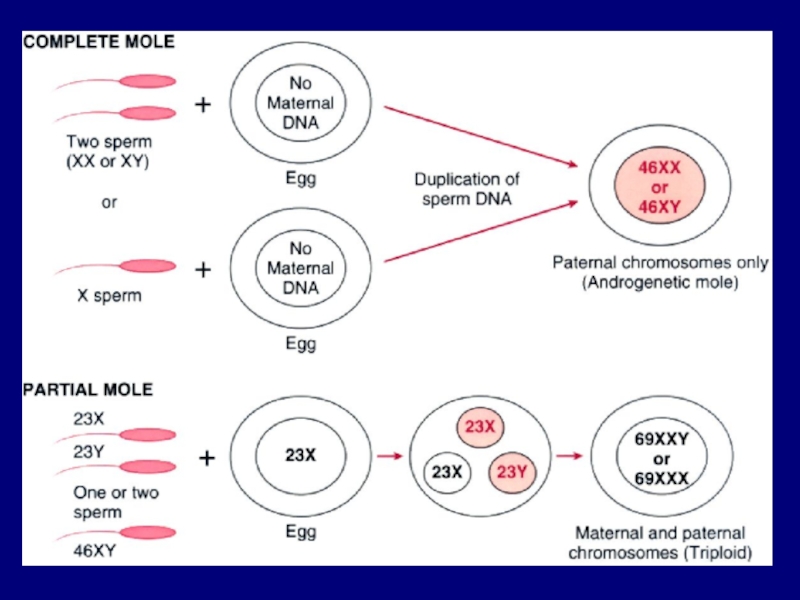
Слайд 18Trophoblastic Disease
1. Hydatidiform Mole - tumor of placental trophoblastic tissue
Complete
or "classic" - In complete hydatidiform mole, grossly swollen chorionic villi, which resemble a bunch of grapes, show varying degrees of trophoblastic proliferation. There is no embryon. The embryon dies at an early stage before the placental circulation has developed, and chorionic villi then contain few blood vessels.

Слайд 19Trophoblastic Disease
1. Hydatidiform Mole - tumor of placental trophoblastic tissue
Partial
- In partial hydatidiform mole two populations of chorionic villi exist, some of which show hydropic swelling. Trophoblastic proliferation is focal and usually less pronounced than in the complete mole. In partial hydatidiform mole, unlike complete mole, there is frequently an associated embryon. The fetus associated with a partial mole usually dies at approximately 10-15 weeks of gestation.

Слайд 20Trophoblastic Disease
1. Hydatidiform Mole - tumor of placental trophoblastic tissue
Invasive
or chorioadenoma destruens - The invasive mole is a hydatidiform mole that has invaded the underlying myometrium. Villi may embolize but not metastasize. The major danger is uterine hemorrhage while the disease is active. Uterine perforation from the locally infiltrative disease is the major complication.

Слайд 22Histologically: , the hydatidiform mole has large avascular grape-like villi and
areas of trophoblastic proliferation. With molar pregnancy, the uterus is large for dates, but no fetus is present. HCG levels are markedly elevated. Patients with a hydatidiform mole are often large for dates and have hyperemesis gravidarum more frequently. Patients may present with bleeding, and may pass some of the grape-like villi.
In partial moles, some villi appear normal, whereas others are swollen. There is minimal trophoblastic proliferation.
In partial moles, some villi appear normal, whereas others are swollen. There is minimal trophoblastic proliferation.

Слайд 23Trophoblastic Disease
2. Choriocarcinoma
This is cancer of the trophoblast. Choriocarcinoma is composed
of a dimorphic population of cytotrophoblast and syncytiotrophoblast, with varying degrees of intermediate trophoblast also present. Typically, the tumor undergoes central hemorrhage with necrosis, viable tumor being confined to the periphery of the mass immediately adjacent to underlying normal tissue.
The tumor metastasizes widely by the hematogenous route to any site, but especially to lung, brain, gastrointestinal tract, and liver. Microscopically, there will be no villi. The pathologist will see cytotrophoblast and syncytiotrophoblast, usually in alternating layers.
The tumor metastasizes widely by the hematogenous route to any site, but especially to lung, brain, gastrointestinal tract, and liver. Microscopically, there will be no villi. The pathologist will see cytotrophoblast and syncytiotrophoblast, usually in alternating layers.

Слайд 24The pathologist determines the histologic diagnosis of the trophoblastic disease, most
importantly, if choriocarcinoma is present. In this hydatidiform mole there is atypical trophoblastic proliferation, but villi are still present. The patient is then followed with serial HCG levels.
Much less common than hydatidiform mole is choriocarcinoma, seen here. Villi are not present. Rather, there is a proliferation of bizarre trophoblastic cells. These tumors are very aggressive. Half of choriocarcinomas arise in preceding hydatidiform moles.
Much less common than hydatidiform mole is choriocarcinoma, seen here. Villi are not present. Rather, there is a proliferation of bizarre trophoblastic cells. These tumors are very aggressive. Half of choriocarcinomas arise in preceding hydatidiform moles.