- Главная
- Разное
- Дизайн
- Бизнес и предпринимательство
- Аналитика
- Образование
- Развлечения
- Красота и здоровье
- Финансы
- Государство
- Путешествия
- Спорт
- Недвижимость
- Армия
- Графика
- Культурология
- Еда и кулинария
- Лингвистика
- Английский язык
- Астрономия
- Алгебра
- Биология
- География
- Детские презентации
- Информатика
- История
- Литература
- Маркетинг
- Математика
- Медицина
- Менеджмент
- Музыка
- МХК
- Немецкий язык
- ОБЖ
- Обществознание
- Окружающий мир
- Педагогика
- Русский язык
- Технология
- Физика
- Философия
- Химия
- Шаблоны, картинки для презентаций
- Экология
- Экономика
- Юриспруденция
Optics of vision. Eye structure презентация
Содержание
- 1. Optics of vision. Eye structure
- 2. EYE STRUCTURE 1 1 – front
- 3. EYE ADAPTATION TO LIGHT & DARKNESS
- 4. Photoreceptors: Rods & Cones Synaptic convergence for
- 7. The cornea, clear liquid of front chamber,
- 9. Presbyopia: ciliary muscles can no longer contract
- 10. Length of Eyeball + Curvature of Cornea Emmetropia: objects focused on retina (normal)
- 11. Normal sight Myopia Myopia (nearsightedness): objects focused in front of retina
- 12. Length of Eyeball + Curvature of Cornea
- 13. concave lens: negative diopter; diffraction Myopia Correction
- 14. Normal sight Farsightedness Hyperopia (farsightedness): objects focused behind retina
- 15. convex lens: positive diopter; refraction Hyperopia Correction
- 16. Normal sight Astigmatism Astigmatism Aspherical cornea: light
- 17. Astigmatism LASER correction of cornea shape

Слайд 2EYE
STRUCTURE
1
1 – front
chamber
2 – yellow spot
An eye has almost
The front clear part of sclera is called cornea.
After the cornea on some distance goes iris, colored with pigment. The aperture in iris is called pupil. Area between cornea & iris is called front chamber – it is filled with liquid – aqueous humor.
Behind the pupil the crystalline lens is situated. Crystalline lens – is an elastic lens-like body.
The rest part of the eye is filled with vitreous humor.
Back part of an eye – the eyeground. The eyeground is covered with retina, which is a complex branching of visual nerve with nerve endings – rods & cones, which are lightsensitive elements of an eye.
2

Слайд 3EYE ADAPTATION TO LIGHT & DARKNESS
This process lasts several hours,
Eye adaptation – is an eye adjustment to the lighting conditions. When an eye first was in a bright lighted conditions then it was placed in the dark, such adaptation is called dark adaptation.
If an eye was in the dark then it was put in the bright lighting conditions such adaptation is called light adaptation. During dark adaptation the sensitivity of an eye increases first very fast then more slowly.

Слайд 4Photoreceptors: Rods & Cones
Synaptic convergence for rods is high (100:1 in
Overall rods out number cones by 20:1, except at the fovea where the cones are concentrated.
Both rods & cones have the same basic structure with an outer segment containing a light sensitive visual pigments in disks, an inner segment containing the cellular organelles and a synaptic region at the base.
Cones function at higher light levels and are responsible for high acuity colour daylight vision.
Rods function at low light levels, and are responsible for monochromic night vision.
The two types of photoreceptor cells found in the retina are rods & cones.
 whereas synaptic")
Слайд 5
Rods and cones..
...are selective light transducers
Dark pigment layer
absorbs stray light
& reduces
Disks in rods & cones are the site of transduction. Disks in cones are pigmented and filter light at different wavelengths
Transduction process
mediated by pigments
in the disks
..example is rod


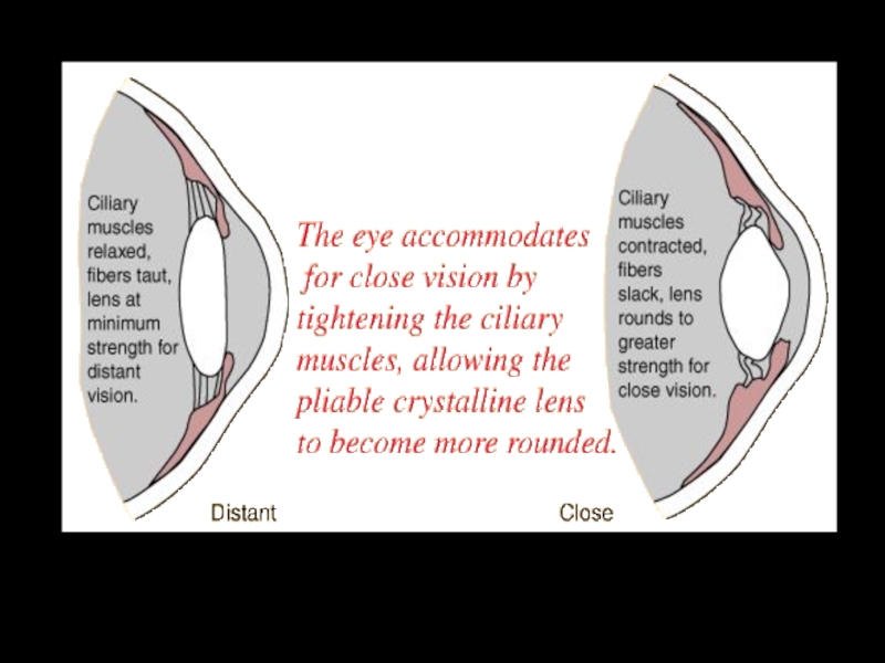
Слайд 7The cornea, clear liquid of front chamber, crystalline lens & vitreous
Eye accommodation

Слайд 9Presbyopia: ciliary muscles can no longer contract as well; lens cannot
Test for Presbyopia
Measurement of ‘near point’:
-measure with ruler and pin
-near point increases with age
-average near points:
-10 yr. old: 7 cm
-40 yr. old: 21 cm
-60 yr. old: 100 cm

")
:objects focused in front of retina")
Слайд 12Length of Eyeball + Curvature of Cornea
Myopia (nearsightedness):
Axial myopia: eyeball too
Refractive myopia: cornea too curved
:Axial myopia: eyeball too long (shown above)Refractive myopia:")
Слайд 13concave lens: negative diopter; diffraction
Myopia Correction
laser surgery: remove corneal tissue in

: objects focused behind retina")
Слайд 15convex lens: positive diopter; refraction
Hyperopia Correction
laser surgery: remove corneal tissue around

Слайд 16Normal sight
Astigmatism
Astigmatism
Aspherical cornea: light at some orientations is focused, while light