- Главная
- Разное
- Дизайн
- Бизнес и предпринимательство
- Аналитика
- Образование
- Развлечения
- Красота и здоровье
- Финансы
- Государство
- Путешествия
- Спорт
- Недвижимость
- Армия
- Графика
- Культурология
- Еда и кулинария
- Лингвистика
- Английский язык
- Астрономия
- Алгебра
- Биология
- География
- Детские презентации
- Информатика
- История
- Литература
- Маркетинг
- Математика
- Медицина
- Менеджмент
- Музыка
- МХК
- Немецкий язык
- ОБЖ
- Обществознание
- Окружающий мир
- Педагогика
- Русский язык
- Технология
- Физика
- Философия
- Химия
- Шаблоны, картинки для презентаций
- Экология
- Экономика
- Юриспруденция
Malignant Lymphomas презентация
Содержание
- 1. Malignant Lymphomas
- 2. Definition Lymphoma means a malignant tumor
- 3. The lymphatic system The lymphatic
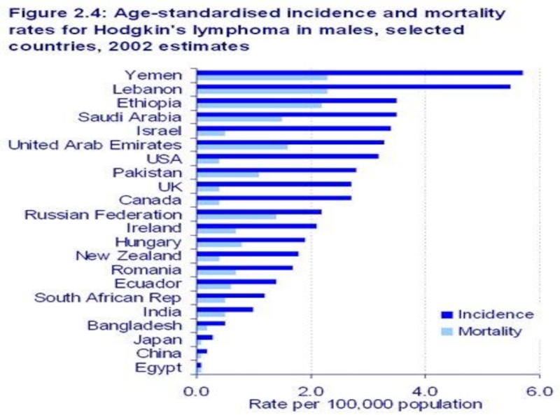
- 4. Epidemiology
- 6. Hodgkin Lymphoma Hodgkin’s disease is one
- 7. Hodgkin's Disease – Reed Stenberg Cell
- 8. Within the latter, four
- 9. Cotswolds staging classification for Hodgkin lymphoma
- 10. Cotswolds staging classification for Hodgkin lymphoma Stage
- 11. All cases are subclassified to indicate
- 12. Hodgkin lymphoma
- 13. Hodgkin lymphoma
- 14. Hodgkin Lymphoma Findings: Subtle soft
- 15. Hodgkin disease Massive involvement of
- 16. Nonenhanced CT scan through
- 17. CT scan Can demonstrate the relationship of
- 18. Comments: 78 y/o man with hepatosplenomegaly.
- 19. Hodgkin Lymphoma This is a liver
- 20. Staging of Hodgkin's lymphoma using FDG-PET
- 22. Positron emission tomography/computed tomography images of Hodgkin’s
- 24. The 301,000 cases of non-Hodgkin Lymphoma (NHL)
- 25. NHLs are slightly more common in developed
- 26. There have been marked increases in the
- 27. REAL: Classification for Non-Hodgkin’s Lymphoma
- 28. BURKITT LYMPHOMA
- 29. . Non-Hodgkin lymphoma. Contrast-enhanced CT shows
- 30. Stenting Through a Portacath for Totally Occluded

Слайд 2Definition
Lymphoma means a malignant tumor of the lymphatic system. There are
two main types of lymphoma
1) Hodgkin's lymphoma
2) Non Hodgkin's lymphoma (NHL)
Most lymphomas are NHL. Only about 1 in 5
are Hodgkin's lymphoma. Hodgkin's disease
was named after the doctor who first
recognised it in 1832 - Thomas Hodgkin. It is
now called Hodgkin's lymphoma.
1) Hodgkin's lymphoma
2) Non Hodgkin's lymphoma (NHL)
Most lymphomas are NHL. Only about 1 in 5
are Hodgkin's lymphoma. Hodgkin's disease
was named after the doctor who first
recognised it in 1832 - Thomas Hodgkin. It is
now called Hodgkin's lymphoma.

Слайд 3The lymphatic system
The lymphatic systemThe lymphatic system is part of
the immune systemThe lymphatic system is part of the immune system, which defends the body against infection. The lymphatic system is a network of small lymph nodesThe lymphatic system is part of the immune system, which defends the body against infection. The lymphatic system is a network of small lymph nodes connected by very thin lymph vessels, which branch into every part of the body except the brain and spinal cord. The major nodes can be found in the neck, armpits, chest, abdomen, pelvis and groin. Other parts of the lymphatic system include the spleenThe lymphatic system is part of the immune system, which defends the body against infection. The lymphatic system is a network of small lymph nodes connected by very thin lymph vessels, which branch into every part of the body except the brain and spinal cord. The major nodes can be found in the neck, armpits, chest, abdomen, pelvis and groin. Other parts of the lymphatic system include the spleen, thymusThe lymphatic system is part of the immune system, which defends the body against infection. The lymphatic system is a network of small lymph nodes connected by very thin lymph vessels, which branch into every part of the body except the brain and spinal cord. The major nodes can be found in the neck, armpits, chest, abdomen, pelvis and groin. Other parts of the lymphatic system include the spleen, thymus and bone marrow.
A clear fluid called lymph flows through the lymph vessels. It contains white blood cells called lymphocytesA clear fluid called lymph flows through the lymph vessels. It contains white blood cells called lymphocytes, special proteins called antibodies, and some waste products. Lymphocytes and antibodies are important parts of your body's immune system.
The lymph fluid passes through the lymph nodes, which filter out bacteria and other harmful things.


Слайд 6Hodgkin Lymphoma
Hodgkin’s disease is one of two common types of
cancers of the lymphatic system. Non-Hodgkin’s lymphoma, the other type, is far more common. Hodgkin’s disease is named after the British physician Thomas Hodgkin, who first described the disease in 1832 and noted characteristics that distinguish it from other lymphomas.
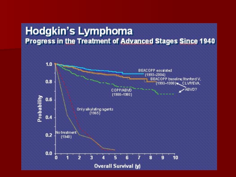
Advances in diagnosis, staging and treatment of Hodgkin’s disease have helped to make this once uniformly fatal disease highly treatable with the potential for full recovery.
Advances in diagnosis, staging and treatment of Hodgkin’s disease have helped to make this once uniformly fatal disease highly treatable with the potential for full recovery.
Sir Thomas Hodgkin (1798-1866)

Слайд 7Hodgkin's Disease –
Reed Stenberg Cell
Presence of typical Reed-Sternberg cell and
reactive component are mandatory for diagnosis of Hodgkin's lymphoma. Characteristics of typical Reed-Sternberg cell : size between 20 - 50 microns; abundant, amphofilic, finely granular/homogenous cytoplasm; two mirror-image nuclei ("owl eyes") each with an eosinophilic nucleolus and a thick nuclear membrane (chromatin is distributed on the inner surface of the nuclear membrane, generating a halo image around the nucleolus). Reed-Sternberg cell has a B-cell origin. (H&E, ob.x40)

Слайд 8 Within the latter, four subtypes have been distinguished:
Hodgkin
Lymphoma
nodular sclerosis,
mixed cellularity,
lymphocyte-rich
lymphocyte-depleted.

Слайд 9
Cotswolds staging classification for Hodgkin lymphoma
Stage I – Involvement of a
single lymph node region (eg, cervical, axillary, inguinal, mediastinal) or lymphoid structure such as the spleen, thymus, or Waldeyer’s ring.
Stage II – Involvement of two or more lymph node regions or lymph node structures on the same side of the diaphragm. Hilar nodes should be considered to be “lateralized” and when involved on both sides, constitute stage II disease. For the purpose of defining the number of anatomic regions, all nodal disease within the mediastinum is considered to be a single lymph node region and hilar involvement constitutes an additional site of involvement. The number of anatomic regions should be indicated by a subscript (eg, II-3).
Stage II – Involvement of two or more lymph node regions or lymph node structures on the same side of the diaphragm. Hilar nodes should be considered to be “lateralized” and when involved on both sides, constitute stage II disease. For the purpose of defining the number of anatomic regions, all nodal disease within the mediastinum is considered to be a single lymph node region and hilar involvement constitutes an additional site of involvement. The number of anatomic regions should be indicated by a subscript (eg, II-3).

Слайд 10Cotswolds staging classification for Hodgkin lymphoma
Stage III – Involvement of lymph
node regions or lymphoid structures on both sides of the diaphragm. This may be subdivided
stage III-1 or III-2: stage III-1 is used for patients with involvement of the spleen or splenic hilar, celiac or portal nodes; and
stage III-2 is used for patients with involvement of the paraaortic, iliac, inguinal, or mesenteric nodes.
Stage IV – Diffuse or disseminated involvement of one or more extranodal organs or tissue beyond that designated E, with or without associated lymph node involvement.
Stage IV – Diffuse or disseminated involvement of one or more extranodal organs or tissue beyond that designated E, with or without associated lymph node involvement.

Слайд 11
All cases are subclassified to indicate the absence (A) or presence
(B) of the systemic symptoms of significant unexplained fever, night sweats, or unexplained weight loss exceeding 10 percent of body weight during the six months prior to diagnosis.
The designation “E” refers to extranodal contiguous extension (ie, proximal or contiguous extranodal disease) that can be encompassed within an irradiation field appropriate for nodal disease of the same anatomic extent. More extensive extranodal disease is designated stage IV.
The designation “E” refers to extranodal contiguous extension (ie, proximal or contiguous extranodal disease) that can be encompassed within an irradiation field appropriate for nodal disease of the same anatomic extent. More extensive extranodal disease is designated stage IV.
 or presence (B) of the systemic")


Слайд 14Hodgkin Lymphoma
Findings:
Subtle soft tissue swelling is present along the
left side of the patient's neck.
The trachea is deviated to the right.
Lung fields are clear.
There is no definite evidence of mediastinal or hilar adenopathy.
Radiographic findings are consistent with a neck mass, subsequently proven to be Hodgkin's Lymphoma.
The trachea is deviated to the right.
Lung fields are clear.
There is no definite evidence of mediastinal or hilar adenopathy.
Radiographic findings are consistent with a neck mass, subsequently proven to be Hodgkin's Lymphoma.

Слайд 15Hodgkin disease
Massive involvement of paratracheal, hilar and subcarinal lymph nodes
as well as two vertebral bodies.

Слайд 16 Nonenhanced CT scan through the mediastinum shows multiple enlarged lymph nodes
in the prevascular space, in the right and left paratracheal region. Nodes in the left paratracheal region cause the trachea to be indented and narrowed on the left side. Note the small, bilateral pleural effusion.

Слайд 17CT scan
Can demonstrate the relationship of the mass to vessels and
other structures.
Can help characterize the lesion.
Can serve as a guide for biopsy.
Can help characterize the lesion.
Can serve as a guide for biopsy.

Слайд 18
Comments:
78 y/o man with hepatosplenomegaly. Splenectomy specimen showed scattered gray-white nodules.
The tumor cells were positive for CD15, CD30, CD20 (partial) and negative for CD45. Spleen is the most common extranodal site of involvement in Hodgkin’s lymphoma. Primary Hodgkin’s lymphoma of spleen is rare.

Слайд 19Hodgkin Lymphoma
This is a liver that is involved with Hodgkin's disease.
The staging of Hodgkin's disease is very important in determining therapy. Thus, it is important to determine whether the patient has only a single lymph node region involved, multiple node regions, or extranodal involvement. This picture could probably suffice for non-Hodgkin's lymphomatous hepatic disease as well.

Слайд 20Staging of Hodgkin's lymphoma using FDG-PET
Maximum intensity projection image of a
staging FDG-PET scan of a 68-year-old man with newly diagnosed Hodgkin's lymphoma. There are markedly FDG-avid lymph nodes in a symmetrical pattern above and below the diaphragm. Note the pathological diffusely increased uptake in the spleen (more than liver uptake). FDG-PET is a more sensitive indicator of diffuse splenic involvement, which is not possible to diagnose in the absence of splenomegaly on CT.
FDG-PET F-18 fluorodeoxyglucose positron emission
FDG-PET F-18 fluorodeoxyglucose positron emission

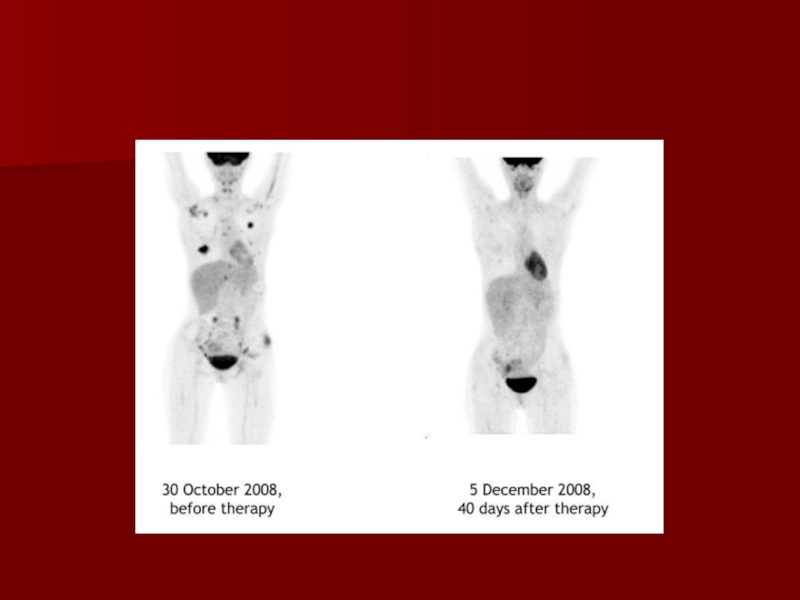
Слайд 22Positron emission tomography/computed tomography images of Hodgkin’s disease staging
Clinical stage
I left axillary disease was upstaged to stage IIIs because of mediastinal nodal and splenic involvement detected on PET/CT.

Слайд 24The 301,000 cases of non-Hodgkin Lymphoma (NHL) that occurred in 2002 (2.8%
of
all cancers) comprise an extremely heteroge-neous group of malignancies displaying distinct
behavioral, prognostic, and epidemiological
characteristics. Advances in molecular biology,
genetics, and immunology have resulted in extensive
changes in the classification of lymphoid
tumors in the last few decades. The
WHO classification74 distinguishes tumors primarily
by cell lineage defined by immunophenotype
and groups together lymphomas and
leukemias, acknowledging that some solid tumors
also pass through circulating leukemic
phases. Three broad categories are now recognized:
B-cell neoplasms, T/NK-cell neoplasms, and Hodgkin disease. Lymphocytic leukemias fall
within the B-cell neoplasm group.
 that occurred in 2002 (2.8% of all cancers)")
Слайд 25NHLs are slightly more common in developed countries (50.5% of cases worldwide),
with rates
highest in Australia and North America, intermediate
in Europe (except eastern Europe) and the
Pacific islands, and relatively low throughout Asia
and eastern Europe. In most African
populations, incidence of NHL is not high overall,
but the relative frequency is above the world
average in sub-Saharan Africa because of the high
incidence of Burkitt lymphoma in children in the
tropical zone of Africa. The relatively high esti-mated incidence in females in central Africa is a consequence of high relative
frequency of such cancers in the few available
datasets from this area.
, with rates highest")
Слайд 26There have been marked increases in the incidence of NHL in many
parts of the world.
While this may in part be due to improved diagnostic
procedures and changes in classification,
there can be little doubt that much of the change
is real and the reasons for it have been the
subject of much debate. The increase is seen in
both sexes across Europe since the 1960s. Increases
of about 1% to 2% per year in incidence
rates in both sexes by period of diagnosis are seen in Australia and, at a lower level, in South America
and Asia. In the United States, the rapid rises
(particularly in younger men) may be partially
attributable to the onset of the AIDS epidemic in
1981, while the declines during the 1990s may be
due in part to a decrease in the incidence of HIV
infection and successful antiretroviral therapies.


Слайд 29.
Non-Hodgkin lymphoma. Contrast-enhanced CT shows multiple low-attenuation nodules replacing most
of them splenic parenchya. Also note renal involvement (arrow) by lymphoma

Слайд 30Stenting Through a Portacath for Totally Occluded Superior Vena Cava in
a Case of Non-Hodgkin’s Lymphoma