- Главная
- Разное
- Дизайн
- Бизнес и предпринимательство
- Аналитика
- Образование
- Развлечения
- Красота и здоровье
- Финансы
- Государство
- Путешествия
- Спорт
- Недвижимость
- Армия
- Графика
- Культурология
- Еда и кулинария
- Лингвистика
- Английский язык
- Астрономия
- Алгебра
- Биология
- География
- Детские презентации
- Информатика
- История
- Литература
- Маркетинг
- Математика
- Медицина
- Менеджмент
- Музыка
- МХК
- Немецкий язык
- ОБЖ
- Обществознание
- Окружающий мир
- Педагогика
- Русский язык
- Технология
- Физика
- Философия
- Химия
- Шаблоны, картинки для презентаций
- Экология
- Экономика
- Юриспруденция
Malaria презентация
Содержание
- 1. Malaria
- 2. MALARIA Infection with pathogenic protozoa exacts an
- 3. Four species are encountered in human disease:
- 4. Parasitology The female mosquito becomes infected after
- 5. Parasitology Here they multiply inside hepatocytes as
- 6. Parasitology Inside the red cells the parasites
- 7. Parasitology A few merozoites develop not into
- 8. Pathogenesis The pathology of malaria is related
- 9. Pathogenesis The female anopheline mosquito becomes infected
- 10. Pathogenesis P. vivax and P. ovale may
- 11. Pathogenesis P. falciparum and P. malariae have
- 12. Effect on red blood cells and capillaries
- 13. Pathogenesis Haemolysis is most severe with P.
- 14. Pathogenesis In P. falciparum malaria, red cells
- 15. Pathogenesis After repeated infections partial immunity develops,
- 16. Pathogenesis Certain genetic traits also confer some
- 17. Clinical features Typical malaria is seen in
- 18. P. vivax and P. ovale malaria In
- 19. P. malariae infection This usually associated with
- 20. P. falciparum infection These are more dangerous
- 21. P. falciparum infection A patient with falciparum
- 22. Complications of malaria due to P. falciparum:
- 23. Complications of malaria due to P. falciparum:
- 24. Clinical features In hyperendemic and in holoendemic
- 25. Diagnosis Malaria should be considered in the
- 26. Laboratory diagnosis To establish the diagnosis, a
- 27. P. vivax
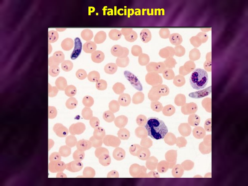
- 28. P. falciparum
- 29. Treatment For many years the standard treatment
- 30. Treatment Alternative agents include mefloquine and halofantrine.
- 31. Treatment Treatment of acute malaria with chloroquine,
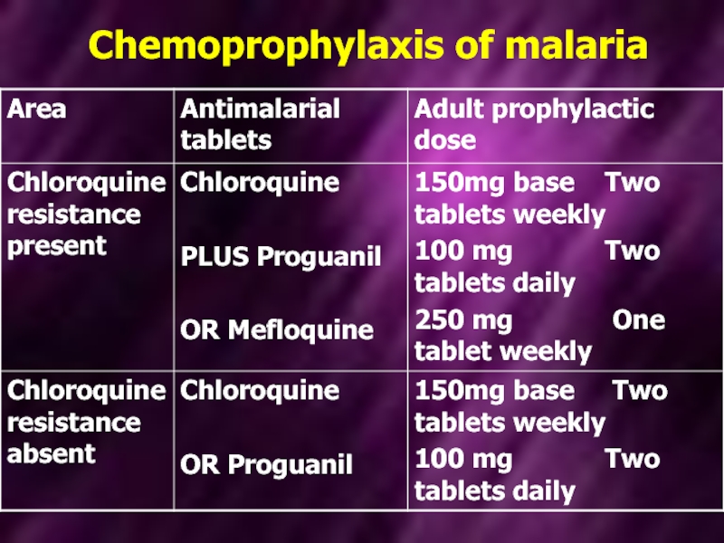
- 32. Prevention Clinical attacks of malaria may be
- 33. Chemoprophylaxis of malaria
- 34. Chemoprophylaxis of malaria Chemoprophylaxis is begun 1
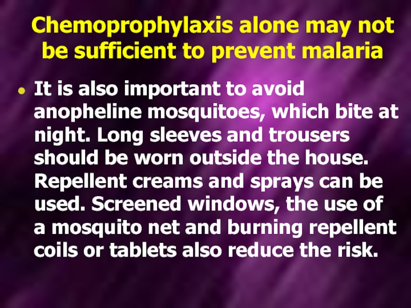
- 35. Chemoprophylaxis alone may not be sufficient to
Слайд 2MALARIA
Infection with pathogenic protozoa exacts an enormous toll of human suffering,
notably, but not exclusively, in the tropics. Numerically the most important of the life-threatening protozoan diseases is malaria. Public health measures and changes in land use have eradicated malaria in most developed countries, although the potential for malaria transmission still exists in many areas. Three hundred million people are infected every year, and over one million die.
Слайд 3Four species are encountered in human disease:
P. vivax and P.
ovale, both of which cause bening tertian malaria (febrile episodes typically occurring at 48-h intervals);
Plasmodium falciparum, which is responsible for most fatalities;
P. malariae, which causes quartan malaria (febrile episodes typically occurring at 72-h intervals);
Слайд 4Parasitology
The female mosquito becomes infected after taking a blood meal containing
gametocytes, the sexual form of the malaria parasite. The developmental cycle in the mosquito usually takes 7-20 days (depending on temperature), culminative sporozoites migrating to the insect’s salivary glands. The sporozoites are inoculated into a new human host, and which are not destroyed by the immune response are rapidly taken up by the liver.
Слайд 5Parasitology
Here they multiply inside hepatocytes as merozoites: this is pre-erythrocytic (or
hepatic) sporogeny. After a few days the infected hepatocytes rupture, releasing merozoites into the blood from where they are rapidly taken up by erythrocytes. In the case of P. vivax and P. ovale, a few parasites remain dormant in the liver as hypnozoites. These may reactivate at any time subsequently, causing relapsing infection.
Слайд 6Parasitology
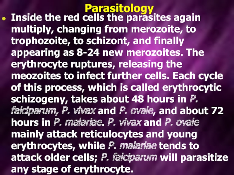
Inside the red cells the parasites again multiply, changing from merozoite,
to trophozoite, to schizont, and finally appearing as 8-24 new merozoites. The erythrocyte ruptures, releasing the meozoites to infect further cells. Each cycle of this process, which is called erythrocytic schizogeny, takes about 48 hours in P. falciparum, P. vivax and P. ovale, and about 72 hours in P. malariae. P. vivax and P. ovale mainly attack reticulocytes and young erythrocytes, while P. malariae tends to attack older cells; P. falciparum will parasitize any stage of erythrocyte.
Слайд 7Parasitology
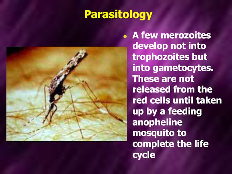
A few merozoites develop not into trophozoites but into gametocytes. These
are not released from the red cells until taken up by a feeding anopheline mosquito to complete the life cycle
Слайд 8Pathogenesis
The pathology of malaria is related to:
anaemia,
cytokine release,
in the
case of P. falciparum, widespread organ damage due to impaired microcirculation.
Слайд 9Pathogenesis
The female anopheline mosquito becomes infected when it feeds on human
blood containing gametocytes, the sexual forms of the malarial parasite. The development in the mosquito takes from 7-20days. Sporozoites inoculated by an infected mosquito disappear from human blood within half an hour and enter the liver. After some days merozoites leave the liver and invade red blood cells, where further asexual cycles of multiplication take place, producing schizonts. Rupture of the schizont releases more merozoites into the blood and causes fever, whose periodicity depends on the species of parasite.
Слайд 10Pathogenesis
P. vivax and P. ovale may persist in liver cells as
dormant forms, hypozoites, capable of developeing into merozoites months or years later. Thus the first attack of clinical malaria may occur long after the patient has left the endemic area, and the disease may relapse after treatment with drugs that kill only the erythrocytic stage of the parasite.
Слайд 11Pathogenesis
P. falciparum and P. malariae have no persistent exoerythrocytic phase but
recrudescences of fever may result from multiplication in the red cells of parasites which have not been eliminated by treatment and immune processes.
Слайд 12Effect on red blood cells and capillaries
Malaria is always accompanied by
haemolysis and in a severe or prolonged attack anaemia may be profound.
The anaemia in malaria is multifactorial:
Haemolysis of infected red cells;
Haemolysis of non-infected red cells;
Dyserythropoiesis;
Splenomegaly causing erythrocyte sequestration and haemodilution;
Deplation of folate stores.
The anaemia in malaria is multifactorial:
Haemolysis of infected red cells;
Haemolysis of non-infected red cells;
Dyserythropoiesis;
Splenomegaly causing erythrocyte sequestration and haemodilution;
Deplation of folate stores.
Слайд 13Pathogenesis
Haemolysis is most severe with P. falciparum, which invades red cells
of all ages but especially young cells. P. vivax and P. ovale invade reticulocytes, and P. malariae normoblasts, so that infections remain lighter.
Слайд 14Pathogenesis
In P. falciparum malaria, red cells containing schizonts adhere to the
lining of capillaries in brain, kidney, liver, lungs. The vessels become congested and the organs anoxic. Rupture of schizonts liberates toxic and antigenic substances which may cause further damage. Thus the main effects of malaria are haemolytic anaemia and, with P. falciparum, widespread organ damage.
Слайд 15Pathogenesis
After repeated infections partial immunity develops, allowing the host to tolerate
parasitaemia with minimal ill effects. This immunity is lost if there is no further infection for a couple of years
Слайд 16Pathogenesis
Certain genetic traits also confer some immunity to malaria. People who
lack the Duffy antigen on the red cell membrane (a common finding in West Africa) are not susceptible to infection with P. vivax.
Certain haemoglobinopathies (including sickle cell trait) also give some protection against the severe effects of malaria: this may account for the persistence of these otherwise harmful mutations in tropical countries.
Certain haemoglobinopathies (including sickle cell trait) also give some protection against the severe effects of malaria: this may account for the persistence of these otherwise harmful mutations in tropical countries.
Слайд 17Clinical features
Typical malaria is seen in non-immune individuals. This includes children
in any area, adults in hypoendemic areas, any visitors from a non-malarious region.
The normal incubation period is 10-21 days. The incubation period may be longer than the pre-erythrocytic cycle and may be up to several month for P. vivax and P. ovale.
The normal incubation period is 10-21 days. The incubation period may be longer than the pre-erythrocytic cycle and may be up to several month for P. vivax and P. ovale.
Слайд 18P. vivax and P. ovale malaria
In many cases the illness starts
with a period of several days of continued fever before the development of classical bouts fever on alternate days. Fever starts with a chill. The patient feels cold and the temperature rises to about 40°C. After half an hour to an hour the hot or flush phase begins. It lasts several hours and gives way to profuse perspiration and gradual fall in temperature. The cycle is repeated 48 hours later. Gradually the spleen and liver enlarge and may become tender. Anaemia develops slowly. Relapses are common in the first 2 years of leaving the malarious area.
Слайд 19P. malariae infection
This usually associated with mild symptoms and bouts of
fever every third day. The cycle is repeated 72 hours later. Parasitaemia may persist for many years with the occasional recrudescence of fever, or without producing any symptoms. P. malariae causes glomerulonephritis and the nephritic syndrome in children.
Слайд 20P. falciparum infection
These are more dangerous than other forms of malaria.
The onset, especially of primary attacks, is often insidious, with malaise, headache and vomiting. Cough and mild diarrhea are common, suggesting influenza. The fever has no particular pattern and does not usually rise quite so high as in the other forms. The cold, hot and sweating stages are seldom found. Jaundice is common due to haemolysis and hepatic dysfunction. The liver and spleen enlarge. Anaemia develops rapidly.
Слайд 21P. falciparum infection
A patient with falciparum malaria, apparently not seriously ill,
may develop serious complications. Children die rapidly without any special symptoms other than fever. Immunity is impaired in pregnancy, and abortion from parasitisation of the maternal side of the placenta is frequent. Splenectomy increases the risk of severe malaria.
Слайд 22Complications of malaria due to P. falciparum:
I. Severe anaemia.
II. Organ damage due to anoxia:
Brain: confusion, coma. This is the most urgent complication and is manifested either by confusion or coma, usually without localizing signs;
Kidneys: oliguria, uraemia (acute tubular necrosis);
Lungs: cough, pulmonary edema;
Intestine: diarrhea;
Liver: jaundice, encephalopathy.
Слайд 23Complications of malaria due to P. falciparum:
III. Intravascular haemolysis. Blackwater fever
is associated with chronic falciparum malaria, most commonly in those who have taken antimalarial treatment irregularly, or are deficient in glucose-6-phosphate dehydrogenase. Haemolysis is unpredictable and severe, destroying uninfected as well as parasitized red cells. The urine is dark or black.
IV. Hypotensive shock.
V. Splenic rupture.
VI. In pregnancy: maternal death, abortion.
IV. Hypotensive shock.
V. Splenic rupture.
VI. In pregnancy: maternal death, abortion.
Слайд 24Clinical features
In hyperendemic and in holoendemic areas malaria may kill up
to 15-20 % of children below the age of 5 years. Pregnancy lowers resistance to malaria. The risks are greatest in the first pregnancy.
Слайд 25Diagnosis
Malaria should be considered in the differential diagnosis of anyone who
presents with a febrile illness in, or having recently left, a malarious area. Falciparum malaria is unlikely to present more than 3 months after exposure, even if the patient has been taking prophylaxis, but vivax malaria may cause symptoms for the first time up to a year after leaving a malarious area.
Слайд 26Laboratory diagnosis
To establish the diagnosis, a drop of peripheral blood is
spread on a glass slide. The smear should not be too thick; a useful criteration is that print should be just visible through it. The smear is allowed to dry thoroughly and stained by Field’s method. This is an aqueous Romanowsky stain, which stains the parasites very rapidly, and haemolyses the red cells, so that the parasites are easy to detect despite the thickness of the film.
Visualization of the parasites in Romanowsky-stained peripheral blood films remains the most reliable method.
Visualization of the parasites in Romanowsky-stained peripheral blood films remains the most reliable method.
Слайд 29Treatment
For many years the standard treatment for acute malaria was chloroquine.
Howewer, resistance to that drug in P. falciparum (and less commonly, in P. vivax) is now widespread and alternative agents often have to be used. The most reliable alternative to chloroquine is quinine (or quinidine). Some antibiotics, including tetracyclines and clindamicin, exhibit anti-malarial activity and are used as an adjunct to quinine therapy.
Слайд 30Treatment
Alternative agents include mefloquine and halofantrine. These drugs are active against
chloroquine-resistant strains, but resistance to them is increasing in prevalence, and both are associated with occasional problems of toxicity.
Слайд 31Treatment
Treatment of acute malaria with chloroquine, quinine or other antimalarials will
not eliminate parasites in the liver. For this purpose the 8-aminoquinoline drug primaquine must be used.
Слайд 32Prevention
Clinical attacks of malaria may be preventable by drugs such as
proguanil which attack the pre-erythrocytic form, or by drugs such as chloroquine or mefloquine after it has entered the erythrocyte. The recommended doses for protection of the non-immune are the next.
Слайд 34Chemoprophylaxis of malaria
Chemoprophylaxis is begun 1 week before entering the malarious
area and is continued until 4 weeks after leaving it.
Слайд 35Chemoprophylaxis alone may not be sufficient to prevent malaria
It is also
important to avoid anopheline mosquitoes, which bite at night. Long sleeves and trousers should be worn outside the house. Repellent creams and sprays can be used. Screened windows, the use of a mosquito net and burning repellent coils or tablets also reduce the risk.
