Lecture on pathological anatomy
for the 3-rd year students
- Главная
- Разное
- Дизайн
- Бизнес и предпринимательство
- Аналитика
- Образование
- Развлечения
- Красота и здоровье
- Финансы
- Государство
- Путешествия
- Спорт
- Недвижимость
- Армия
- Графика
- Культурология
- Еда и кулинария
- Лингвистика
- Английский язык
- Астрономия
- Алгебра
- Биология
- География
- Детские презентации
- Информатика
- История
- Литература
- Маркетинг
- Математика
- Медицина
- Менеджмент
- Музыка
- МХК
- Немецкий язык
- ОБЖ
- Обществознание
- Окружающий мир
- Педагогика
- Русский язык
- Технология
- Физика
- Философия
- Химия
- Шаблоны, картинки для презентаций
- Экология
- Экономика
- Юриспруденция
Liver diseases презентация
Содержание
- 1. Liver diseases
- 2. The morphologic alterations that cause liver failure
- 3. Hepatosis It is metabolic liver dysfunction, that
- 4. Massive hepatic necrosis The mechanism is direct
- 5. Hepatitis This term is used
- 6. Hepatotropic viruses infect primarily hepatocytes. The known
- 7. HBV and HCV are mostly
- 8. CLINICAL SYMPTOMS 1. Asymptomatic infection (serologic evidence
- 9. Acute Viral Hepatitis Sporadic
- 10. 1. Liver is enlarged, reddened liver; greenish
- 11. 4. Regenerative changes: hepatocyte proliferation 5. Sinusoidal
- 12. Fulminant Viral Hepatitis Fulminant hepatitis
- 13. Microscopically: - the coagulative-liquefactive necrosis of cells;
- 14. Chronic Hepatitis Chronic hepatitis
- 15. CPH is characterized clinically by: -
- 16. In contrast, in chronic
- 17. Chronic Active Hepatitis In contrast
- 18. Active destruction of hepatocytes at the interface
- 19. AUTOIMMUNE CHRONIC HEPATITIS Chronic hepatitis unrelated
- 20. Liver cirrhosis Cirrhosis is a result of
- 21. According to cause: Alcoholic liver disease (ALD)
- 22. According to morphology: Micronodular (less then 3
- 23. CLINICAL SYMPTOMS Cirrhosis has many possible manifestations.
- 24. Spider angiomata or spider nevi are vascular
- 25. Hypogonadism, a decrease in sex hormones manifest
- 26. Portal hypertension Liver
- 27. Outcomes Liver coma Bleeding from varicose-enlarged veins Ascites-peritonitis Portal hypertension Portal-vein thrombosis Liver carcinoma
Слайд 1Liver Diseases
ZAPORIZHZHIAN STATE MEDICAL UNIVERSITY
The department of pathological anatomy and forensic
medicine
Слайд 2The morphologic alterations that cause liver failure fall into 4 categories:
Hepatic
dysfunction without overt necrosis (Hepatosis) - Hepatocytes may be viable but unable to perform normal metabolic function, as with Reye syndrome, tetracycline toxicity, and acute fatty liver of pregnancy
Chronic liver disease - this is the most common route to hepatic failure and is the endpoint of relentless chronic hepatitis ending in cirrhosis.
Massive hepatic necrosis
Cirrhosis
Chronic liver disease - this is the most common route to hepatic failure and is the endpoint of relentless chronic hepatitis ending in cirrhosis.
Massive hepatic necrosis
Cirrhosis
Слайд 3Hepatosis
It is metabolic liver dysfunction, that is characterized
by dystrophic changers of
hepatocytes without
inflammation and disorders of bile excresion.
Types: - holestatic (stasis of bile)
- pigmental
- fatty
Morphology:
- hepatomegaly
- in liver cells: bile, pigmental or fatty depositions
- enlargement of spleen
Outcomes: 1) total restitution; 2) cirrhosis, 3) acute liver insufficiency; 4) liver cancer
Слайд 4Massive hepatic necrosis
The mechanism is direct toxic damage to hepatocytes or
a
variable combination of toxicity and inflammation with
immune-mediated hepatocyte destruction.
Reasons of development :
- drug- or toxin-induced (antituberculosis drugs, antidepressants),
- industrial chemicals such as carbon tetrachloride,
- mushroom poisoning,
- hepatitis
Morphological stages:
1. Yellow dystrophy – (during 2 weeks after poisoning) centrolobular necrosis, liver is yellow-brownish, flabby.
2. Red dystrophy – total destruction of hepatocytes, liver is small and red.
Outcomes: post-necrotic cirrhosis
Слайд 5Hepatitis
This term is used to describe the presence of
inflammation in the liver and to describe a syndrome in which the patient demonstrates laboratory evidence of liver cell necrosis, often immediately preceded or accompanied by malaise, fever, and jaundice.
Hepatitis may be caused by:
- a large number of infectious organisms (hepatotropic viruses & other infectious agents - e.g., tuberculosis, infectious mononucleosis, and cytomegalovirus infection),
- therapeutic agents,
- toxins,
- alcohol,
-autoimmune reactions.
Hepatitis may be caused by:
- a large number of infectious organisms (hepatotropic viruses & other infectious agents - e.g., tuberculosis, infectious mononucleosis, and cytomegalovirus infection),
- therapeutic agents,
- toxins,
- alcohol,
-autoimmune reactions.
Слайд 6Hepatotropic viruses infect primarily hepatocytes. The known hepatotropic viruses include:
hepatitis A
virus (HAV),
hepatitis В virus (HBV),
the hepatitis В-associated delta virus (D virus - HDV),
hepatitis С virus,
and hepatitis E virus;
The last two were formerly referred to as non-A, non-B hepatitis viruses
hepatitis В virus (HBV),
the hepatitis В-associated delta virus (D virus - HDV),
hepatitis С virus,
and hepatitis E virus;
The last two were formerly referred to as non-A, non-B hepatitis viruses
Слайд 7 HBV and HCV are mostly often transmitted by the
parenteral route:
- blood transfusions (high risk of transmission with hemodialysis and transplantation);
- infusions of plasma, fibrinogen, or other blood fractions;
- contaminated hypodermic needles (a particular problem among IV drug addicts);
- dental and surgical instruments;
razors.
Transmission may also occur accidentally to health care personnel. Spread by close personal contact may account for the so-called vertical transmission from mothers who have HBV infection to their infants during the perinatal period.
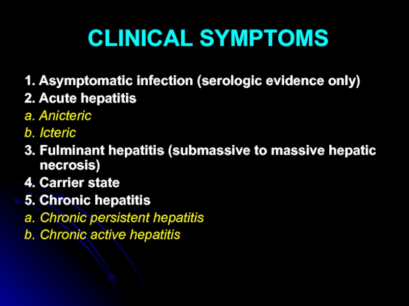
Слайд 8CLINICAL SYMPTOMS
1. Asymptomatic infection (serologic evidence only)
2. Acute hepatitis
a. Anicteric
b. Icteric
3.
Fulminant hepatitis (submassive to massive hepatic necrosis)
4. Carrier state
5. Chronic hepatitis
a. Chronic persistent hepatitis
b. Chronic active hepatitis
4. Carrier state
5. Chronic hepatitis
a. Chronic persistent hepatitis
b. Chronic active hepatitis
Слайд 9Acute Viral Hepatitis
Sporadic attacks of acute hepatitis caused
by all of
the hepatotropic viruses are clinically indistinguishable but most often caused by HAV. When caused by HBV or HCV sporadic cases can be related to these agents only by virologic or serologic criteria.
Acute hepatitis can be divided into four stages:
1) an incubation period,
2) a preicteric stage,
3) an icteric stage,
4) convalescence.
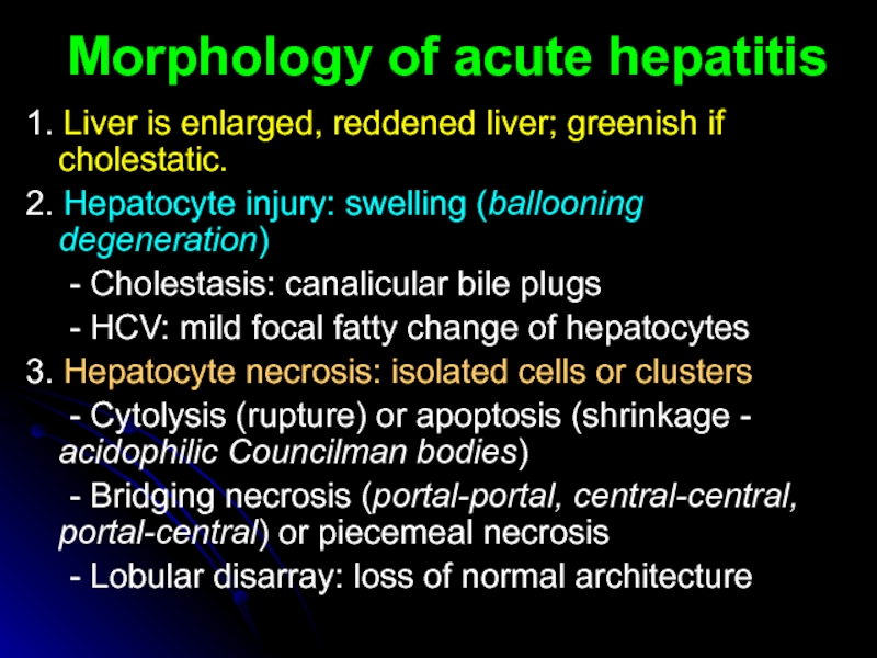
Слайд 101. Liver is enlarged, reddened liver; greenish if cholestatic.
2. Hepatocyte
injury: swelling (ballooning degeneration)
- Cholestasis: canalicular bile plugs
- HCV: mild focal fatty change of hepatocytes
3. Hepatocyte necrosis: isolated cells or clusters
- Cytolysis (rupture) or apoptosis (shrinkage - acidophilic Councilman bodies)
- Bridging necrosis (portal-portal, central-central, portal-central) or piecemeal necrosis
- Lobular disarray: loss of normal architecture
Morphology of acute hepatitis
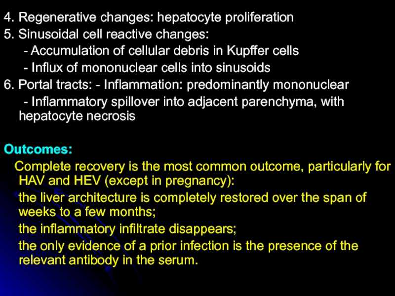
Слайд 114. Regenerative changes: hepatocyte proliferation
5. Sinusoidal cell reactive changes:
- Accumulation of
cellular debris in Kupffer cells
- Influx of mononuclear cells into sinusoids
6. Portal tracts: - Inflammation: predominantly mononuclear
- Inflammatory spillover into adjacent parenchyma, with hepatocyte necrosis
Outcomes:
Complete recovery is the most common outcome, particularly for HAV and HEV (except in pregnancy):
the liver architecture is completely restored over the span of weeks to a few months;
the inflammatory infiltrate disappears;
the only evidence of a prior infection is the presence of the relevant antibody in the serum.
Слайд 12Fulminant Viral Hepatitis
Fulminant hepatitis with submassive to massive necrosis
of the liver is, fortunately, one of the infrequent expressions of viral hepatitis.
MORPHOLOGY
The anatomic changes in the liver depend on the severity of the necrotizing process and duration of survival of the patient. The extent of destruction is extremely variable, as the term "submassive to massive" implies.
Early the liver is normal in size,
Later, as the necrotic areas are resorbed, it is transformed into a shrunken, red-green limp organ with a wrinkled capsule that may weigh as little as 500 gm.
With submassive necrosis and survival, regeneration of hepatocytes may produce irregular, firm nodules that bulge above the surrounding surface and may be stained yellow-brown to green, depending on the degree of cholestasis.
Слайд 13Microscopically:
- the coagulative-liquefactive necrosis of cells;
- entire lobules are destroyed, with
preservation of bile ducts and collapse of the reticulin framework;
- little inflammatory infiltrate accompanies this extensive destruction;
- if the patient survives, irregular nodules of regeneration appear;
- little residual scarring;
if necrosis has been extensive, pseudolobules of functional hepatocytes separated by bands of fibrous tissue may be formed, obviously, massive necrosis and early death preclude scarring.
Fulminant hepatitis carries a poor prognosis. The prognosis is best for those with HAV, better in HBV/HDV than in HBV, and poorest with HCV. Fortunately, with the increasing availability of liver transplantation some of these unhappy victims may be rescued.
Слайд 14Chronic Hepatitis
Chronic hepatitis is defined as the continuation
of hepatic inflammation and necrosis for longer than six months. It presents:
- with evidence of persistent biochemical and symptomatic abnormalities following an episode of acute hepatitis
- the milder forms are detected only during routine laboratory testing.
Traditionally two histologic forms of chronic hepatitis have been distinguished:
chronic persistent hepatitis (CPH), in which there is minimal necrosis and the course is usually benign,
chronic active hepatitis (САН), which is characterized by progressive liver destruction often leading to cirrhosis, chronic liver failure, and death.
Слайд 15CPH is characterized clinically by:
- persistence of elevated levels of
liver aminotransferase enzymes (AST and ALT) and alkaline phosphatase;
- usually there are no symptoms, but some patients may experience relapsing and remitting episodes of malaise, loss of appetite, nausea, and sometimes mild jaundice.
Two morphologic patterns exist:
In one, tabular architecture is nearly normal but there is a dense portal infiltrate consisting of lymphocytes admixed with plasma cells and macrophages, with no necrosis of hepatocytes. This pattern is most typical of CPH ("usual" CPH).
Chronic Persistent Hepatitis
Слайд 16 In contrast, in chronic "lobular" hepatitis: the portal
tracts are only mildly inflamed and random hepatocytes within the lobules demonstrate the same degenerative and necrotic changes that occur in acute hepatitis.
Important features of both the usual form of CPH and chronic lobular hepatitis are that the limiting plate (the delimitation between portal connective tissue and periportal hepatocytes) is preserved; unlike full-blown САН, piecemeal necrosis and fibrosis are minimal or nonexistent.
Important features of both the usual form of CPH and chronic lobular hepatitis are that the limiting plate (the delimitation between portal connective tissue and periportal hepatocytes) is preserved; unlike full-blown САН, piecemeal necrosis and fibrosis are minimal or nonexistent.
Слайд 17Chronic Active Hepatitis
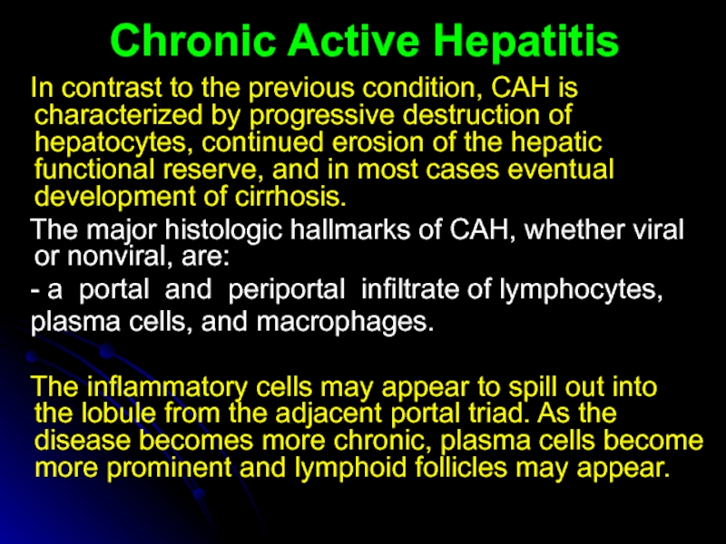
In contrast to the previous condition, САН
is characterized by progressive destruction of hepatocytes, continued erosion of the hepatic functional reserve, and in most cases eventual development of cirrhosis.
The major histologic hallmarks of САН, whether viral or nonviral, are:
- a portal and periportal infiltrate of lymphocytes,
plasma cells, and macrophages.
The inflammatory cells may appear to spill out into the lobule from the adjacent portal triad. As the disease becomes more chronic, plasma cells become more prominent and lymphoid follicles may appear.
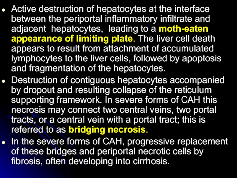
Слайд 18Active destruction of hepatocytes at the interface between the periportal inflammatory
infiltrate and adjacent hepatocytes, leading to a moth-eaten appearance of limiting plate. The liver cell death appears to result from attachment of accumulated lymphocytes to the liver cells, followed by apoptosis and fragmentation of the hepatocytes.
Destruction of contiguous hepatocytes accompanied by dropout and resulting collapse of the reticulum supporting framework. In severe forms of САН this necrosis may connect two central veins, two portal tracts, or a central vein with a portal tract; this is referred to as bridging necrosis.
In the severe forms of САН, progressive replacement of these bridges and periportal necrotic cells by fibrosis, often developing into cirrhosis.
Слайд 19AUTOIMMUNE CHRONIC HEPATITIS
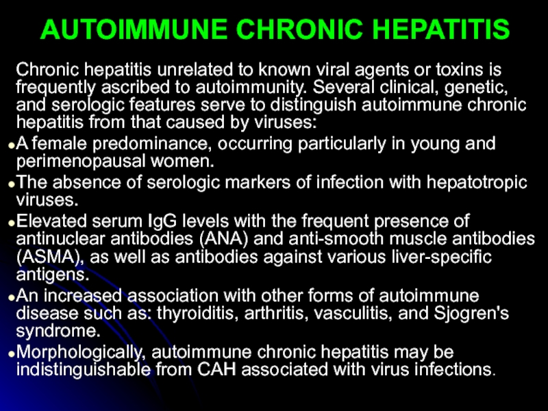
Chronic hepatitis unrelated to known viral agents or
toxins is frequently ascribed to autoimmunity. Several clinical, genetic, and serologic features serve to distinguish autoimmune chronic hepatitis from that caused by viruses:
A female predominance, occurring particularly in young and perimenopausal women.
The absence of serologic markers of infection with hepatotropic viruses.
Elevated serum IgG levels with the frequent presence of antinuclear antibodies (ANA) and anti-smooth muscle antibodies (ASMA), as well as antibodies against various liver-specific antigens.
An increased association with other forms of autoimmune disease such as: thyroiditis, arthritis, vasculitis, and Sjogren's syndrome.
Morphologically, autoimmune chronic hepatitis may be indistinguishable from CAH associated with virus infections.
Слайд 20Liver cirrhosis
Cirrhosis is a result of advanced liver disease. It is
characterized by replacement of liver tissue by fibrosis (scar tissue) and regenerative nodules (lumps that occur due to attempted repair of damaged tissue). These changes lead to loss of liver function and development of portal hypertension and liver failure
Слайд 21According to cause:
Alcoholic liver disease (ALD) Alcoholic cirrhosis,
Non-alcoholic
steatohepatitis (NASH),
Viral hepatitis (B, C),
Primary biliary cirrhosis,
Primary sclerosing cholangitis,
Autoimmune hepatitis,
Hereditary hemochromatosis,
Wilson's disease,
Cardiac cirrhosis,
Hepatotoxic drugs or toxins,
Cryptogenic
Viral hepatitis (B, C),
Primary biliary cirrhosis,
Primary sclerosing cholangitis,
Autoimmune hepatitis,
Hereditary hemochromatosis,
Wilson's disease,
Cardiac cirrhosis,
Hepatotoxic drugs or toxins,
Cryptogenic
Classification of liver cirrhosis
Слайд 22According to morphology:
Micronodular (less then 3 mm),
Macronodular (more then 3 mm),
Mixed,
Septal
According
to morphogenesis:
Post-necrotic,
Portal,
Mixed,
Biliary
Post-necrotic,
Portal,
Mixed,
Biliary
Слайд 23CLINICAL SYMPTOMS
Cirrhosis has many possible manifestations. These signs and symptoms may
be either as a direct result of the failure of liver cells or secondary to the resultant portal hypertension. There are also some manifestations which causes are nonspecific but may occur in cirrhosis. Likewise, the absence of any does not rule out the possibility of cirrhosis.
Слайд 24Spider angiomata or spider nevi are vascular lesions consisting of a
central arteriole surrounded by many smaller vessels (hence the name "spider"), occur due to an increase in estradiol.
Palmar erythema is a reddening of palms at the thenar and hypothenar eminences also as a result of increased estrogen
Gynecomastia, or increase in breast gland size in men that is not cancerous, is caused by increased estradiol and can occur in up to 2/3 of patients.
Palmar erythema is a reddening of palms at the thenar and hypothenar eminences also as a result of increased estrogen
Gynecomastia, or increase in breast gland size in men that is not cancerous, is caused by increased estradiol and can occur in up to 2/3 of patients.
Liver dysfunction
Слайд 25Hypogonadism, a decrease in sex hormones manifest as impotence, infertility, loss
of sexual drive, and testicular atrophy, can result from primary gonadal injury or suppression of hypothalamic/pituitary function. Hypogonadism is associated with cirrhosis due to alcoholism and hemochromatosis.
Liver size can be enlarged, normal, or shrunken in patients with cirrhosis.
Ascites, accumulation of fluid in the peritoneal cavity (space in the abdomen), gives rise to flank dullness (needs about 1500 ml to detect flank dullness).
Jaundice is yellow discoloration of the skin and mucous membranes (with the eye being especially noticeable) due to increased bilirubin (at least 2–3 mg/dL or 30 mmol/L).
Слайд 26Portal hypertension
Liver cirrhosis increases resistance to blood flow and
higher pressure in the portal venous system, resulting in portal hypertension. Effects of portal hypertension include:
Splenomegaly (increase in size of the spleen) is found in 35% to 50% of patients.
Esophageal varices result from collateral portal blood flow through vessels in the stomach and esophagus (a process called Portacaval anastomosis). When these blood vessels become enlarged, they are called varices and are more likely to burst.
Caput medusa are dilated periumbilical collateral veins due to portal hypertension. Blood from the portal venous system may be shunted through the periumbilical veins and ultimately to the abdominal wall veins, manifesting as a pattern that may resemble the head of Medusa.
