- Главная
- Разное
- Дизайн
- Бизнес и предпринимательство
- Аналитика
- Образование
- Развлечения
- Красота и здоровье
- Финансы
- Государство
- Путешествия
- Спорт
- Недвижимость
- Армия
- Графика
- Культурология
- Еда и кулинария
- Лингвистика
- Английский язык
- Астрономия
- Алгебра
- Биология
- География
- Детские презентации
- Информатика
- История
- Литература
- Маркетинг
- Математика
- Медицина
- Менеджмент
- Музыка
- МХК
- Немецкий язык
- ОБЖ
- Обществознание
- Окружающий мир
- Педагогика
- Русский язык
- Технология
- Физика
- Философия
- Химия
- Шаблоны, картинки для презентаций
- Экология
- Экономика
- Юриспруденция
Лимфопролиферативные заболевания презентация
Содержание
- 1. Лимфопролиферативные заболевания
- 2. Patients with hematological malignancies in Belarus (adults) (2007).
- 3. Limphoproliferative diseases
- 4. B-cell lymphopoiesis
- 5. B-cell malignan-cies
- 6. T-cell differen-tiation stages
- 7. Lymphopoiesis in lymph nodes.
- 8. B-cell malignancies
- 9. Morphology of leukocytes
- 10. Acute leukemia. Originated from bone marrow (>25%
- 11. Acute leukemia (WHO classification, 2008). Mixed
- 12. Cytogenetic and genetic features of ALL.
- 13. Chronic lymphocytic leukemia (WHO classification, 2008).
- 14. Chronic lymphocytic leukemia (WHO classification, 2008).
- 15. Adverse prognostic factors of CLL Diffuse infiltration
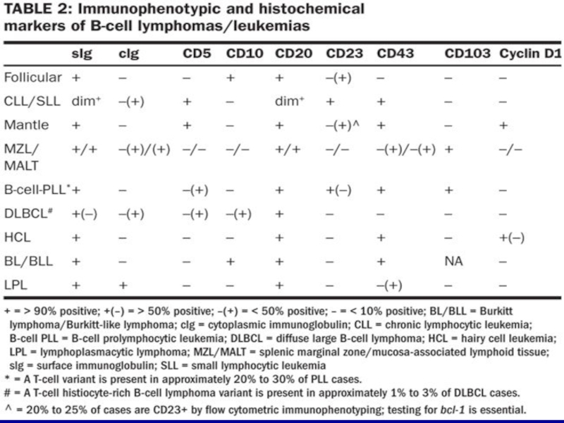
- 16. Typical B cell phenotype in CLL
- 17. Strategy for CLL therapy. First line of
- 18. Types of lymphomas.
- 19. Hodgkin Lymphoma et al. (WHO, 2008). Hodgkin
- 20. Histological diagnosis of HD. The Reed–Sternberg
- 21. The adverse prognostic factors for HD
- 22. Stages and Therapy of HD Therapy strategy:
- 23. Non-Hodgkin lymphoma Causes The many different forms
- 26. Cytogenetic analysis for B-cell malignancies t(11;14)
- 27. Diagnosis of DLBCL by MicroArray technique:
- 28. Burkitt’s lymphoma (rare type of NHL) (endemic= EBV positive)
- 29. Immunophenotypic diagnosis of Burkitt’s lymphoma The cells
- 30. T (8,14) in Burkitt’s lymphoma
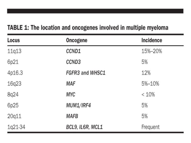
- 31. Path from Normal plasma cells through Monoclonal Gammopathy of Undetermined Significance to Multiple Myeloma.
- 32. Plasma cell malignancies
- 33. Morphology of malignant plasma cells in blood (H&E staining)
- 34. Immunophenotyping of Plasma Cells
- 36. Multiple Myeloma diagnosis and therapy. Diagnosis: Roentgen
- 37. Serum paraprotein detection
- 38. M-protein and diseases. More than 50% of
- 39. Waldenstrom macroglobulinemia: pathogenesis Immunophenotype of BM
- 40. Diagnosis and Therapy of WM.
- 41. Light chain Disease (Bence-Jones proteins). A Bence
- 42. (Bence-Jones protein in serum/urine (up) and serum (down))
- 43. HEAVY CHAIN DISEASE Heavy chain disease is a
- 44. Secondary immunodeficiency in lymphoproliferative diseases. 1. Lymphoadenopathy

 (2007).")


Слайд 10Acute leukemia.
Originated from bone marrow (>25% blasts).
Usually monoclonal disease.
Lineage committed
B and T or myeloid malignant cells are estimated by immunophenotyping (FAB classif. 1996 classif.)
Cytogenetic abnormalities (WHO classif. 2001,2008).
Fusion genes as markers of disease diagnosis and prognosis.
.Usually monoclonal disease. Lineage committed morphology (FAB classif.) B")
Слайд 11Acute leukemia
(WHO classification, 2008).
Mixed phenotype acute leukemia (T or B-
B lymphoblastic leukemia/lymphoma with t(9:22)(q34;q11.2); BCR-ABL1.
B lymphoblastic leukemia/lymphoma with t (v;11q23); MLL rearranged.
B lymphoblastic leukemia/lymphoma with t(12;21)(p13;q22) TEL-AML1 (ETV6-RUNX1)
B lymphoblastic leukemia/lymphoma with hyperdiploidy.
B lymphoblastic leukemia/lymphoma with hypodiploidy.
B lymphoblastic leukemia/lymphoma with t(5;14)(q31;q32); IL-3-IgH
B lymphoblastic leukemia/lymphoma with t (1;19)(q23;p13.3); TCF-PBX1
T lymphoblastic leukemia/lymphoma.
.Mixed phenotype acute leukemia (T or B- myeloid, NK-cell…)B lymphoblastic leukemia/lymphoma")

Слайд 13Chronic lymphocytic leukemia
(WHO classification, 2008).
Mature B-cell neoplasms
Chronic lymphocytic leukemia/small lymphocytic
B-cell prolymphocytic leukemia,
Splenic marginal zone lymphoma,
Hairy cell leukemia,
Lymphoplasmacytic lymphoma,
Waldenstrom macroglobulinemia,
Heavy chain diseases,
Plasma cell myeloma,
-MALT lymphoma,
Follicular lymphoma,
Diffuse large B-cell lymphoma,
Plasmablastic lymphoma,
Burkitt lymphoma.
.Mature B-cell neoplasmsChronic lymphocytic leukemia/small lymphocytic lymphoma,B-cell prolymphocytic leukemia,Splenic marginal")
Слайд 14Chronic lymphocytic leukemia
(WHO classification, 2008).
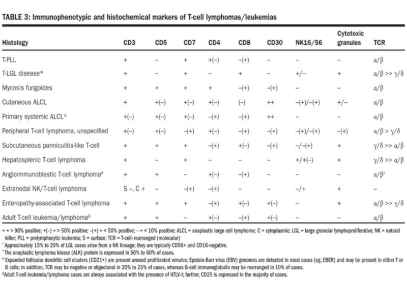
Mature T-cell and NK-cell neoplasms:
T-cell prolymphocytic
T-cell large granular lymphocytic leukemia,
Aggressive NK-cell leukemia,
Adult T-cell leukemia/lymphoma,
Mycosis fungoides,
Sezary syndrome,
Primary cutaneous CD30+ T cell lymphoproliferative disorders,
Peripheral T-cell lymphoma,
Anaplastic large cell lymphoma…
.Mature T-cell and NK-cell neoplasms:T-cell prolymphocytic leukemia,T-cell large granular lymphocytic")
Слайд 15Adverse prognostic factors of CLL
Diffuse infiltration of bone marrow by lymphocytes;
Advanced
Male gender;
Deletions in chr.17p (p53!) or 11q (ATM !) (5-10% of pts for each) ;
High serum level of beta-2 – microglobulin;
Increased fraction of prolymphocytes in PB;
>20% of ZAP-70-positive cells, >30% CD38+ cells;
No rearangement in IgH V region.
Favorable prognostic factors
No diffuse infiltration of bone marrow by lymphocytes;
Deletion in chr.13 q (50% of pts);
<20% of ZAP-70-positive cells, <30% CD38+ cells;
Mutations in IgH V region.


Слайд 17Strategy for CLL therapy.
First line of therapy: Fludarabine, Cyclophosphamine, Rituximabe
Chemotherapy, MABs such as alemtuzumab (directed against CD52) and ofatumumab (directed against CD20) are also used.
Stem cell transplantation – rare.
Survival:
Subclinical “disease” can be identified in 3,5% of normal adults and up to 7% of individuals over the age of 70.
Survival rate depends on subtypes (6-8 years to 22 years).
.Chemotherapy, MABs such as alemtuzumab (directed")

Слайд 19Hodgkin Lymphoma et al. (WHO, 2008).
Hodgkin lymphoma:
- classical Hodgkin lymphoma,
-
Histiocytic and dendritic cell neoplasms:
- histiocytic sarcoma,
- Langerhans cell histiocytic,
- Follicular dendritic cell sarcoma,…
Posttranplantation lymphoproliferative disorders:
-plasmacytic hyperplasia,
-Infectious mononucleous-like PTLD,
-polymorphic PTLD,
- monomorphic PTLD (B- and T/NK-cell types),…
.Hodgkin lymphoma: - classical Hodgkin lymphoma, - Lymphocyte-rich classical Hodgkin lymphoma,")
Слайд 20Histological diagnosis of HD.
The Reed–Sternberg cells are identified as large often

Слайд 21The adverse prognostic factors for HD
Age ≥ 45 years
Stage IV disease
Hemoglobin
Lymphocyte count < 600/µl or < 8%
Male gender
Albumin < 40 g/l
White blood count ≥ 15,000/µl

Слайд 22Stages and Therapy of HD
Therapy strategy: radiation therapy +/- chemotherapy.
Prognosis: The
Stage I is involvement of a single lymph node region (I) (mostly the cervical region) or single extralymphatic site (IIe);
Stage II is involvement of two or more lymph node regions on the same side of the diaphragm (II) or of one lymph node region and a contiguous extralymphatic site (IIe);
Stage III is involvement of lymph node regions on both sides of the diaphragm, which may include the spleen (IIIs) and/or limited contiguous extralymphatic organ or site (IIIe, IIIes);
Stage IV is disseminated involvement of one or more extralymphatic organs

Слайд 23Non-Hodgkin lymphoma
Causes
The many different forms of lymphoma likely have different causes.
Infectious agents like Epstein-Barr virus, Human T-cell leukemia virus, Helicobacter pylori, HHV-8 and HIV infection.
Chemicals, like diphenylhydantion, dioxin, and phenoxyherbicides.
Medical treatments like radiation therapy and chemotherapy. Genetic diseases, like Klinefelter ‘s syndrome, Chediak-Higashi syndrome, ataxia-telangiectasia syndrome
Autoimmune diseases, like Sjogren’s syndrome, celiac sprue, rheumatoid arthritis and systemic lupus erythematosis

Слайд 26Cytogenetic analysis for B-cell malignancies
t(11;14) is mainly found in mantle
 is mainly found in mantle cell lymphoma, but also")
Слайд 27Diagnosis of DLBCL by MicroArray technique: Germinal center B cell

 (endemic= EBV positive)")
Слайд 29Immunophenotypic diagnosis of Burkitt’s lymphoma
The cells of BL typically express monotypic
A remarkable feature of BL is the high growth fraction (> 95%) as demonstrated by Ki-67. The leukemic cells of BL express a mature immunophenotype that distinguishes it from precursor B-cell acute lymphoblastic leukemia (ALL).

 in Burkitt’s lymphoma")
Слайд 31Path from Normal plasma cells through Monoclonal Gammopathy of Undetermined Significance


")

Слайд 36Multiple Myeloma diagnosis and therapy.
Diagnosis: Roentgen + BM biopsy+..
Therapy: chemotherapy, BMT.
Survival:


Слайд 38M-protein and diseases.
More than 50% of patients with serum M protein

Слайд 39Waldenstrom macroglobulinemia: pathogenesis
Immunophenotype of
BM cells in WM
Ig
CD19 - Positive
CD20 - Positive
CD52 - Positive
Surface IgM - Positive
CD79b - Positive
CD11c - Usually negative
CD25 - Positive
CD23 - Usually negative
CD38 - Dim positive
FMC7 - Usually dim positive
CD22 - May be positive
CD5 - Negative
CD10 - Negative
CD27 - Dim positive
CD75 - Usually negative
CD138 - Usually negative
Bcl2 - Dim positive
Bcl6 - Usually absent
PAX5+ - Dim positive
CD45 (RA) - Usually positive


Слайд 41Light chain Disease
(Bence-Jones proteins).
A Bence Jones protein is a monoclonal globulion protein
.A Bence Jones protein is a monoclonal globulion protein or immunoglobulin light chain")
 and serum (down))")
Слайд 43HEAVY CHAIN DISEASE
Heavy chain disease is a form of paraproteinemia with a proliferation of
There are four forms:
alpha chain disease (Seligmann's disease)
gamma chain disease (Franklin's disease)
mu chain disease
delta chain disease

Слайд 44Secondary immunodeficiency in lymphoproliferative diseases.
1. Lymphoadenopathy (decreased lymphocyte proliferation to mitogens,
2. Autoimmunity (autoantibodies, amyloidosis, renal and liver failure, coagulopathy, vasculitis).
.2.")