- Главная
- Разное
- Дизайн
- Бизнес и предпринимательство
- Аналитика
- Образование
- Развлечения
- Красота и здоровье
- Финансы
- Государство
- Путешествия
- Спорт
- Недвижимость
- Армия
- Графика
- Культурология
- Еда и кулинария
- Лингвистика
- Английский язык
- Астрономия
- Алгебра
- Биология
- География
- Детские презентации
- Информатика
- История
- Литература
- Маркетинг
- Математика
- Медицина
- Менеджмент
- Музыка
- МХК
- Немецкий язык
- ОБЖ
- Обществознание
- Окружающий мир
- Педагогика
- Русский язык
- Технология
- Физика
- Философия
- Химия
- Шаблоны, картинки для презентаций
- Экология
- Экономика
- Юриспруденция
Laboratory tests in Rheumatology презентация
Содержание
- 1. Laboratory tests in Rheumatology
- 2. Introduction In rheumatic disease lab test contribute
- 3. Utility of Lab Tests Aims of lab
- 4. Diagnostic vs. Evaluative Tests Need to determine
- 5. Blood Panel - Hemoglobin Anemia of chronic
- 6. Blood panel - WBC White blood cells
- 7. Platelets Thrombocytosis can accompany active phases of
- 8. Examples What CBC abnormalities do you expext in this patient?
- 9. Biochemical testing- liver Synthetic activity
- 10. Kidney function tests Connective tissue diseases and
- 11. Uric acid Commonly included in the workup
- 12. Acute-phase reactants Are not specific for rheumatic
- 13. Acute phase reactants Produced by hepatocytes upon
- 14. Example What lab abnormalities do you expect in this patient?
- 15. Example What lab abnormalities do you expect in this patient?
- 16. Serologic testing Testing for autoantibodies is frequently
- 17. Rheumatoid Factor Autoantibodies directed against Fc– chains
- 18. Rheumatoid Factor Not specific for Rheumatoid Arthritis
- 19. Antibodies to citrullinated protein and peptide ACPA-
- 20. Anti-nuclear Antibodies (ANA) Immunoglobulins directed against structures
- 21. ANA ANAs do not correlate with disease
- 22. ANA Low titres (
- 23. ANA detection and measurement IIF - the
- 24. ANA patterns In the homogeneous staining pattern,
- 25. homogenous nucleolar speckled cenromere
- 26. ELISA method Solid phase assays - enzyme-linked
- 27. Advantages and Disadvantages The major advantage of
- 28. Advantages and Disadvantages The number of autoantigens
- 29. Anti-dsDNA antibodies Antibodies that target DNA
- 30. Anti-histone antibodies Found in 95% of patients
- 31. Anti-Sm and anti-RNP antibodies “extractable” (ENA) Produce
- 32. Anti-Sm and anti-RNP antibodies “extractable” (ENA) Anti-Sm
- 33. Anti-Ro (SS-A) and anti-La (SS-B) antibodies (ENAs)
- 34. Anti-Ro (SS-A) and anti-La (SS-B) antibodies Produce
- 35. Anticentomere and anti-SCL-70 Anticentromere antibodies (ACA) produce
- 36. Antineutrophil cytoplasmic antibodies - ANCA Subgroup of
- 37. ANCA c-ANCA is seen in 90% of
- 38. Complement The most frequent clinical parameters used
- 39. Antiphospholipid antibodies (APLA) Anti-cardiolipin antibodies (ELIZA) IgG
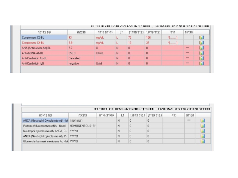
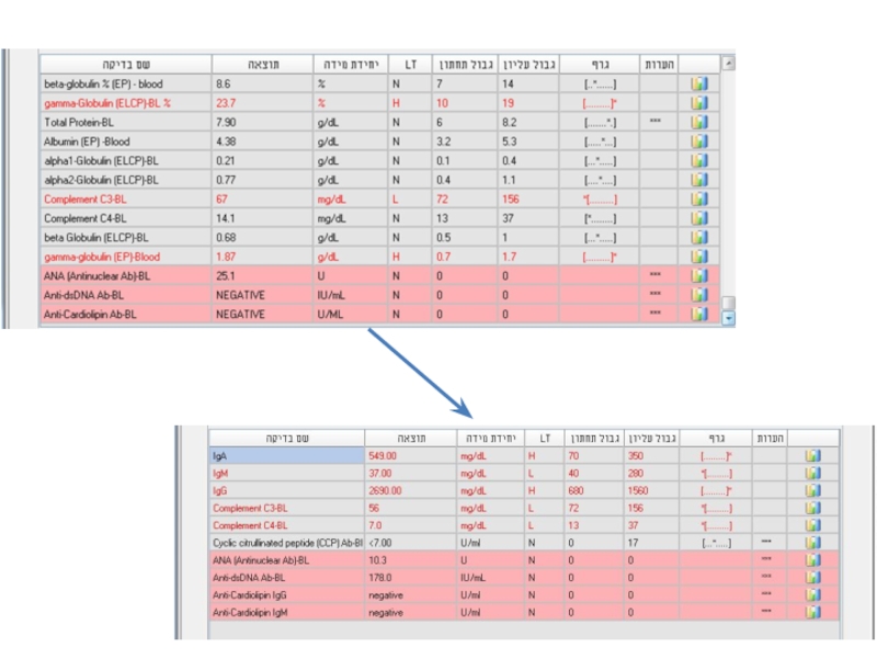
- 40. Examples 24y woman presents with weakness, nausea, ptechia and echymozes
- 41. Laboratory analyses
- 42. Laboratory analyses
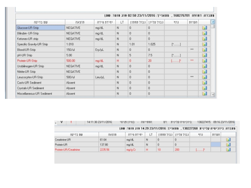
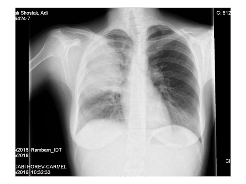
- 45. Examples 28y old woman presents with cough, fever, dyspnea, fatigue
- 46. Laboratory analyses
- 47. Laboratory analyses
- 49. Laboratory analyses
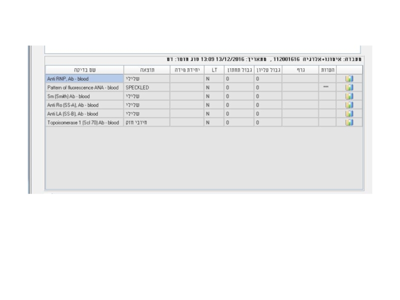
- 50. Example 42y old woman with SLE presents with fatigue, hair loss and new digital ulcer
- 51. Examples
- 53. Examples
- 54. Examples 42y old man presents with weakness,

Слайд 2Introduction
In rheumatic disease lab test contribute to diagnosis
Laboratory investigation should be
guided by clinical picture
Measurement of biomarkers can be useful to monitor treatment efficacy and safety
Stratification of patients to predict prognosis
Measurement of biomarkers can be useful to monitor treatment efficacy and safety
Stratification of patients to predict prognosis

Слайд 3Utility of Lab Tests
Aims of lab test:
1. Identification of pathological process
in the body & evaluation of its severity
2. Support or negation of specific diagnosis
3. Follow up of disease & complications
4. Detection of adverse reactions of drug therapy
Interpretation of lab tests should be done only in relation to certain clinical context.
Without the clinical picture most lab tests are useless.
2. Support or negation of specific diagnosis
3. Follow up of disease & complications
4. Detection of adverse reactions of drug therapy
Interpretation of lab tests should be done only in relation to certain clinical context.
Without the clinical picture most lab tests are useless.

Слайд 4Diagnostic vs. Evaluative Tests
Need to determine which test is appropriate
Diagnostic tests
accurately distinguish a group of patients with a specific disease from a non-disease group
Evaluative tests monitor disease activity over time
Evaluative tests monitor disease activity over time

Слайд 5Blood Panel - Hemoglobin
Anemia of chronic disease – usually normocytic and
normochromic, but sometimes hypochromic microcytic
Should be differentiated from iron deficiency
Macrocytic anemia – not common in rheumatology, except for methotrexate treatment
Hemolytic anemia – LDH, Bilirubin, haptoglobin
Due to Gastrointestinal bleeding (acute or chronic)
Should be differentiated from iron deficiency
Macrocytic anemia – not common in rheumatology, except for methotrexate treatment
Hemolytic anemia – LDH, Bilirubin, haptoglobin
Due to Gastrointestinal bleeding (acute or chronic)

Слайд 6Blood panel - WBC
White blood cells – neutrophils, lymphocytes, eosinophils:
Neutrophils are
acute phase reactants
Neutropenia – in patients undergoing immunosupressive treatment
Neutropenia can be associated with splenomegaly
Lymphopenia – active phases of SLE
Eosinophilia – Churg-Strauss (EGPA)
Neutropenia – in patients undergoing immunosupressive treatment
Neutropenia can be associated with splenomegaly
Lymphopenia – active phases of SLE
Eosinophilia – Churg-Strauss (EGPA)

Слайд 7Platelets
Thrombocytosis can accompany active phases of autoimmune diseases – RA (APR)
Thrombocytopenia
–
can be related to the presence of antithrombocyte antibodies, as in SLE
Drug induced toxicity
can be related to the presence of antithrombocyte antibodies, as in SLE
Drug induced toxicity
Thrombocytopenia – can be related")

Слайд 9 Biochemical testing- liver
Synthetic activity (albumin, coagulation factors, Glucose, Bil)
Liver
enzymes – hepatocellular, cholestatic
Should be ordered before and after initiation of treatment (NSAIDS, DMARDS, including MTX, biological)
Should be ordered before and after initiation of treatment (NSAIDS, DMARDS, including MTX, biological)
Liver enzymes – hepatocellular, cholestaticShould")
Слайд 10Kidney function tests
Connective tissue diseases and systemic vasculitides are frequently associated
with kidney involvement – vascular/glomerular/tubular-interstitial
Creatinine/ Creatinine Clearance provide sufficient information
Urinalysis – always part of investigation (hematuria, leukocyturia, proteins)
Monitor for adverse effects of treatment
Creatinine/ Creatinine Clearance provide sufficient information
Urinalysis – always part of investigation (hematuria, leukocyturia, proteins)
Monitor for adverse effects of treatment

Слайд 11Uric acid
Commonly included in the workup of patients with arthritis
Elevated in
90% of patients with Gout
Healthy population can also have increased levels of uric acid
Important to monitor urate lowering therapy – goal of <5-6 mg/dL
Healthy population can also have increased levels of uric acid
Important to monitor urate lowering therapy – goal of <5-6 mg/dL

Слайд 12Acute-phase reactants
Are not specific for rheumatic disorders
AP response occurs in
a variety of inflammatory conditions – infection, trauma, malignancy.
The most widely used APR
ESR – erythrocyte sedimentation rate
CRP
Ferritin
The most widely used APR
ESR – erythrocyte sedimentation rate
CRP
Ferritin

Слайд 13Acute phase reactants
Produced by hepatocytes upon stimulation by cytokines (IL-1, IL
-6, TNF – tumor necrosis factor)
Examples – CRP, fibrinogen, ferritin, haptoglobin, ceruloplasmin, amyloid protein A, complement (C3, immunoglobilins
ESR and CRP are useful for monitoring the level of inflammation, however sometimes are not sensitive enough and sometimes are “slow” and should not guide the clinical decisions
Examples – CRP, fibrinogen, ferritin, haptoglobin, ceruloplasmin, amyloid protein A, complement (C3, immunoglobilins
ESR and CRP are useful for monitoring the level of inflammation, however sometimes are not sensitive enough and sometimes are “slow” and should not guide the clinical decisions


Слайд 16Serologic testing
Testing for autoantibodies is frequently used in the diagnoses of
rheumatic conditions and sometimes for monitoring of disease activity.
An adjunct to diagnosis and management rather than precise clinical guide.
An adjunct to diagnosis and management rather than precise clinical guide.

Слайд 17Rheumatoid Factor
Autoantibodies directed against Fc– chains of IgG molecules
Laboratories test only
for IgM RF
The main immunoglobilin classes of RF that can be easily detected are IgA and IgM RF
IgM RF is produced in many chronic inflammatory conditions – endocarditis, hepatitis B/C, tuberculosis, Idiopathic pulmonary fibrosis, mixed connective tissue disease, SLE, cryoglobulinemia
Can be present in 5% of normal elderly population

Слайд 18Rheumatoid Factor
Not specific for Rheumatoid Arthritis
The main indication for RF testing
– suspicion for RA and Sjogren syndrome
The specificity of RF increases with higher titers
Higher titers are associated with more aggressive and erosive disease
The specificity of RF increases with higher titers
Higher titers are associated with more aggressive and erosive disease

Слайд 19Antibodies to citrullinated protein and peptide ACPA- antigens
Citrullination of proteins (arginine
– citrullin) occurs as the result of synovial inflammation and inflammation induced apoptosis
RA patients react to such modified proteins by creating ACPAs
ACPAs are especially prevalent in RF patients but can be found in 25% of RF negative patients
ACPAs predict later development of erosive RA
Helpful in discriminating between RA and psoriasis with erosive arthritis
RA patients react to such modified proteins by creating ACPAs
ACPAs are especially prevalent in RF patients but can be found in 25% of RF negative patients
ACPAs predict later development of erosive RA
Helpful in discriminating between RA and psoriasis with erosive arthritis
 occurs as")
Слайд 20Anti-nuclear Antibodies (ANA)
Immunoglobulins directed against structures within the cell ( i.e.
DNA, ribonuclear proteins, histones, and centromere)
Screening tool
Titer/ pattern
Found in a variety of autoimmune diseases such as SLE, MCTD, JRA, scleroderma, Sjogren’s syndrome in high titres (>1:320)
Almost always present in SLE (95-98%)
High titer increases the likelihood that the presence of ANA is related to autoimmune disease
Screening tool
Titer/ pattern
Found in a variety of autoimmune diseases such as SLE, MCTD, JRA, scleroderma, Sjogren’s syndrome in high titres (>1:320)
Almost always present in SLE (95-98%)
High titer increases the likelihood that the presence of ANA is related to autoimmune disease
Immunoglobulins directed against structures within the cell ( i.e. DNA, ribonuclear proteins, histones,")
Слайд 21ANA
ANAs do not correlate with disease activity
Consider using as a screening
test in only symptomatic patients (arthritis, rash, serositis, proteinuria)
Must measure ANAs in patients with JIA (esp. oligoarticular) to assess risk of uveitis
Must measure ANAs in patients with JIA (esp. oligoarticular) to assess risk of uveitis

Слайд 22ANA
Low titres (
bacterial endocarditis, HIV)
Drugs (hydralazine, INH, dilantin, tegretol, ETX, PCN, and sulfas)
Neoplasias (lymphoma)
It is sensitive but not specific
~ 10% of the population have a positive low titer ANA and can be asymptomatic
As one ages, ANA titers increase
Drugs (hydralazine, INH, dilantin, tegretol, ETX, PCN, and sulfas)
Neoplasias (lymphoma)
It is sensitive but not specific
~ 10% of the population have a positive low titer ANA and can be asymptomatic
As one ages, ANA titers increase

Слайд 23ANA detection and measurement
IIF - the indirect immunofluorescence test is the
most widely used assay for the detection of ANA and remains the reference method of choice for the detection of these antibodies
Nuclear staining patterns include: homogeneous, speckled, centromere, and nucleolar
Nuclear staining patterns include: homogeneous, speckled, centromere, and nucleolar

Слайд 24ANA patterns
In the homogeneous staining pattern, the entire nucleus is diffusely
stained. EX: Antibodies to histone proteins, DNA, and DNA-histone complexes.
In the speckled staining pattern, fine or coarse speckles are seen throughout the nucleus. Ex: Antibodies against U1 RNP, Sm, and La antigens.
The centromere pattern - anti centromere
The nucleolar pattern refers to homogeneous or speckled staining of the nucleolus; Ex: fibrillarin, RNA polymerase I and III, Th, PM-Scl, and RNA helicase.


Слайд 26ELISA method
Solid phase assays - enzyme-linked immunoabsorbant assays (ELISA)
A panel of
purified native or recombinant autoantigens is prepared and each antigen is immobilized on a solid surface
The panel of antigens used in solid phase assays may include all or some of the following: Ro, La, Sm, U1 RNP, Scl-70, PM-Scl, Jo-1, centromere, histone, ribosomal P, and DNA.
Diluted human serum is incubated with the immobilized antigen and, as with the indirect immunofluorescence assay, a secondary antibody is used to detect bound autoantibodies.
A panel of purified native or recombinant")
Слайд 27Advantages and Disadvantages
The major advantage of indirect immunofluorescence is the large
number of autoantibodies that can be detected.
Some autoantigens may not be present in the HEp-2 cell substrate
The Ro60 antigen, (SLE, Sjogren’s)
Anti-ribosomal P antibodies (SLE)
Some autoantigens may not be present in the HEp-2 cell substrate
The Ro60 antigen, (SLE, Sjogren’s)
Anti-ribosomal P antibodies (SLE)

Слайд 28Advantages and Disadvantages
The number of autoantigens that are included in solid
phase (ELIZA) assays is limited compared with the number that are present in the HEp-2 cell substrate. As an example, most solid phase assays do not contain antigens found in the nucleolus; patients with autoantibodies directed against these structures will have a falsely negative solid phase ANA result
 assays is")
Слайд 29Anti-dsDNA antibodies
Antibodies that target DNA
Produce homogenous pattern in ANA IIF
Positive
result for anti-dsDNA screening should be confirmed by additional assays
Anti-dsDNA antibody testing is very specific (95%), but less sensitive (70%) for SLE
Are associated with disease activity in lupus nephritis
Anti-dsDNA antibody testing is very specific (95%), but less sensitive (70%) for SLE
Are associated with disease activity in lupus nephritis

Слайд 30Anti-histone antibodies
Found in 95% of patients with drug-induced lupus syndrome
Seen with:
Procainamide
Quinidine
Hydralazine
Phenytoin
or other anti-epileptics

Слайд 31Anti-Sm and anti-RNP antibodies
“extractable” (ENA)
Produce coarse speckled pattern in ANA IIF
The
nucleoli are spared
Anti-Sm antibodies are almost exclusive for SLE patients, not sensitive (10-40%)
RNP antibodies are part of diagnosis of Mixed Connective Tissue Disease (a syndrome of arthritis, myositis, Raynauds’ and sclerodactly)
RNP antibodies are not specific for MCTD
Anti-Sm antibodies are almost exclusive for SLE patients, not sensitive (10-40%)
RNP antibodies are part of diagnosis of Mixed Connective Tissue Disease (a syndrome of arthritis, myositis, Raynauds’ and sclerodactly)
RNP antibodies are not specific for MCTD
Produce coarse speckled pattern in ANA IIFThe nucleoli are sparedAnti-Sm")
Слайд 32Anti-Sm and anti-RNP antibodies
“extractable” (ENA)
Anti-Sm antibodies generally remain positive, even when
a patient has entered remission. The titer of anti-dsDNA antibodies may fall into the normal range when a patient’s disease is quiescent
Anti-U1 RNP antibodies may be found in 3 to 69 percent of patients with SLE. High levels of anti-U1 RNP antibodies are always present in patients with mixed connective tissue disease (MCTD)
Anti-U1 RNP antibodies may be found in 3 to 69 percent of patients with SLE. High levels of anti-U1 RNP antibodies are always present in patients with mixed connective tissue disease (MCTD)
Anti-Sm antibodies generally remain positive, even when a patient has")
Слайд 33Anti-Ro (SS-A) and anti-La (SS-B) antibodies
(ENAs)
Two sets of names assigned by
two different groups; first seen in Sjogren’s patients and then seen in SLE patients
Anti Ro/SS-A antibodies seen in:
5-15% of normals
50% of Sjogren’s patients
30% of SLE patients (many have negative ANA or subacute cutaneous lupus)
Correlates with active nephritis and cytopenias
Anti Ro/SS-A antibodies seen in:
5-15% of normals
50% of Sjogren’s patients
30% of SLE patients (many have negative ANA or subacute cutaneous lupus)
Correlates with active nephritis and cytopenias
 and anti-La (SS-B) antibodies (ENAs)Two sets of names assigned by two different groups;")
Слайд 34Anti-Ro (SS-A) and anti-La (SS-B) antibodies
Produce fine speckled pattern in ANA
IIF with staining of the nucleoli as well
Are part of the classification criteria for Sjogren syndrome, but are also frequent in SLE patients
Are associated with cutaneous lupus and photosensitivity
Associated with neonatal lupus and congenital heart block
Are part of the classification criteria for Sjogren syndrome, but are also frequent in SLE patients
Are associated with cutaneous lupus and photosensitivity
Associated with neonatal lupus and congenital heart block
 and anti-La (SS-B) antibodiesProduce fine speckled pattern in ANA IIF with staining of")
Слайд 35Anticentomere and anti-SCL-70
Anticentromere antibodies (ACA) produce a typical pattern in ANA
IIF by staining the centromere region of the chromosomes – this pattern is pathognomonic
The presence of anti-Scl-70 antibodies should be confirmed using ELIZA
These two antibodies are associated with distinct clinical pictures and are mutually exclusive
Anti-Scl-70 antibodies (also known as anti-topoisomerase I) are associated with increased risk of pulmonary fibrosis in both limited and diffuse cutaneous systemic sclerosis
 produce a typical pattern in ANA IIF by staining the")
Слайд 36Antineutrophil cytoplasmic antibodies - ANCA
Subgroup of neutrophil specific antibodies
Commonly directed to
myeloperoxidase (MPO) - and proteinase 3 (PR3)
P-ANCA – perinuclear staining (MPO)
C-ANCA – cytoplasmic staining (PR3)
Positive result on IIF should be confirmed using ELIZA
ANCA is useful in diagnosis of ANCA – associated vasculitides (GPA, EGPA, microscopic polyangiitis)
C-ANCA – GPA
P-ANCA – EGPA, microscopic polyangiitis
P-ANCA – perinuclear staining (MPO)
C-ANCA – cytoplasmic staining (PR3)
Positive result on IIF should be confirmed using ELIZA
ANCA is useful in diagnosis of ANCA – associated vasculitides (GPA, EGPA, microscopic polyangiitis)
C-ANCA – GPA
P-ANCA – EGPA, microscopic polyangiitis
 - and")
Слайд 37ANCA
c-ANCA is seen in 90% of GPA (Wegener’s granulomatosis)
p-ANCA is associated
with microscopic polyangiitis, EGPA (Churg-Strauss), and Ulcerative Colitis
Consider vasculitis (ANCA) if patient has:
Fever of unknown origin
Palpable purpura, vasculitis urticaria, or dermal necrosis
Mononeuritis multiplex
Unexplained arthritis, myositis, or serositis
Unexplained pulmonary, CV or renal disease
Consider vasculitis (ANCA) if patient has:
Fever of unknown origin
Palpable purpura, vasculitis urticaria, or dermal necrosis
Mononeuritis multiplex
Unexplained arthritis, myositis, or serositis
Unexplained pulmonary, CV or renal disease
p-ANCA is associated with microscopic polyangiitis, EGPA")
Слайд 38Complement
The most frequent clinical parameters used for judging complement activation –
C3, C4
C3, C4 are APR
C3, C4 consumption is associated with immune complexes diseases
In SLE low levels reflect disease activity and in lupus nephritis normalization is associated with better outcomes
C3, C4 are APR
C3, C4 consumption is associated with immune complexes diseases
In SLE low levels reflect disease activity and in lupus nephritis normalization is associated with better outcomes

Слайд 39Antiphospholipid antibodies (APLA)
Anti-cardiolipin antibodies (ELIZA)
IgG – better related to procoagulant activity
compared to IgM/IgA
β2glycoprotein-1 IgG and IgM
Lac - functional assay for the lupus anticoagulant (LA) phenomenon (prolonged aPTT/dRVVT) not corrected by control plasma but shortened by adding excess phospholipid
β2glycoprotein-1 IgG and IgM
Lac - functional assay for the lupus anticoagulant (LA) phenomenon (prolonged aPTT/dRVVT) not corrected by control plasma but shortened by adding excess phospholipid
Anti-cardiolipin antibodies (ELIZA)IgG – better related to procoagulant activity compared to IgM/IgAβ2glycoprotein-1 IgG")


Слайд 54Examples
42y old man presents with weakness, loss of weight, puffy painfull
hands, “hardening” of skin and hyperpigmentation