- Главная
- Разное
- Дизайн
- Бизнес и предпринимательство
- Аналитика
- Образование
- Развлечения
- Красота и здоровье
- Финансы
- Государство
- Путешествия
- Спорт
- Недвижимость
- Армия
- Графика
- Культурология
- Еда и кулинария
- Лингвистика
- Английский язык
- Астрономия
- Алгебра
- Биология
- География
- Детские презентации
- Информатика
- История
- Литература
- Маркетинг
- Математика
- Медицина
- Менеджмент
- Музыка
- МХК
- Немецкий язык
- ОБЖ
- Обществознание
- Окружающий мир
- Педагогика
- Русский язык
- Технология
- Физика
- Философия
- Химия
- Шаблоны, картинки для презентаций
- Экология
- Экономика
- Юриспруденция
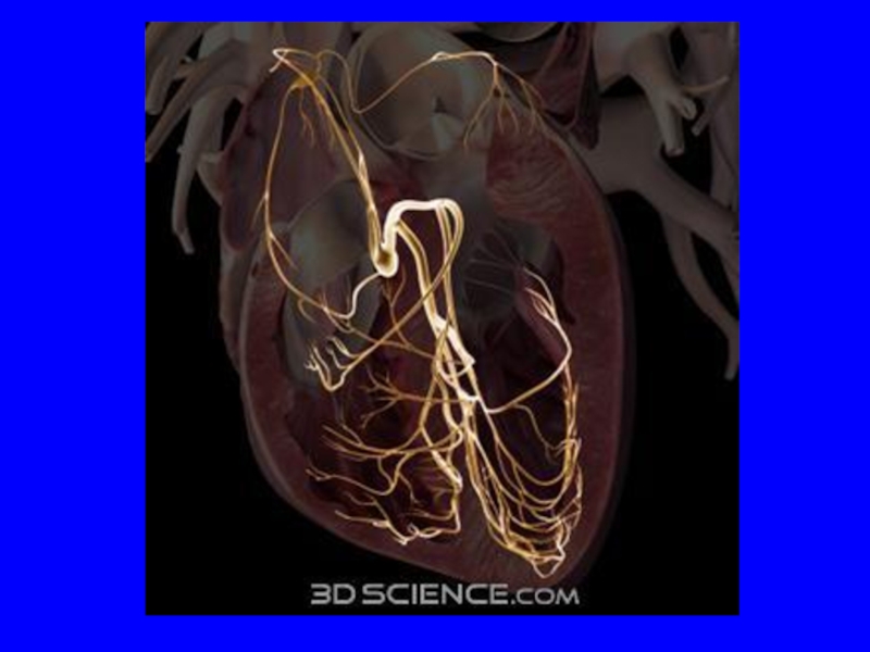
Кровоснабжение проводящей системы сердца презентация
Содержание

Слайд 2
A. Right & Left Coronary Arteries
B. Branches of Right Coronary
Artery
1. Main
2. Posterior Descending Branch
3. Sinus Node branch
C. Branches of Left Coronary Artery
1. Main
2. Left Anterior Descending (LAD)
3. Circumflex
D. Blood Supply of Cardiac Conduction System
1. Sinus Node Artery Supplies Blood to Sinus Node
a. Origin of Sinus Node Artery
2. AV Nodal Artery Supplies Blood to the AV node
a. Origin of AV Nodal Artery
3. LAD supplies the right and left bundle branches
1. Main
2. Posterior Descending Branch
3. Sinus Node branch
C. Branches of Left Coronary Artery
1. Main
2. Left Anterior Descending (LAD)
3. Circumflex
D. Blood Supply of Cardiac Conduction System
1. Sinus Node Artery Supplies Blood to Sinus Node
a. Origin of Sinus Node Artery
2. AV Nodal Artery Supplies Blood to the AV node
a. Origin of AV Nodal Artery
3. LAD supplies the right and left bundle branches

Слайд 3
Diagram showing normal aortic origin and initial distribution of four major
coronary arteries: left anterior descending (LAD), left circumflex (LC), left main (LM), and right (R). AV= aortic valve; NC= noncoronary; P=posterior.


Слайд 5
Diagram illustrating the principal arteries and veins on (A) the anterior
surface of the heart and (B) the posterior and inferior surfaces of the heart. Part of the right atrial appendage has been resected to show the proximal right coronary artery. IN B the heart is shown more vertically oriented to expose the inferior surface.
 the anterior surface of the heart")
Слайд 6
Diagram showing myocardial perfusion patterns of major epicardial coronary arteries as
viewed from three tomographic cuts: four-chamber view, cross-sectional view, and parasternal view, and parasternal long-axis. A= anterior; ALPM= anterolateral papillary muscle; LC= left circumflex; LAD= left anterior descending; LM= left main; LV= left ventricle; LVFW= left ventricular free wall; P= posterior; PMPM= posteromedial papillary muscle; RV= right ventricle; RVFW= right ventricular free wall; RVOFT= right ventricular outflow tract.

Слайд 7
Diagram showing arterial blood supply of the cardiac conduction system. The
nodal artery (NA) arises from the posterior descending artery (PD). The bulk of the arterial blood supply to the right and left bundle branches comes from the left anterior descending artery (LAD).
 arises")

Слайд 10
Schematic diagram of blood supply to cardiac conduction system. The first
septal branch of the left anterior descending (LAD) coronary artery supplies a critical portion of the interventricular conduction system (red oval). AV, atrioventricular; marginal a., marginal artery; PDA, posterior descending artery; RCA, right coronary artery; SA, sinoatrial. Reproduced, with permission, from Harthorne JW, Pohost. GM, Electrical therapy of cardiac dysrhythmias. In Levine, HJ (ed) Clinical Cardiovascular Physiology, New York, Grune and Stratton: 1976. pp. 853–882