Inflammatory reaction of an organism презентация

Лейкоцитарная фаза воспаления (в коже) Leucocytic phase of inflammation (in skin) Гистамин histamine Разрушение бактерий и тканевых структур Destruction of bacteria and damaged tissue Oedema zona Leucocytic infiltration

Слайд 1Inflammatory reaction of an organism is carried out by a loose

collagen tissue together with leukocytes of blood

At damage of a tissue labrocytes react the first and throw out the granules in tissue,
histamin increases permeability of capillaries for blood plasma and leukocytes.
Plasma collects in a tissue and creates an inflammatory oedema which separates
the inflammatory focus from healthy tissues and does not allow to extend to products
of disintegration on an organism.

Neutrophils come the first to the center of damage. They throw out in a tissue much
oxygen superoxide, lysosomal enzyms and contents of specific granules, and then
perish. Oxygen superoxide and lysocim destroy bacteria, cation-proteins increase an
oedema, and lysosomal enzymes even more destroy the damaged tissue. This phase
of an inflammation refers to leucocytic (or the sharp period).

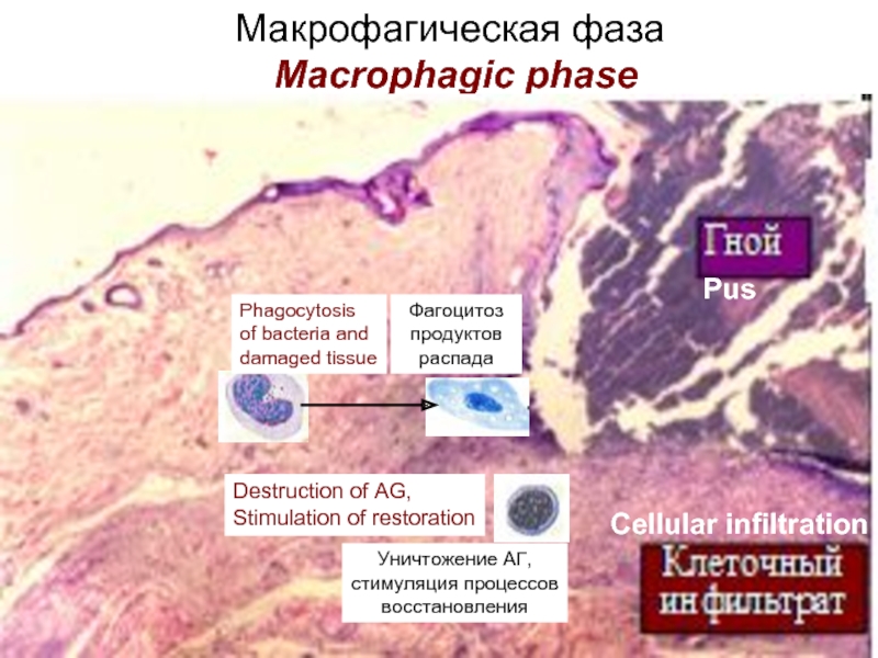
Then the macrophagic phase develops. Macrophages come to the center of damage
and phagocyte bacteria and products of disintegration of tissue, completely clearing
from them the inflammatory focus.

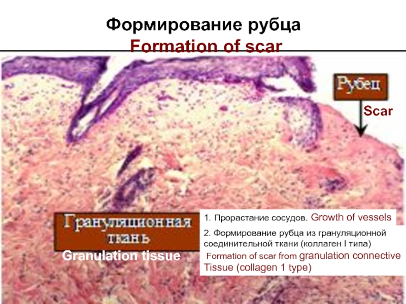
The fibroblastic (reparative) phase begins. Fibroblasts are actively made multiple
copies, form around of the cleared site of damage a fibroblastic capsule and secrete of
fundamantal substance and collagen of 1 type of which at once form thick fibres, filling
with them defect of a tissue. Vessels grow into a capsule, the granulation tissue is
formed of thick, rough collagenic fibres and vessels. Within the next 6 months rough
scar is gradually reconstructed, thick fibres from collagen of 1 type are replaced with
more thin fibres from collagen 3 types
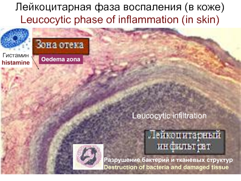
Слайд 2Лейкоцитарная фаза воспаления (в коже) Leucocytic phase of inflammation (in skin)

Гистамин
histamine
Разрушение бактерий

и тканевых структур
Destruction of bacteria and damaged tissue

Oedema zona

Leucocytic infiltration


Слайд 3Макрофагическая фаза Macrophagic phase
Фагоцитоз
продуктов
распада
Уничтожение АГ,
стимуляция процессов
восстановления
Phagocytosis
of bacteria and


damaged tissue

Pus

Cellular infiltration

Destruction of AG,
Stimulation of restoration


Слайд 4Фибробластическая (репаративная) фаза Fibroblastic (reparative) phase

Образование капсулы из фибробластов.

Formation of capsule from fibroblasts

2. Секреция основного вещества и волокон.
Secretion of fundamantal substance and fibers

Surface of wound

Fibroblastic capsule


Слайд 5Формирование рубца Formation of scar
1. Прорастание сосудов. Growth of vessels
2.

Формирование рубца из грануляционной
соединительной ткани (коллаген І типа)
Formation of scar from granulation connective
Tissue (collagen 1 type)

Scar

Granulation tissue


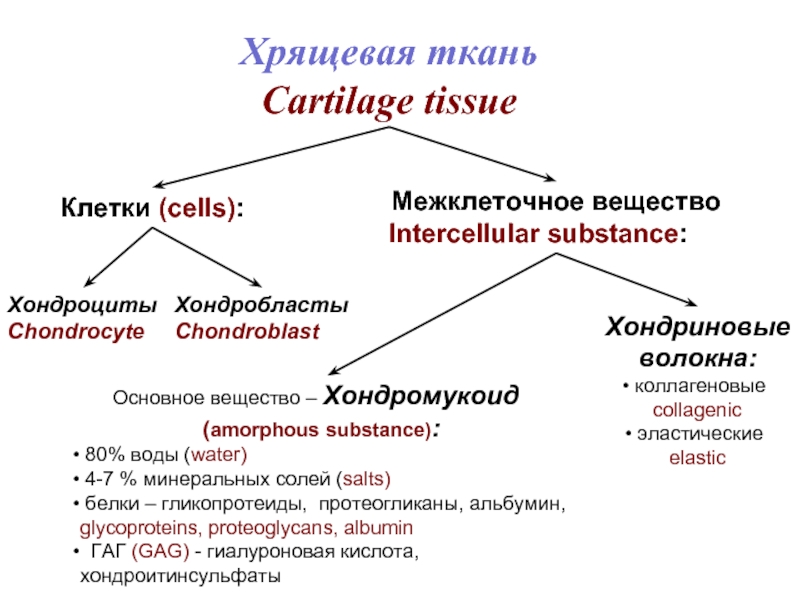
Слайд 6Хрящевая ткань
Cartilage tissue
Клетки (cells):
Межклеточное вещество
Intercellular substance:
Хондробласты
Chondroblast
Хондроциты
Chondrocyte
Основное вещество –

Хондромукоид
(amorphous substance):
80% воды (water)
4-7 % минеральных солей (salts)
белки – гликопротеиды, протеогликаны, альбумин,
glycoproteins, proteoglycans, albumin
ГАГ (GAG) - гиалуроновая кислота, хондроитинсульфаты

Хондриновые
волокна:
коллагеновые
collagenic
эластические
elastic


Слайд 7

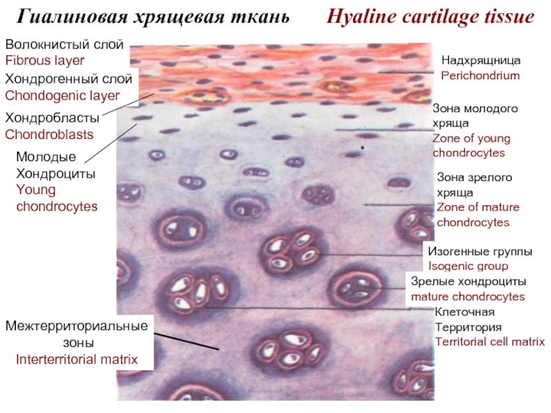
Гиалиновая хрящевая ткань Hyaline cartilage tissue
Надхрящница
Perichondrium
Зона молодого
хряща
Zone of

young
chondrocytes

Зона зрелого
хряща
Zone of mature
chondrocytes

Изогенные группы
Isogenic group

Зрелые хондроциты
mature chondrocytes

Клеточная
Территория
Territorial cell matrix

Межтерриториальные
зоны
Interterritorial matrix

Волокнистый слой
Fibrous layer

Хондрогенный слой
Chondogenic layer

Хондробласты
Chondroblasts

Молодые
Хондроциты
Young
chondrocytes


Слайд 8
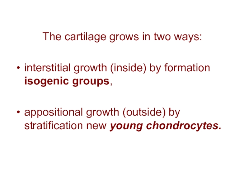
The cartilage

grows in two ways:

interstitial growth (inside) by formation isogenic groups,

appositional growth (outside) by stratification new young chondrocytes.

Слайд 9

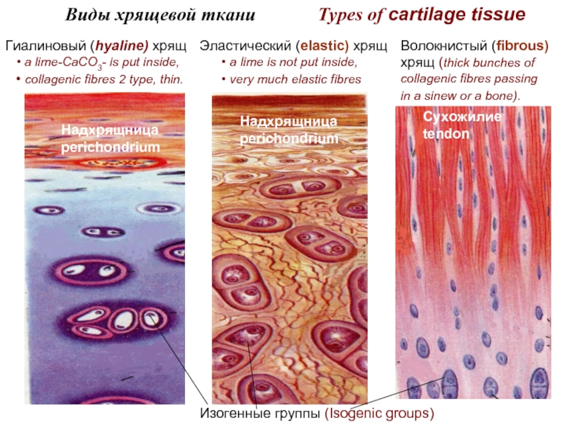
Виды хрящевой ткани

Types of cartilage tissue

Гиалиновый (hyaline) хрящ

Эластический (elastic) хрящ

Волокнистый (fibrous)
хрящ (thick bunches of
collagenic fibres passing
in a sinew or a bone).

Надхрящница
perichondrium

Надхрящница
perichondrium

Сухожилие
tendon

Изогенные группы (Isogenic groups)

a lime-CaCO3- is put inside,
collagenic fibres 2 type, thin.

a lime is not put inside,
very much elastic fibres


Обратная связь

Если не удалось найти и скачать презентацию, Вы можете заказать его на нашем сайте. Мы постараемся найти нужный Вам материал и отправим по электронной почте. Не стесняйтесь обращаться к нам, если у вас возникли вопросы или пожелания:

Email: Нажмите что бы посмотреть 

Что такое ThePresentation.ru?

Это сайт презентаций, докладов, проектов, шаблонов в формате PowerPoint. Мы помогаем школьникам, студентам, учителям, преподавателям хранить и обмениваться учебными материалами с другими пользователями.


Для правообладателей

Яндекс.Метрика